

Robotic-assisted excision of left para-aortic paraganglioma: a novel approach
- PMID: 39839207
- PMCID: PMC11750046
- DOI: 10.1093/jscr/rjae842
Robotic-assisted excision of left para-aortic paraganglioma: a novel approach
Abstract
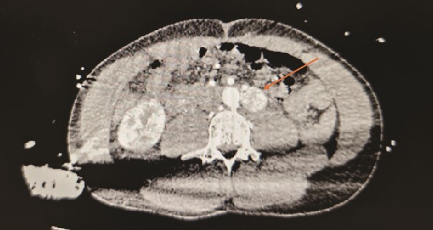
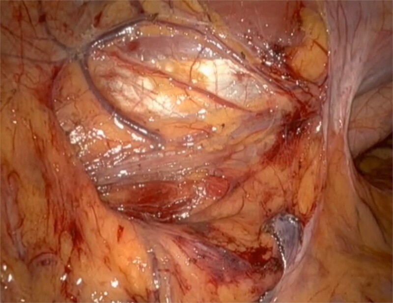
Paragangliomas, a type of extra-adrenal tumour, albeit rare, are dangerous due to their high metastatic potential and risk of hypertensive crisis from massive catecholamine release. It typically presents with sympathetic overdrive symptoms such as diaphoresis, headache, and palpitation, accompanied by substantially high plasma metanephrines level and mass on contrasted computed tomography abdomen and pelvis, whilst some are found incidentally. In this report, we discuss a case of an extra-adrenal lesion located near susceptible major structures with extensive vascularisation, in a patient with near-death experience. Complete excision of the pulsatile mass with minimal bleeding and no complications, was made possible utilizing the da Vinci Robotic System. Robotic surgery, being a part of a multidisciplinary approach, leads to better patient outcomes and shorter hospitalisations. Moreover, it offers enhanced dexterity and improved depth perception compared to conventional methods. However, further studies are needed to validate its application in standard practice.
Keywords: daVinci; extra-adrenal; paraganglioma; pheochromocytoma; retroperitoneal; robotic.
Published by Oxford University Press and JSCR Publishing Ltd. © The Author(s) 2025.
Conflict of interest statement
None declared.
Figures


References
-
- Handa A, Dash SC, Solanki N, et al. . Robotic excision of interaortocaval paraganglioma: a case report with the literature review. African J Urol 2021;27:162. 10.1186/s12301-021-00264-5. - DOI
-
- Lenders JW, Kerstens MN, Amar L, et al. . Genetics, diagnosis, management and future directions of research of phaeochromocytoma and paraganglioma: a position statement and consensus of the working group on endocrine hypertension of the European Society of Hypertension. J Hypertens 2020;38:1443–56. 10.1097/HJH.0000000000002438. - DOI - PMC - PubMed
Publication types
LinkOut - more resources
Full Text Sources
