Spontaneous bilateral thyroid follicular cell carcinoma (subtype: compact cellular carcinoma) with C-cell complexes in a male beagle
- PMID: 39839717
- PMCID: PMC11745505
- DOI: 10.1293/tox.2024-0072
Spontaneous bilateral thyroid follicular cell carcinoma (subtype: compact cellular carcinoma) with C-cell complexes in a male beagle
Abstract
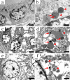
We report the features of spontaneous bilateral thyroid follicular cell carcinoma in a 10-year-old male beagle. Necropsy revealed bilateral masses on the trachea, corresponding to the left and right sides of the thyroid gland. The masses were elastic, encapsulated, and distinct, with no connecting tumor tissues between them. Histologically, the tumor cells exhibited a predominant sheet-like growth pattern in both masses, and small follicular structures containing colloids were observed. Immunohistochemically, >50% of the tumor cells were positive for thyroglobulin. In the sheet-like growth area, all tumor cells were positive for cytokeratin and approximately 50% of them were positive for vimentin. The tumor cells were negative for calcitonin and parathormone. Electron microscopy of the tumor cells revealed colloid droplets and lysosomes in the cytoplasm, which are characteristics of follicular cells of the thyroid gland, although they were abnormally shaped and smaller in size compared to the normal cells. Many calcitonin-positive C cells were observed in the nodule area without a capsule in the left mass and were scattered within the tumor in the right mass. C cells were found individually and were negative for Ki-67 expression. Therefore, each of these cells was deemed to be derived from an individual C-cell complex. Based on these morphological features, the tumor was diagnosed as spontaneous bilateral thyroid follicular cell carcinoma of the compact cellular carcinoma subtype. This is the first report of electron microscopic findings and co-expression of cytokeratin and vimentin in thyroid follicular cell carcinoma in beagles.
Keywords: C-cell complex; beagles; compact cellular carcinoma; follicular cell carcinoma; thyroid glands.
©2025 The Japanese Society of Toxicologic Pathology.
Conflict of interest statement
The authors declare no Conflicts of Interest
Figures


Similar articles
-
Co-expression of vimentin and 19S-thyroglobulin in follicular cells located in the C-cell complex of dog thyroid gland.J Histochem Cytochem. 1995 Nov;43(11):1097-106. doi: 10.1177/43.11.7560892. J Histochem Cytochem. 1995. PMID: 7560892
-
Thyroglobulin and calcitonin immunoreactivity in canine thyroid carcinomas.Vet Pathol. 1984 Mar;21(2):168-73. doi: 10.1177/030098588402100206. Vet Pathol. 1984. PMID: 6375099
-
Thyroid-like follicular carcinoma of the kidney: A report of two cases and literature review.Oncol Lett. 2014 Jun;7(6):1796-1802. doi: 10.3892/ol.2014.2027. Epub 2014 Apr 3. Oncol Lett. 2014. PMID: 24932236 Free PMC article.
-
Spindle cell variant of follicular thyroid carcinoma: An extremely unusual case and review of the literature.Diagn Cytopathol. 2022 Nov;50(11):E333-E338. doi: 10.1002/dc.25018. Epub 2022 Jul 22. Diagn Cytopathol. 2022. PMID: 35866458 Review.
-
[Clinicopathologic characteristics of thyroid-like follicular carcinoma of the kidney: an analysis of five cases and review of literature].Zhonghua Bing Li Xue Za Zhi. 2016 Oct 8;45(10):687-691. doi: 10.3760/cma.j.issn.0529-5807.2016.10.004. Zhonghua Bing Li Xue Za Zhi. 2016. PMID: 27760609 Review. Chinese.
References
-
- Wucherer KL, and Wilke V. Thyroid cancer in dogs: an update based on 638 cases (1995–2005). J Am Anim Hosp Assoc. 46: 249–254. 2010. - PubMed
-
- Grant M. Jubb, Kennedy, and Palmer’s Pathology of Domestic Animals, 5th ed. Elsevier, Philadelphia. 366–407. 2007.
-
- Klein MK, Powers BE, Withrow SJ, Curtis CR, Straw RC, Ogilvie GK, Dickinson KL, Cooper MF, and Baier M. Treatment of thyroid carcinoma in dogs by surgical resection alone: 20 cases (1981–1989). J Am Vet Med Assoc. 206: 1007–1009. 1995. - PubMed
-
- Théon AP, Marks SL, Feldman ES, and Griffey S. Prognostic factors and patterns of treatment failure in dogs with unresectable differentiated thyroid carcinomas treated with megavoltage irradiation. J Am Vet Med Assoc. 216: 1775–1779. 2000. - PubMed
-
- Rosol TJ, Meuten DJ. Tumors of the endocrine glands. In: Tumors in Domestic Animals, 5th ed. DJ Meuten (ed). John Wiley & Sons, Ames. 791–815. 2017.

