. 2025 Jan 7:14:1366467.
doi: 10.3389/fonc.2024.1366467. eCollection 2024.

An intraoperative nomogram for predicting secondary margin positivity in breast conserving surgery utilizing frozen section analysis

Affiliations

An intraoperative nomogram for predicting secondary margin positivity in breast conserving surgery utilizing frozen section analysis

Cheng Li et al. Front Oncol. .

Abstract

Background: Breast conserving surgery (BCS) is a standard treatment for breast cancer. Intraoperative frozen section analysis (FSA) is widely used for margin assessment in BCS. In addition, FSA-assisted excisional biopsy is still commonly practiced in many developing countries. The aim of this study is to develop a predictive model applicable to BCS with FSA-assisted excisional biopsy and margin assessment, with a focus on predicting the risk of secondary margin positivity in re-excision procedures following positive initial margins. This may reduce surgical complications and healthcare costs associated with multiple re-excisions and FSAs for recurrent positive margins.

Methods: Patients were selected, divided into training and testing sets, and their data were collected. The Least Absolute Shrinkage and Selection Operator (LASSO) was used to identify significant variables from the training set for model building. Model performance was evaluated using Receiver Operating Characteristic (ROC) curves, calibration curves, and Decision Curve Analyses (DCAs). An optimal threshold identified by the Youden index was validated using sensitivity, specificity, positive predictive value (PPV), and negative predictive value (NPV).

Results: The study included 348 patients (256 in the training set, 92 in the testing set). No significant statistical differences were found between the sets. LASSO identified six variables to construct the model and corresponding nomogram. The model showed good discrimination (mean area under the curve (AUC) values of 0.79 in the training set and 0.83 in the testing set), calibration (Hosmer-Lemeshow test results (p-values 0.214 in the training set, 0.167 in testing set)) and clinical utility. The optimal threshold was set at 97 points in the nomogram, yielding a sensitivity of 0.66 (0.54-0.77), specificity of 0.80 (0.74-0.85), PPV of 0.56 (0.47-0.64) and NPV of 0.86 (0.82-0. 90) for the training set, and a sensitivity of 0.65 (0.46-0.84), specificity of 0.88 (0.79-0.95), PPV of 0.68 (0.53-0.85) and NPV of 0.87 (0.81-0.93) for the testing set, demonstrating the model's effectiveness in both sets.

Conclusions: This study successfully developed a novel predictive model for secondary margin positivity applicable to BCS with FSA-assisted excisional biopsy and margin assessment. It demonstrates good discriminative ability, calibration, and clinical utility.

Keywords: breast conserving surgery; frozen section analysis; intraoperative decision making; margin assessment; nomogram predictive model; surgical margin positivity.


Conflict of interest statement

The authors declare that the research was conducted in the absence of any commercial or financial relationships that could be construed as a potential conflict of interest.

Figures
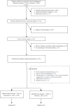
Figure 1
Figure 1
Flow chart of patient inclusion and exclusion.
Figure 2
Figure 2
Variable selection process using the Least Absolute Shrinkage and Selection Operator (LASSO) algorithm in the training set. (A) shows the LASSO cross-validation curve of the binomial deviance, a measure of model fit, plotted against the log-transformed penalty parameter, log λ. The mean binomial deviance is indicated by red dots, and its standard error by the surrounding error bars, for various values of log λ. At the top of the plot, the enumerated non-zero coefficients for the corresponding log λ values indicate the number of significant predictors retained during regularization. (B) shows the progression of the coefficients of the predictors within the LASSO as log λ varies. Individual paths are marked by unique colors, each representing the change in coefficient magnitude as log λ increases. The blue vertical line marks the log λ within one standard error of the minimum deviation, while the red vertical line indicates the log λ associated with the model’s minimum deviation. Predictors with non-zero coefficients at the log λ of the red line are identified for inclusion in the final logistic regression model.
Figure 3
Figure 3
Nomogram for predicting the risk of secondary margin positivity in re-excision procedures after initial margin positivity in breast conserving surgery. Assign risk points to each variable by aligning them with the corresponding position on the “Points scale, then sum these points to determine the overall risk score, which is located on the “Total Points” axis. This score translates directly to a risk percentage on the “Risk” axis, which indicates the likelihood of secondary margin positivity during re-excision after initial positive margins in breast conserving surgery. In this nomogram, a total score of 97 (corresponding to a risk of 0.3) serves as the threshold for distinguishing between low and high risk of secondary margin positivity. Non-dense breast' represents breast density categories A and B, and 'Dense breasts' represents breast density categories C and D, as defined by the ACR BI-RADS 5th edition. “Initial positive margin count” represents the number of positive margins identified during initial margin examination in breast-conserving surgery. A positive margin is characterized by the detection of atypical hyperplasia (including atypical cells, atypical ductal hyperplasia, and atypical lobular hyperplasia) or the presence of in situ and invasive carcinoma at the surgical margin. NST, Invasive Carcinoma of No Special Type; DCIS, Ductal Carcinoma In Situ; ILC, Invasive Lobular Carcinoma.
Figure 4
Figure 4
Receiver Operating Characteristic (ROC) curves for model performance evaluation. (A) ROC curve in the training set and (B) ROC curve in the testing set. The light blue shaded areas delineate the 95% confidence intervals for the mean ROC curves, derived from bootstrap sampling methods.
Figure 5
Figure 5
Calibration curve for model performance evaluation in training and testing sets. The dotted line indicates a perfectly calibrated model, where the predicted probabilities exactly match the actual probabilities. The closer the model’s calibration line is to the dotted line, the better the model is calibrated.
Figure 6
Figure 6
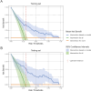
Decision Curve Analysis (DCA) for model performance evaluation. (A) DCA curve in the training set and (B) DCA curve in the testing set. DCA evaluates the clinical utility of the model by comparing the net benefit of model-based interventions to strategies of intervention for all or none. The 95% confidence interval shade area are derived from bootstrap sampling methods. At the optimal risk threshold, indicated by the red dashed line, the model shows a significantly higher net benefit compared to the “intervention for all” and “intervention for none” strategies, indicating robust clinical utility.

Similar articles

References

    1. Siegel RL, Miller KD, Wagle NS, Jemal A. Cancer statistics, 2023. CA: A Cancer J Clin. (2023) 73:17–48. doi: 10.3322/caac.21763 - DOI - PubMed
    1. Sung H, Ferlay J, Siegel RL, Laversanne M, Soerjomataram I, Jemal A, et al. . Global cancer statistics 2020: GLOBOCAN estimates of incidence and mortality worldwide for 36 cancers in 185 countries. CA: A Cancer J Clin. (2021) 71:209–49. doi: 10.3322/caac.21660 - DOI - PubMed
    1. Cochrane RA, Valasiadou P, Wilson ARM, Al-Ghazal SK, Macmillan RD. Cosmesis and satisfaction after breast-conserving surgery correlates with the percentage of breast volume excised. Br J Surg. (2003) 90:1505–9. doi: 10.1002/bjs.4344 - DOI - PubMed
    1. Houssami N, Macaskill P, Luke Marinovich M, Morrow M. The association of surgical margins and local recurrence in women with early-stage invasive breast cancer treated with breast-conserving therapy: A meta-analysis. Ann Surg Oncol. (2014) 21:717–30. doi: 10.1245/s10434-014-3480-5 - DOI - PMC - PubMed
    1. Riedel F, Hennigs A, Hug S, Schaefgen B, Sohn C, Schuetz F, et al. . Is mastectomy oncologically safer than breast-conserving treatment in early breast cancer. Breast Care. (2017) 12:385–90. doi: 10.1159/000485737 - DOI - PMC - PubMed

LinkOut - more resources