. 2024 Dec 19;8(1):40-46.
doi: 10.1093/abt/tbae032. eCollection 2025 Jan.

A bioluminescent reporter bioassay for in-process assessment of chimeric antigen receptor lentiviral vector potency

Affiliations

A bioluminescent reporter bioassay for in-process assessment of chimeric antigen receptor lentiviral vector potency

Julia K Gilden et al. Antib Ther. .

Abstract

Background: Chimeric antigen receptor (CAR)-T-cell therapy is a breakthrough in the field of cancer immunotherapy, wherein T cells are genetically modified to recognize and attack cancer cells. Delivery of the CAR gene is a critical step in this therapy and is usually achieved by transducing patient T cells with a lentiviral vector (LV). Because the LV is an essential component of CAR-T manufacturing, there is a need for simple bioassays that reflect the mechanism of action (MOA) of the LV and can measure LV potency with accuracy and specificity. Common methods for LV quantification may overestimate functional titer and lack a functional readout of LV MOA.

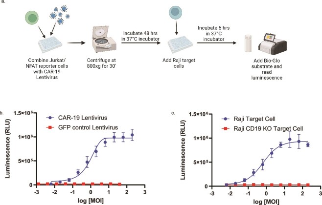
Methods: We developed a bioluminescent reporter bioassay using Jurkat T cells stably expressing a luciferase reporter under the control of an nuclear factor of activated T cells (NFAT) response element and tested its suitability for measuring LV potency.

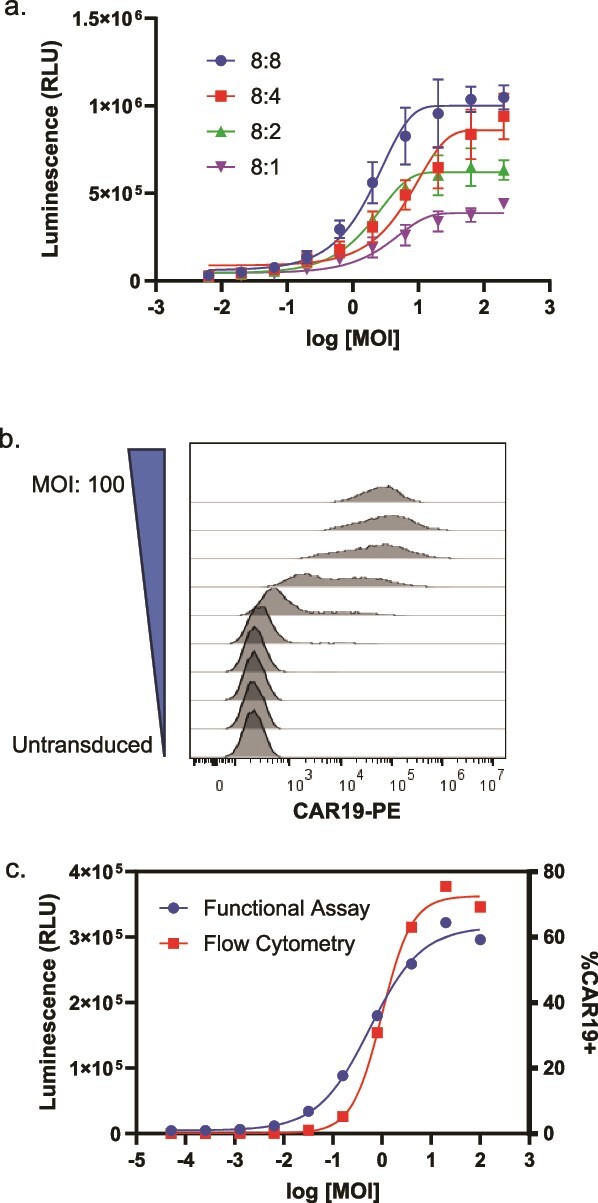
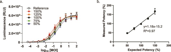
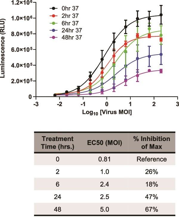
Results: Jurkat reporter cells can be transduced with CAR LV and combined with target cells, yielding a luminescent signal that is dependent on the identity and potency of the LV used. Bioluminescence was highly correlated with CAR expression. The assay is stability indicating and suitable for use in drug development and quality control settings.

Conclusions: We have developed a simple bioassay for potency testing of CAR LV. The bioassay represents a significant improvement over other approaches to LV quantification because it reflects the MOA of the LV and selectively detects fully functional viral particles, making it ideal for inclusion in a matrix of in-process quality control assays for CAR LV.

Keywords: CAR-T; bioassay; lentivirus; potency assay.


Conflict of interest statement

The authors are employees of Promega Corp.

Figures

Figure 1
Figure 1
NFAT-Luc2 activity stimulated by multiple T-cell activation signals. (a) Jurkat/NFAT-Luc2 cells were incubated with α-CD3 antibody crosslinked with goat α-mouse IgG antibody at the indicated concentrations. (b) Jurkat/NFAT-Luc2 cells were incubated with or without Raji target cells and the indicated concentrations of blinatumomab. The curves were compared using an extra-sum-of-squares F test and were determined to be different (P > .0001) (c) Jurkat/NFAT-Luc2 cells were transiently transfected with varying concentrations of α-CD19-CAR or α-CD20-CAR plasmid DNA, balanced with carrier DNA (pGEM-3z) such that all conditions received the same total amount of DNA. One day after transfection, cells were incubated with or without Raji target cells. In all panels, assays were incubated for 6 h at 37°C, and luciferase activity was detected using the Bio-Glo™ Luciferase Assay System. Data are representative of at least three independent experiments with n = 3 technical replicates. Error bars indicate standard deviation. For (c), a one-way ANOVA was performed separately for a-CD19 and a-CD20 conditions. Dunnett’s multiple comparison post-testing found a significant difference between the carrier DNA only (0:1) condition and the CAR DNA conditions in the presence of Raji cells (P < .0001) and no differences in the absence of Raji cells. Post-testing for linear trend found a relationship between CAR DNA quantity and assay response in the presence of Raji cells (P < .0001).
Figure 2
Figure 2
NFAT-Luc2 activity induced by CAR-19 LV and target cell coculture. (a) Schematic representation of assay workflow. (b) Jurkat/NFAT-Luc2 cells were transduced with CAR-19 or GFP control LV at the indicated MOI and incubated with wild-type Raji target cells. Only CAR-19 LV transduction led to a dose-dependent luminescent signal. (c) Jurkat/NFAT-Luc2 cells were transduced with CAR-19 LV and incubated with wild-type or CD19-knockout Raji target cells. A luminescent response was observed only with target cells expressing CD19. The curves within each panel were compared using extra-sum-of-squares F tests and were determined in each case to be different (P > .0001). Data are representative of at least two independent experiments with n = 3 technical replicates. Error bars indicate standard deviation. MOI = multiplicity of infection.
Figure 3
Figure 3
Correlation between flow cytometry and functional bioassay. (a) Jurkat/NFAT-Luc2 cells were transduced with CAR-19 LV and then combined with Raji target cells at the indicated E:T ratios. Luminescence was measured after a 6-h incubation at 37°C. The upper asymptotes and EC50s of the curves were compared using extra-sum-of-squares F tests, which indicated differences among the curves (P < .0001). Post hoc testing revealed a weak linear relationship between the E:T ratio and upper asymptote (R^2 = 0.56) and no relationship between E:T ratio and IC50 (R^2 = 0.03, slope not significantly different from 0; data not shown.) (b) Jurkat/NFAT-Luc2 cells were transduced with CAR-19 LV at the indicated MOI and stained for flow cytometry to measure surface expression of α-CD19 CAR. (c) Overlay of flow cytometry and functional bioassay data generated in parallel in a single experiment. Error bars indicate a standard deviation of n = 3 replicates.
Figure 4
Figure 4
Prequalification of LV potency assay. Mock potency samples were prepared with relative potencies between 50% and 150% of the reference standard and then used in the LV potency assay. JMP17® software was used to fit 4PL curves, test for parallelism (F test), and calculate the measured relative potency of each sample. (a) Representative data from one of three independent experiments. Error bars indicate a standard deviation of n = 3 replicates. (b) Analysis of assay linearity based on pooled average recovery from all three experiments.
Figure 5
Figure 5
LV potency assay is stability indicating. CAR-19 LV was incubated at 37°C for the indicated times and then used to transduce Jurkat/NFAT-Luc2 cells, which were then incubated with Raji target cells for 6 h at 37°C. Luciferase activity was detected using the Bio-Glo™ Luciferase Assay System. Data were analyzed for 4PL fit, and the upper asymptotes and EC50s of the curves were compared using extra-sum-of-squares F tests, which indicated differences among the curves (P < .0001). F tests did not detect differences between the hill slopes or lower asymptotes.

Similar articles

References

    1. Jayaraman J, Mellody MP, Hou AJ. et al. CAR-T design: elements and their synergistic function. EBioMedicine 2020;58:102931. 10.1016/j.ebiom.2020.102931. - DOI - PMC - PubMed
    1. Maher J, Brentjens RJ, Gunset G. et al. Human T-lymphocyte cytotoxicity and proliferation directed by a single chimeric TCRzeta/CD28 receptor. Nat Biotechnol 2002;20:70–5. 10.1038/nbt0102-70. - DOI - PubMed
    1. Finney HM, Akbar AN, Lawson AD. Activation of resting human primary T cells with chimeric receptors: costimulation from CD28, inducible costimulator, CD134, and CD137 in series with signals from the TCR zeta chain. J Immunol 2004;172:104–13. 10.4049/jimmunol.172.1.104. - DOI - PubMed
    1. Shirasu N, Shibaguci H, Kuroki M. et al. Construction and molecular characterization of human chimeric T-cell antigen receptors specific for carcinoembryonic antigen. Anticancer Res 2010;30:2731–8. - PubMed
    1. Honikel MM, Olejniczak SH. Co-stimulatory receptor signaling in CAR-T cells. Biomolecules 2022;12. 10.3390/biom12091303. - DOI - PMC - PubMed

LinkOut - more resources