. 2024 Dec 16;8(1):26-39.
doi: 10.1093/abt/tbae031. eCollection 2025 Jan.

Humanized anti-CD11d monoclonal antibodies suitable for basic research and therapeutic applications

Affiliations

Humanized anti-CD11d monoclonal antibodies suitable for basic research and therapeutic applications

Eoin N Blythe et al. Antib Ther. .

Abstract

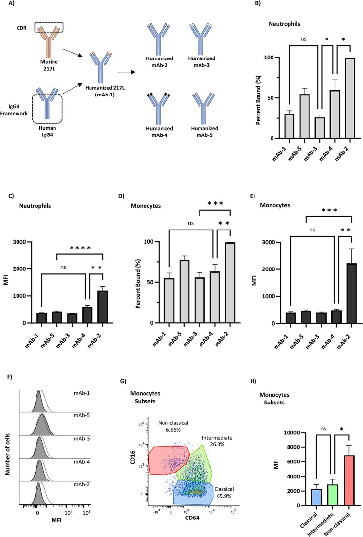
Background: Immunomodulatory agents targeting the CD11d/CD18 integrin are in development for the treatment of several pathophysiologies including neurotrauma, sepsis, and atherosclerosis. Murine anti-human CD11d therapeutic antibodies have successfully improved neurological and behavioral recovery in rodent neurotrauma models. Here, we present the progression of CD11d-targeted agents with the development of humanized anti-CD11d monoclonal antibodies.

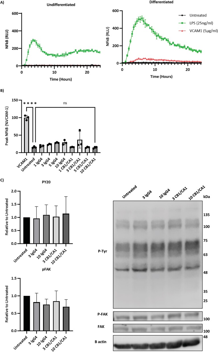
Methods: Primary human leukocytes and the THP-1 monocytic cell line were used to determine the binding of the CD11d antibodies, determine binding affinities, and assess outside-in signaling induced by CD11d antibody binding. In addition, a rat model of spinal cord injury was employed to demonstrate that the humanized monoclonal antibodies retained their therapeutic function in vivo. These determinations were made using a combination of flow cytometry, western blotting, immunohistochemistry, biochemical assays, and a locomotor behavioral assessment.

Results: Flow cytometric analysis demonstrated that the humanized anti-CD11d clones bind both human monocytes and neutrophils. Using a THP-1 model, the humanized anti-CD11d-2 clone was then determined to bind both the active and inactive CD11d/CD18 conformations without inducing inflammatory cell signaling. Finally, an investigation using anti-CD11d-2 as a detection tool uncovered a mismatch between total and surface-level CD11d and CD18 expression that was not altered by CK2 inhibition.

Conclusions: By developing humanized anti-CD11d monoclonal antibodies, new tools are now available to study CD11d biology and potentially treat inflammation arising from acute neurotrauma via CD11d targeting.

Keywords: CD11dCD18; integrin; monoclonal antibody; therapeutic antibody.


Conflict of interest statement

The authors share the following patents with Eli Lilly & Company:

Figures

Figure 1
Figure 1
Creation of humanized anti-CD11d monoclonal antibodies. (A) Conceptual diagram of the components that were combined to create the humanized anti-CD11d clones. The murine 217 L clone CDR sequence was isolated and inserted into a human IgG4 framework. Four variants of the 217 L CDR sequence were subsequently made and inserted into the same human IgG4 framework. (B) Percent-bound and (C) quantified MFI flow cytometric analysis of anti-CD11d clone binding to primary human neutrophils (N = 5). (D) Percent-bound and (E) quantified MFI flow cytometric analysis of anti-CD11d antibodies binding to primary human monocytes. (F) Nonspecific binding of the anti-CD11d clones (shaded histogram) and IgG4 isotype control antibody (white histogram with black outline) to CD11d Jurkat T cells. (G) Gating strategy identifying primary human monocyte subsets. (H) MFI analysis of CD11d expression among primary human monocyte subsets as determined by the humanized anti-CD11d-2 clone (N = 4). Error bars represent standard error. Significance was calculated by one-way ANOVA and Tukey’s multiple comparisons test (*P < .05), (**P < .01), (***P < .001), and (****P < .0001).
Figure 2
Figure 2
Humanized anti-CD11d clones improve biochemical and behavioral recovery in a rat SCI model. (A) Conceptual diagram of spinal cord compression injury and treatment with therapeutic anti-CD11d antibodies. (B) Spinal cord lesion homogenates from SCI rats treated with either anti-CD11d 1–5 or IgG4 isotype control antibody were assayed for myeloperoxidase as a surrogate for neutrophil infiltration (N = 6). Note an uninjured control was only performed for the 24-h timepoint. Error bars represent standard error. Significance was calculated by one-way ANOVA and Tukey’s multiple comparisons test (*P < .05), (**P < .01), (***P < .001), (****P < .0001). (C) BBB open-field locomotor scores in anti-CD11d-3 treated rats (N = 9) and IgG4 isotype control-treated rats (N = 10). Error bars represent standard error. Two-way ANOVA and Newman–Keuls post hoc test demonstrated a significant treatment effect (P = .0029), a significant effect of time (P < .0001), and a significant interaction of treatment and time (P = .0006). The only time that is not significant is at 1 week when a difference is not expected. From 2 to 10 weeks, they are all significant at either P < .01 or P < .05. Note that there were no data obtained in week 9. This statistical analysis was performed on only the BBB scores from the anti-CD11d-3-treated and IgG4-treated rats. The 217 L–treated rats were a smaller group of rats added in to illustrate the degree of recovery was similar to the anti-CD11d-3-treated group.
Figure 3
Figure 3
Humanized anti-CD11d-2 binding dynamics in a THP-1 model. (A) Flow cytometry analysis gated on live THP-1 cells differentiated with 100 nM PMA for up to 72 h (N = 3). (B) Immunohistochemistry of 100 nM PMA–differentiated THP-1 Luc2 cells for 72 h and then stained in the presence of human TruStain FcX block. Images are a representation of multiple fields of view (n = 5) across several independent repeats (N = 3). (C) Binding dynamics of anti-CD11d-2 to endogenous CD11d on 100 nM PMA–differentiated THP-1 cells as determined by flow cytometry (N = 3). Blocking and cell surface staining occurred live to allow for conformation change in HBSS, HBSS + 1 mM EDTA, or HBSS + 1 mM Mn2+. Cells were subsequently fixed for analysis. The binding curve is presented using a break in the x-axis. Error bars represent standard error. Significance was calculated by one-way ANOVA and Tukey’s multiple comparisons test (*P < .05), (**P < .01), (****P < .0001). (D) Predicted computational CD11d I-domain (residues 148–336) from AlphaFold (AF-Q13349-F1-v4). The ribbon diagram highlights the predicted α7-helix, MIDAS motif, and ligand binding site. The electrostatic diagram highlights the negatively charged MIDAS motif for divalent cation binding. Both the ribbon diagram and electrostatic diagram are of the same position in space.
Figure 4
Figure 4
Absence of humanized anti-CD11d-2 inducing pro-inflammatory β2 integrin signaling. (A) NF-κB expression was detected by a luciferase assay in THP-1 Luc2 cells. Following a 48-h culture in the presence or absence of 100 nM PMA, THP-1 Luc2 cells were collected and blocked with 5% HSA. Blocked THP-1 Luc2 cells were dropped onto untreated wells, LPS (25 ng/ml) containing wells, or VCAM-1 (5 μg/ml) coated wells. The plates were incubated and NF-κB expression was measured in triplet every hour for 24 h (N = 3). (B) Luciferase NF-κB assay following anti-CD11d-2 stimulation in 4-h PMA differentiated THP-1 Luc2 cells. Cells blocked by 5% HSA were dropped onto plates coated with VCAM-1 (5 μg/ml) or various concentrations (μg/ml) of antibodies. The plates were incubated and NF-κB expression was measured in triplet every hour for 24 h (N = 3). Peak NF-κB expression was calculated as the mean value between 4- and 8-h poststimulation and normalized to VCAM-1 (N = 3). Significance was calculated by one-way ANOVA and Tukey’s multiple comparisons test (P < .05), (****P < .0001). (C) Western blot analysis of 72-h 100 nM PMA–differentiated THP-1 cells stimulated with soluble anti-CD11d-2 (μg/ml) or IgG4 isotype control (μg/ml). Blots were performed in duplicate and normalized to untreated wells (N = 3). The representative images are from the same blot that was probed first for P-Tyr and FAK (Supplemental Fig. 3A), then re-probed for P-FAK and β-actin (Supplemental Fig. 3B). Monochrome cropped singlet rows of the same columns are displayed for each protein target. Error bars represent standard error. Significance was calculated by one-way ANOVA and Tukey’s multiple comparisons test (P < .05).
Figure 5
Figure 5
Modulation of β2 integrin expression by CK2 inhibition. Flow cytometric analysis of β2 integrin expression in THP-1 cells treated with combinations of 100 nM PMA and CGS-CK2-1 inhibitor (5 μg/ml) for 48 h. Surface level and internal level integrin expression were recorded to determine the surface level only (white) and total (grey) β2 integrin expression. Error bars represent standard error, (N = 3). Significance between surface and total expression within a treatment group was calculated by a two-way ANOVA and Tukey’s multiple comparisons (**P < .01), (***P < .001), (****P < .0001). Additional total β2 integrin expression analysis was then performed between treatment groups. Error bars represent standard error, (N = 3). Significance in levels of total expression between treatment groups was calculated by one-way ANOVA and Tukey’s multiple comparisons (**P < .01), (***P < .001), and (****P < .0001).

Similar articles

References

    1. Blythe EN, Weaver LC, Brown A. et al. β2 integrin CD11d/CD18: from expression to an emerging role in staged leukocyte migration. Front Immunol 2021;12:4471. 10.3389/fimmu.2021.775447. - DOI - PMC - PubMed
    1. Gris D, Marsh DR, Oatway MA. et al. Transient blockade of the CD11d/CD18 integrin reduces secondary damage after spinal cord injury, improving sensory, autonomic, and motor function. J Neurosci 2004;24:4043–51. 10.1523/JNEUROSCI.5343-03.2004. - DOI - PMC - PubMed
    1. Geremia NM, Bao F, Rosenzweig TE. et al. CD11d antibody treatment improves recovery in spinal cord-injured mice. J Neurotrauma 2012;29:539–50. 10.1089/neu.2011.1976. - DOI - PMC - PubMed
    1. Utagawa A, Bramlett HM, Daniels L. et al. Transient blockage of the CD11d/CD18 integrin reduces contusion volume and macrophage infiltration after traumatic brain injury in rats. Brain Res 2008;1207:155–63. 10.1016/j.brainres.2008.02.057. - DOI - PMC - PubMed
    1. Shultz SR, Bao F, Weaver LC. et al. Treatment with an anti-CD11d integrin antibody reduces neuroinflammation and improves outcome in a rat model of repeated concussion. J Neuroinflammation 2013;10:26. 10.1186/1742-2094-10-26. - DOI - PMC - PubMed

LinkOut - more resources