. 2025 Jan 7:15:1504556.
doi: 10.3389/fphar.2024.1504556. eCollection 2024.

Uvaol attenuates TGF-β1-induced epithelial-mesenchymal transition in human alveolar epithelial cells by modulating expression and membrane localization of β-catenin

Affiliations

Uvaol attenuates TGF-β1-induced epithelial-mesenchymal transition in human alveolar epithelial cells by modulating expression and membrane localization of β-catenin

Liliane Patrícia Gonçalves Tenório et al. Front Pharmacol. .

Abstract

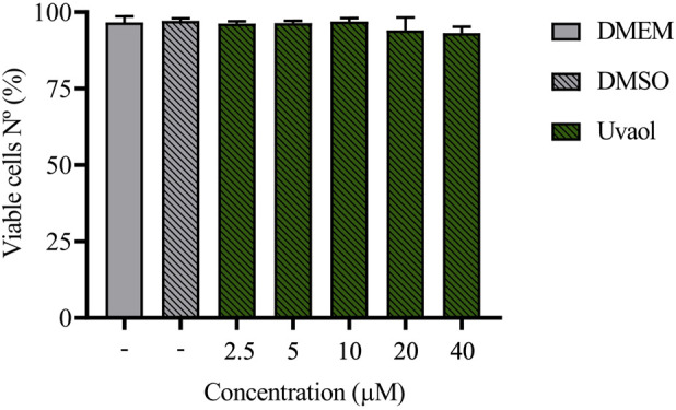
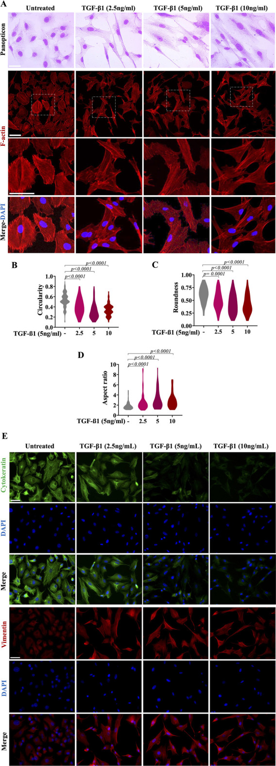
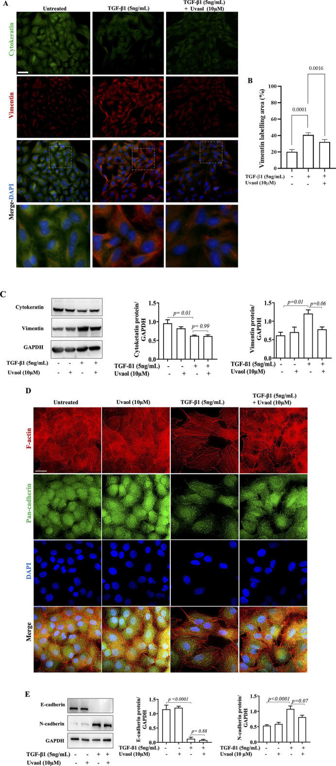
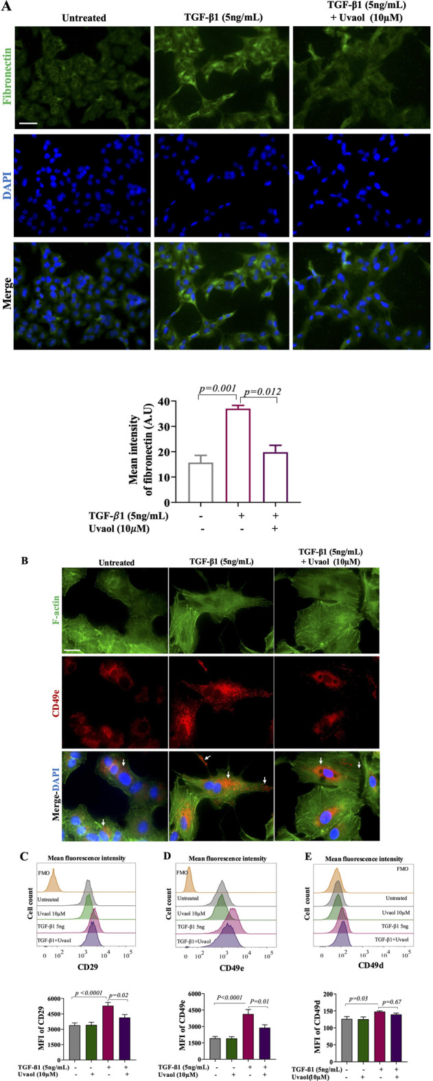
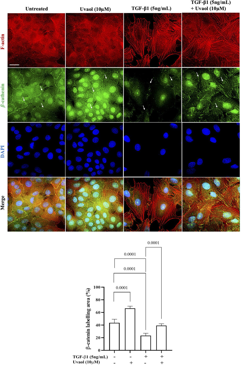
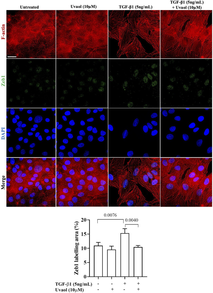
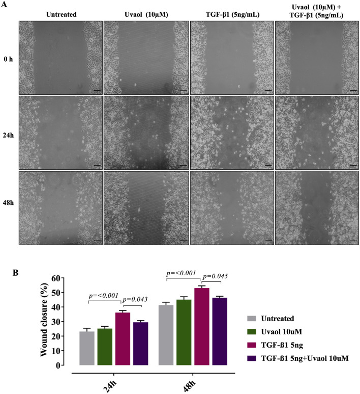
The epithelial-mesenchymal transition (EMT) is a biological process in which epithelial cells change into mesenchymal cells with fibroblast-like characteristics. EMT plays a crucial role in the progression of fibrosis. Classical inducers associated with the maintenance of EMT, such as TGF-β1, have become targets of several anti-EMT therapeutic strategies. Natural products from the pentacyclic triterpene class have emerged as promising elements in inhibiting EMT. Uvaol is a pentacyclic triterpene found in olive trees (Olea europaea L.) known for its anti-inflammatory, antioxidant, and antiproliferative properties. Yet, its effect on the TGF-β1-induced EMT in alveolar epithelial cells is unknown. The present study aimed to investigate the impact of uvaol upon TGF-β1-induced EMT in a cultured A549 human alveolar epithelial cell line, a classic in vitro model for studies of EMT. Changes in cell shape were measured using phase-contrast and confocal microscopy, whereas protein expression levels were measured using immunofluorescence, flow cytometry, and Western blotting. We also performed wound scratch experiments to explore its effects on cell migration. Uvaol had no significant cytotoxic effects on A549 cells. By contrast, the changes in the cell morphology consistent with TGF-β1-induced EMT were largely suppressed by treatment with uvaol. In addition, increased contents of mesenchymal markers, namely, vimentin, N-cadherin, and fibronectin in TGF-β1-induced A549 cells, were downregulated by uvaol treatment. Furthermore, the TGF-β1-induced migration of A549 cells was significantly suppressed by uvaol. Mechanistically, uvaol prevented the nuclear translocation of β-catenin and reduced the TGF-β1-induced levels of ZEB1 in A549 cells. These results provide compelling evidence that uvaol inhibits EMT by regulating proteins related to the mesenchymal profile in human alveolar epithelial cells, likely by modulating β-catenin and ZEB1 levels.

Keywords: TGF-β1; cell migration; epithelial-mesenchymal transition; uvaol; β-catenin.


Conflict of interest statement

The authors declare that the research was conducted in the absence of any commercial or financial relationships that could be construed as a potential conflict of interest.

Figures

FIGURE 1
FIGURE 1
Treatment with uvaol does not alter the viability of A549 cells. Cells were treated with uvaol (2.5–40 µM), vehicle (0.01% DMSO) or culture medium alone (DMEM) for 48 h. Cell viability was determined by FVS labelling and analysed by flow cytometry. Bar graphs represent mean values ± S.E.M.
FIGURE 2
FIGURE 2
TGF-β1 induces morphological and molecular changes in A549 alveolar epithelial cells. Morphologic changes consistent with EMT were observed in TGF-β1-treated cells for 48 h, compared with those untreated cells (control, cells that were not exposed to TGF-β1, cultured with medium DMEM) maintained the cobblestone morphology characteristic of epithelial cells. The phenotypic changes (transition towards a fibroblast-like phenotype) in TGF-β1-treated cells were compared to untreated cells and evaluated by light microscopy and panoptic staining [upper micrographs in panel (A)]. Lower micrographs revealed phalloidin staining of A549 cells incubated for 48 h as indicated without TGF-β1 (untreated) or treated with TGF-β1 at 2.5 ng/mL, 5 ng/mL or 10 ng/mL Phalloidin (red) was used to detect F-actin, while DAPI (blue) was used to decorate the nuclei. Scale bar, 100 µm. The violin plots reveal the distribution of circularity (B), roundness (C) and aspect ratio (D) in A549 cells treated with medium (DMEM) alone or TGF-β1 for 48 h. Morphometric analyses from violin plots were performed using ImageJ FiJi software, and statistically significant differences between groups are indicated by One-way ANOVA followed by the Kruskal–Wallis test. p values are shown in each graph. Panel (E) depicts immunofluorescence analyses of EMT-related intermediate filament proteins in A549 alveolar epithelial cells. Cells show a decrease in cytokeratin and, in parallel, an increase in vimentin after TGF-β1-treatment. Phenotypic marker changes were consistent with EMT in TGF-β1-treated A549 cells for 48 h, compared with those untreated cells (control cells cultured with medium DMEM). DAPI counterstaining was used to detect nuclei (blue). Scale bar = 100 µm.
FIGURE 3
FIGURE 3
Uvaol prevents morphological changes induced by TGF-β1 on A549 alveolar epithelial cells. Bright-field microscopy showing the effect of uvaol (10 µM) on morphologic changes consistent with EMT in TGF-β1-stimulated A549 cells for 48 h. Untreated cells were those not exposed to TGF-β1 cultured with DMEM. In panel (A), phalloidin staining of A549 cells was incubated for 48 h as indicated without TGF-β1 (untreated) or treated with uvaol (10 µM) and stimulated with TGF-β1 (5 ng/mL). Phalloidin (red) was used to detect F-actin, while DAPI (blue) was used to decorate nuclei. Scale bar, 100 µm. The violin plots illustrate the morphometric parameters, namely, circularity (B), roundness (C) and aspect ratio (D) in A549 cells treated with medium (DMEM) or uvaol and stimulated with TGF-β1 for 48 h. Morphometric analyses of violin plots were performed using ImageJ FiJi software, and statistically significant differences between groups are indicated by One-way ANOVA followed by the Kruskal–Wallis test. p values are shown in each graph.
FIGURE 4
FIGURE 4
Effect of uvaol on the expression of epithelial-mesenchymal transition biomarkers. A549 cells were treated with TGF-β1 with or without uvaol co-treatment for 48 h. Protein levels of cytokeratin (green) and vimentin (red) was determined by immunofluorescence staining (A). DAPI (blue) was used to detect nuclei. Scale bar: 20 μm. The contents of vimentin was determined by immunofluorescence staining (B). The contents of cytokeratin and vimentin was determined by Western blotting (C). Phalloidin (red) was used to detect F-actin and pan-cadherin (green) was determined by immunofluorescence staining (D). Protein levels of E-cadherin and N-cadherin was determined by Western blotting (E). Quantification of the western blots was performed after 3 independent experiments, and data are expressed as the means ± SEM. Bar graphs show statistical significance between groups, determined by one-way ANOVA followed by Tukey’s post-test. p values are shown in each graph.
FIGURE 5
FIGURE 5
Uvaol modulates fibronectin production and adhesion receptors expression in TGF-β1-stimulated A549 cells. Cells were treated with TGF-β1 with or without uvaol co-treatment for 48 h. Fibronectin contents were ascertained by immunofluorescence staining (A). DAPI (blue) was used to detect nuclei. Scale bar: 100 µm. In (B), phalloidin (green) was used to detect F-actin, and CD49e (red) was identified by immunofluorescence staining (white arrows). Scale bar, 50 µm.The histogram plots and bar charts represent the mean fluorescence intensity (MFI) of integrin receptor subunits CD29 (C), CD49e (D) and CD49d (E), determined by flow cytometry. Data are from 4 independent experiments. In the bar graph, data were expressed as the means ± SEM. Bar graphs show statistical significance between groups, determined by one-way ANOVA followed by Tukey’s post-test considering p values showed in each graph. AU = arbitrary unit.
FIGURE 6
FIGURE 6
Uvaol attenuates the pronounced loss of β-catenin in the subcellular region adjacent to the plasma membrane, induced by TGF-β1 in A549 cells. Cells were treated with TGF-β1 (5 ng/mL), with or without co-treatment with uvaol, for 48 h. Immunolocalization of β-catenin at adherens junctions (indicated by white arrows) was performed through co-labeling of β-catenin (green) and F-actin (red) in A549 cells. In untreated and uvaol-treated cells, β-catenin is localized adjacent to the plasma membrane and in the nuclear/perinuclear region of A549 cells. In cells treated with TGF-β1 alone, β-catenin at the membrane was drastically lost, primarily localizing in higher concentration in the nuclear/perinuclear region (white arrows) compared to untreated control (DMEM medium). Simultaneous treatment with TGF-β1 and uvaol maintained β-catenin adjacent to the plasma membrane, thereby preventing total loss and exacerbated nuclear translocation of β-catenin induced by TGF-β1. Scale bar: 20 μm.
FIGURE 7
FIGURE 7
Uvaol decreases ZEB1 contents in the nuclear region of A549 cells treated with TGF-β1. Cells were treated with TGF-β1 (5 ng/mL) with or without uvaol co-treatment for 48 h. Immunolocalization of nuclear/cytoplasmic ZEB1 (indicated by white arrows) was performed by co-labeling ZEB1 (green) and F-actin (red) in A549 cells. Scale bar: 20 µm. DAPI (blue) was used to decorate nuclei. 20 μm. The bar graph shows the total labeling of nuclear/cytoplasmic ZEB1 localization, data were expressed as mean ± SEM. The bar graphs show statistical significance between the groups, determined by one-way ANOVA followed by Tukey’s post-test considering the p-values presented in each graph.
FIGURE 8
FIGURE 8
Uvaol prevents the TGF-β1-induced migration of A549 alveolar cells. Cells were treated with TGF-β1 (5 ng/mL) with or without uvaol co-treatment for 24 and 48 h. The migration was measured by the scratch assay. Panel (A) depicts representative images of the horizontal migration assay showing the closure of the wound area induced with a tip from pipette on a monolayer of A549 cells and evaluated at 0, 24 and 48 h after treatment with uvaol and stimulation with TGF-β1 (5 ng/mL). In panel (B) the bar graphs represent the quantitation of the cell migration at 24 and 48 h after treatment with uvaol or TGF-β1. Statistical significance among groups was determined by one-way ANOVA followed by Tukey’s post-test. p values are shown in each graph.

Similar articles

References

    1. Agra L. C., Lins M. P., Da Silva Marques P., Smaniotto S., Bandeira De Melo C., Lagente V., et al. (2016). Uvaol attenuates pleuritis and eosinophilic inflammation in ovalbumin-induced allergy in mice. Eur. J. Pharmacol. 780, 232–242. 10.1016/j.ejphar.2016.03.056 - DOI - PubMed
    1. Akhmetshina A., Palumbo K., Dees C., Bergmann C., Venalis P., Zerr P., et al. (2012). Activation of canonical Wnt signalling is required for TGF-β-mediated fibrosis. Nat. Commun. 3, 735. 10.1038/NCOMMS1734 - DOI - PMC - PubMed
    1. Allouche Y., Warleta F., Campos M., Sánchez-Quesada C., Uceda M., Beltrán G., et al. (2011). Antioxidant, antiproliferative, and pro-apoptotic capacities of pentacyclic triterpenes found in the skin of olives on MCF-7 human breast cancer cells and their effects on DNA damage. J. Agric. Food Chem. 59 (1), 121–130. 10.1021/JF102319Y - DOI - PubMed
    1. Anwar S., Malik J. A., Ahmed S., Kameshwar V. A., Alanazi J., Alamri A., et al. (2022). Can natural products targeting EMT serve as the future anticancer therapeutics? Molecules 27 (22), 7668. 10.3390/MOLECULES27227668 - DOI - PMC - PubMed
    1. Bonel-Pérez G. C., Pérez-Jiménez A., Gris-Cárdenas I., Parra-Pérez A. M., Lupiáñez J. A., Reyes-Zurita F. J., et al. (2020). Antiproliferative and pro-apoptotic effect of uvaol in human hepatocarcinoma HepG2 cells by affecting G0/G1 cell cycle arrest, ROS production and AKT/PI3K signaling pathway. Mol. Basel, Switz. 25 (18), 4254. 10.3390/MOLECULES25184254 - DOI - PMC - PubMed

LinkOut - more resources