. 2025 Mar;12(10):e2410951.
doi: 10.1002/advs.202410951. Epub 2025 Jan 22.

A Baicalin-Based Functional Polymer in Dynamic Reversible Networks Alleviates Osteoarthritis by Cellular Interactions

Affiliations

A Baicalin-Based Functional Polymer in Dynamic Reversible Networks Alleviates Osteoarthritis by Cellular Interactions

Yili Yang et al. Adv Sci (Weinh). 2025 Mar.

Abstract

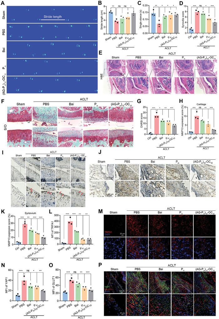
Osteoarthritis (OA) is increasingly recognized as a whole-organ disease predominantly affecting the elderly, characterized by typical alterations in subchondral bone and cartilage, along with recurrent synovial inflammation. Despite the availability of various therapeutics and medications, a complete resolution of OA remains elusive. In this study, novel functional hydrogels are developed by integrating natural bioactive molecules for OA treatment. Specifically, baicalin (Bai) is combined with 2-hydroxyethyl acrylate (HEA) to form a polymerizable monomer (HEA-Bai) through esterification, which is subjected to reversible addition-fragmentation chain transfer (RAFT) polymerization to produce Bai-based polymer (Pm). These macromolecules are incorporated into Schiff-base hydrogels, which demonstrate excellent mechanical properties and self-healing performance. Notably, the Bai-based formulations are taken up by fibroblast-like synoviocytes (FLSs), where they regulate glycolysis. Mechanistically, inhibition of yes-associated protein 1 (YAP1) by the formulations suppressed the FLSs glycolysis and reduced the secretion of inflammatory factors, including interleukin 1β (IL-1β), IL-6, and IL-8. Furthermore, the functional hydrogel (AG-Pm)-OC, severing as a lubricant and nutrient, prolonged joint retention of Bai, thereby reducing cartilage degradation and synovial inflammation. Meanwhile, (AG-Pm)-OC alleviated joint pain by targeting the YAP1 signaling and inhibiting macrophage recruitment and polarization. Taken together, this flavonoid-based injectable hydrogel exhibits enhanced biocompatibility and efficacy against OA.

Keywords: Bai‐based polymer; Schiff‐base hydrogel; anti‐inflammation; glycolysis; osteoarthritis.


Conflict of interest statement

The authors declare no conflict of interest.

Figures

Scheme 1
Scheme 1
Schematic representation of the synthesis of HEA‐Bai, Pm, AG, AG‐Pm, OC, and (AG‐Pm)‐OC and the investigation of the bioactivies of Bai‐based functional formulations.
Figure 1
Figure 1
Characterization of Bai‐based monomer HEA‐Bai, macromolecular Pm, and precursor AG‐Pm. A) High‐performance liquid chromatography (HPLC) analysis showing the conversion of Bai to HEA‐Bai. B) High‐resolution mass spectrometry (HRMS) reveals the accurate molecular weight of HEA‐Bai. C) Hydrogen nuclear magnetic resonance (1H NMR) spectra of Pm. D) Size exclusion chromatography (SEC) analysis demonstrating the structural uniformity of Pm. E) UV–vis spectroscopy determining the Bai content in Pm. F) In vitro cumulate release profile of Bai from Pm at pH 6.8 and 7.4. G) Ninhydrin assay results were analyzed by UV–vis spectroscopy. H) Fourier transform infrared spectroscopy (FT‐IR) spectra of Bai, Pm, AG, and AG‐Pm.
Figure 2
Figure 2
Rheological study, mechanical properties, and degradation of (AG‐Pm)‐OC hydrogels. A) Strain amplitude sweep mode of (AG‐Pm)10‐OC10. B) Frequency amplitude sweep mode of (AG‐Pm)10‐OC10. C) Alternating strain amplitude sweep mode of (AG‐Pm)10‐OC10. D) Shear‐thinning test of (AG‐Pm)10‐OC10, inserted with a macroscopic image. E) Friction test of (AG‐Pm)10‐OC10 and (AG‐Pm)5‐OC10 under the loading of 5 N at 1 Hz. F) Compression test of (AG‐Pm)10‐OC10 and (AG‐Pm)5‐OC10. G) Tensile test of (AG‐Pm)10‐OC10 and (AG‐Pm)5‐OC10. H) Swelling behavior of (AG‐Pm)10‐OC10 and (AG‐Pm)5‐OC10. I) Enzymatic degradation of (AG‐Pm)10‐OC10 and (AG‐Pm)5‐OC10 with Collagenase Type I. J) Hydrolysis degradation of (AG‐Pm)10‐OC10 and (AG‐Pm)5‐OC10 at pH 6.8. K) Hydrolysis degradation of (AG‐Pm)10‐OC10 and (AG‐Pm)5‐OC10 at pH 7.4. L) In vitro cumulate release profile of Bai from (AG‐Pm)10‐OC10 at pH 6.8 and pH 7.4.
Figure 3
Figure 3
Pm regulates the secretion of inflammatory factors via YAP1‐mediated glycolysis in FLSs. A) Representative fluorescence images of FLSs treated with Pm at 5, 10, 15, and 17.5 µg mL−1 for 12 h. Scale bars = 20 µm. B) Representative fluorescence images of FLSs treated with Pm at 15 µg mL−1 for 4, 8, 12, and 16 h. Scale bars = 20 µm. C–E) Expression levels of IL‐1β (C), IL‐6 (D), and IL‐8 (E) in FLSs treated with Bai and Pm at 15 µg mL−1 for 6, 12, and 24 h. F–H) Seahorse metabolic flux measuring ECAR (F), glycolysis (G), and glycolytic capacity (H) in FLSs treated with Bai and Pm in the presence or absence of IL‐1β for 24 h. I) YAP1 expression levels in FLSs treated with Pm at 5, 10, and 15 µg mL−1 for 24 h. J) YAP1 expression levels in FLSs treated with Bai and Pm at 15 µg mL−1 for 24 h. Data are presented as mean ± SD. n.s.: not significant, * p < 0.05, ** p < 0.01, and *** p < 0.001. One‐way ANOVA for (C–E and G–J).
Figure 4
Figure 4
Joint retention, biodistribution, and co‐localization of Bai‐based formulations. A) Illustration of OA mice model which undergoes a series of treatments at different times and subsequent analysis. B) Representative images showing the time course of radiant efficiency in OA knee joints following a 10‐day intra‐articular injection of DiD‐labeled Bai, Pm, and (AG‐Pm)10‐OC10. C) Quantitative analysis of radiant efficiency in (B). D) Quantitative analysis of AUC in (C). E) Biodistribution of Bai‐based formulations in OA knee joints 24 h after a single injection. F) Biodistribution of Bai‐based formulations in major organs of OA mice 24 h after a single injection. G,H) Quantitative analysis of relative cellular uptake (G) and representative image of the fluorescence intensity (H) of DiD‐labeled (AG‐Pm)10‐OC10 in OA knee joints, 7 and 14 days after a single injection. Scale bars = 100 and 20 µm. Data are presented as mean ± SD. n.s.: not significant, * p < 0.05, and ** p < 0.01. Student's t‐test for (G) and one‐way ANOVA for (D).
Figure 5
Figure 5
Bai‐based formulations mitigate OA progression via the YAP1/GLUT1 axis. A–C) Gait analysis of OA mice for footprints (A), stride length (B), and single stance (C), treated with Bai‐based formulations after six‐week ACLT surgery. D,E) Synovitis score (D) and representative H&E staining images (E) of OA knee joints following treatments with Bai, Pm, and (AG‐Pm)10‐OC10 for 6 weeks. Scale bars = 200 µm. F,G) Representative S/O staining images (F) and OARSI score (G) of knee joint severity following treatments with Bai, Pm, and (AG‐Pm)10‐OC10 for 6 weeks. Scale bars = 20 µm. H,I) Quantitative analysis (H) and representative immunohistochemical (IHC) images (I) of MMP13 in cartilage. Scale bars = 100 and 20 µm. J,K) Representative IHC images (J) and quantitative analysis (K) of MMP13 in synovial tissues. Scale bars = 100 and 20 µm. L,M) Quantitative analysis of TRPA1 fluorescence intensity (L) and representative fluorescence images of TRPA1 (red) in DRG tissues (J) following treatments with Bai, Pm, and (AG‐Pm)10‐OC10 for 6 weeks. Scale bars = 20 µm. N–P) Quantitative analysis of YAP1 (N) and GLUT1 (O) and representative fluorescence images (P) of YAP1 (green) and GLUT1 (red) following treatments with Bai, Pm, and (AG‐Pm)10‐OC10 for 6 weeks. Scale bars = 100 and 20 µm. Data are presented as mean ± SD. n.s.: not significant, * p < 0.05, ** p < 0.01, and *** p < 0.001. One‐way ANOVA for (B,C,D,G,H,K,L,N,O).
Figure 6
Figure 6
Bai‐based formulations regulate macrophage polarization. A) Representative fluorescence images of RAW264.7 cells treated with Pm at 5, 10, 15, and 17.5 µg mL−1 for 12 h. Scale bars = 20 µm. B) Representative fluorescence images of RAW264.7 cells treated with Pm at 15 µg mL−1 for 4, 8, 12, and 16 h. Scale bars = 20 µm. C,D) Representative fluorescence images of iNOS in RAW264.7 cells (C) and quantitative analysis of MFI of iNOS (D) after Bai or Pm treatments for 24 h (15 µg mL−1). Scale bars = 10 µm. E) Expression levels of iNOS in RAW264.7 cells treated with Bai and Pm at 15 µg mL−1 for 6, 12, and 24 h. F,G) Representative fluorescence images of CD206 (red) expression in synovial tissues (F) and quantitative analysis of MFI of CD206 (G) following treatments with Bai, Pm, and (AG‐Pm)10‐OC10 for 6 weeks. Scale bars = 20 µm. H,I) Quantitative analysis of MFI of iNOS (H) and representative fluorescence images of iNOS (green) expression in synovial tissues (I) following treatments with Bai, Pm, and (AG‐Pm)10‐OC10 for 6 weeks. Scale bars = 100 and 20 µm. Data are presented as mean ± SD. n.s.: not significant, * p < 0.05, ** p < 0.01, and *** p < 0.001. One‐way ANOVA for (D,E,G,H).

Similar articles

Cited by

References

    1. a) Obeidat A. M., Wood M. J., Adamczyk N. S., Ishihara S., Li J., Wang L., Ren D., Bennett D. A., Miller R. J., Malfait A.‐M., Miller R. E., Nat. Commun. 2023, 14, 2479; - PMC - PubMed
    2. b) Wang L., Chen X., Wang S., Ma J., Yang X., Chen H., Xiao J., Adv. Healthcare Mater. 2024, 13, 2302833;
    3. c) Fu W., Vasylyev D., Bi Y., Zhang M., Sun G., Khleborodova A., Huang G., Zhao L., Zhou R., Li Y., Liu S., Cai X., He W., Cui M., Zhao X., Hettinghouse A., Good J., Kim E., Strauss E., Leucht P., Schwarzkopf R., Guo E. X., Samuels J., Hu W., Attur M., Waxman S. G., Liu C.‐j., Nature 2024, 625, 557. - PMC - PubMed
    1. Van Linthout S., Miteva K., Tschöpe C., Cardiovasc. Res. 2014, 102, 258. - PubMed
    1. Lv Z., Xu X., Sun Z., Yang Y. X., Guo H., Li J., Sun K., Wu R., Xu J., Jiang Q., Ikegawa S., Shi D., Cell Death Dis. 2021, 12, 504. - PMC - PubMed
    1. Louwe P. A., Badiola Gomez L., Webster H., Perona‐Wright G., Bain C. C., Forbes S. J., Jenkins S. J., Nat. Commun. 2021, 12, 1770. - PMC - PubMed
    1. a) Koo J. H., Guan K.‐L., Cell Metab. 2018, 28, 196; - PubMed
    2. b) Janczi T., Fehrl Y., Kinne R. W., Böhm B., Burkhardt H., Rheumatology 2022, 62, 850. - PubMed

LinkOut - more resources