. 2025 Mar;12(10):e2407900.
doi: 10.1002/advs.202407900. Epub 2025 Jan 22.

Targeting MAPK14 by Lobeline Upregulates Slurp1-Mediated Inhibition of Alternative Activation of TAM and Retards Colorectal Cancer Growth

Affiliations

Targeting MAPK14 by Lobeline Upregulates Slurp1-Mediated Inhibition of Alternative Activation of TAM and Retards Colorectal Cancer Growth

Mingxia Zhao et al. Adv Sci (Weinh). 2025 Mar.

Abstract

Colorectal cancer (CRC) usually creates an immunosuppressive microenvironment, thereby hindering immunotherapy response. Effective treatment options remain elusive. Using scRNA-seq analysis in a tumor-bearing murine model, it is found that lobeline, an alkaloid from the herbal medicine lobelia, promotes polarization of tumor-associated macrophages (TAMs) toward M1-like TAMs while inhibiting their polarization toward M2-like TAMs. Additionally, lobeline upregulates mRNA expression of secreted Ly-6/UPAR-related protein 1 (Slurp1) in cancer cells. The inhibitory effects of lobeline on tumor load and TAM polarization are almost completely eliminated when Slurp1-deficient MC38 cells are subcutaneously injected into mice, suggesting that lobeline exerts an antitumor effect in a Slurp1-dependent manner. Furthermore, using target-responsive accessibility profiling, MAPK14 is identified as the direct target protein of lobeline. Mechanistically, upon binding to MAPK14 in colon cancer cells, lobeline prevents nuclear translocation of MAPK14, resulting in decreased levels of phosphorylated p53. Consequently, negative transcriptional regulation of SLURP1 by p53 is suppressed, leading to enhanced transcription and secretion of SLURP1. Finally, combination therapy using lobeline and anti-PD1 exhibits stronger antitumor effects. Taken together, these findings suggest that remodeling the immunosuppressive microenvironment using small-molecule lobeline may represent a promising therapeutic strategy for CRC.

Keywords: MAPK14; Slurp1; TAMs polarization; colorectal cancer; lobeline.


Conflict of interest statement

The authors declare no conflict of interest.

Figures
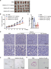
Figure 1
Figure 1
Lobeline reduces tumor load in MC38 xenograft C57BL/6 mice and colorectal cancer organoids. C57BL/6 mice were subcutaneously injected with 1 × 10⁶ MC38 CRC cells, and treated with different doses of lobeline (12.5, 25, and 50 mg kg−1, i.p. every day) when the tumor grew to 100–150 mm3. A) Representative images. B) Mean tumor volumes of different groups. C) Tumor weight. D) IHC staining of PCNA. Scale bar = 50 µm. After 3 days of subculture, human colon cancer organoids were incubated with lobeline (30 µmol L−1) for 48 h, following which images were captured. N = 6 mice each group in (A–D). E) Organoid volume, scale bar = 50 µm. F) HE staining of organoid sections. scale bar = 100 µm. Data are presented as mean ± SEM. p‐values are determined by a two‐tailed Student's t‐test. * p < 0.05, ** p < 0.01 versus PBS.
Figure 2
Figure 2
ScRNA‐seq analysis revealed that lobeline retarding the growth of transplanted CRC in mice. A) Schematic diagram of the scRNA‐seq experiment. The mice were subcutaneously injected with 1 × 106 MC38 cancer cells, and treated with PBS or lobeline (25 mg kg−1 i.p. every day) when the tumors grew between 100–150 mm3. Nine days later, the tumors were removed, and then a single‐cell suspension was prepared for testing. Scale bar=5 mm. N =6 mice. B) UMAP (uniform manifold approximation and projection) for dimension reduction of 21226 cells clustered into 3 major clusters (middle) and 20 minor clusters (right) in two groups (left). C) 3D pie chart of the major cluster proportion in PBS and lobeline. D) Bar plots showing the percentage (%) of cell types in PBS and lobeline. E) Heatmap showing the classification of cell subsets and selected marker genes. Red: high expression; blue: low expression. F–H) GO analysis for immune cells, cancer cells, and fibroblasts. Data are represented as mean ± SEM. P‐values are determined by a two‐tailed Student's t‐test, * p < 0.05. ** p < 0.01.
Figure 3
Figure 3
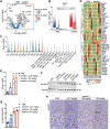
SLURP1 transcription and translation are promoted by lobeline. A) Volcano plot showing transcriptome dynamics between PBS and lobeline. Significantly differentially expressed genes are colored according to experimental groups. B) Raincloud plots show expression differences for Slurp1 between PBS and lobeline. C) Violin plots show the mRNA of Slurp1 in minor clusters. D) Heatmap showing the different genes in cancer cell subclusters between PBS and lobeline. Mice were subcutaneously injected with 1 × 106 MC38 cancer cells and subcutaneously injected with PBS or different doses of lobeline (12.5, 25, and 50 mg kg−1). E) qPCR for Slurp1 expression in tumors. F,G) Protein levels of SLURP1 were analyzed by Western blot. GAPDH was shown as the loading control. H) The expression of SLURP1 in tumor tissues was detected by IHC staining. N = 6 mice each group in (E–H). Scale bar = 50 µm. p‐values are determined by a two‐tailed Student's t‐test, ** p < 0.01.
Figure 4
Figure 4
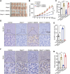
Slurp1 knockout reverses the amelioration of lobeline on tumor load. Wild‐type mice were injected with Slurp1 knockout MC38 cells or control cells subcutaneously (1 × 106), and treated with lobeline (25 mg kg−1, i.p. every day) when the tumor grew to 100–150 mm3. A) Tumor photos. B) Tumor growth curve. C) Tumor weight. N = 6 mice in each group in (A–C). D–G) IHC staining of SLURP1 and PCNA. N = 3 in (D–G). Scale bar = 50 µm. n = 6 per group. p‐values are determined by two‐way ANOVA with Student׳s t‐test, ** p < 0.01.
Figure 5
Figure 5
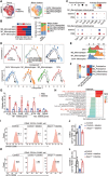
Colon cancer cells‐derived SLURP1 is involved in the process of lobeline regulating macrophage polarization. A) The UMAP projection of myeloid subclusters in PBS and lobeline. B) The percentage (%) of myeloid cell subpopulation in PBS and lobeline. C) Dot plots showing M1 and M2 macrophages‐related genes across the myeloid subtypes. D) Pseudo‐time trajectory analysis of myeloid cells based on groups and subtypes, pseudo‐time (above), and faceted plots showing cell types in pseudo‐time series (under). E) Ridge plots showing the differentiation of monocyte and macrophage subtypes along the pseudo‐time. F) Heat map of macrophage polarization and anti‐inflammatory function. G) qPCR for M1 and M2 macrophages‐related gene expression in tumors. N = 3 in (G). H) Representative KEGG pathways enriched in myeloid cells. Pathways associated with macrophage polarization are highlighted in red. I–L) Histograms showing the number of M1 and M2 macrophages detected by flow cytometry in mice tumors. N = 3 each group in (I–L). All data are expressed as mean ± SEM from 3 independent experiments. p‐values are determined by Student׳s t‐test (G) and two‐way ANOVA (I‐L), * p < 0.05, ** p < 0.01.
Figure 6
Figure 6
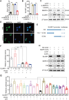
MAPK14 is the target protein of lobeline. A) The Flow chart shows the process of searching for lobeline target protein by TRAP experiment. B) Volcano map showing lobeline binding proteins. FC = Fold change (ratio of lobeline to DMSO). C) Isothermal titration calorimetry (ITC) results of lobeline and MAPK14‐WT protein. D) Docking prediction of the binding sites of lobeline and MAPK14 (PDBID: 5ETI). E–I) 293T cells transfected with MAPK14 (WT) or point mutant plasmids for 48 h and the protein lysate was taken for MST detection. J) EGFP‐N1 was transfected as a control.
Figure 7
Figure 7
MAPK14 inhibits SLURP1 transcription and translation via p53. MC38 cells were transfected with si‐MAPK14 and then treated with or without 30 nmol L−1 lobeline for 48 h. A) Slurp1 mRNA level was detected by qPCR. B) Cell supernatant was collected for ELISA assay to detect the secretion level of SLURP1.N= 3 in (A,B). C) MAPK14 and SLURP1 protein levels were detected by Western blot. D) After the MC38 cells were treated with lobeline (30 nmol L−1) for 48 h, the position relationship between MAPK14 and the nucleus was detected by immunofluorescence staining. E,F) A schematic diagram of human Slurp1 promoter and its truncates cloned to the luciferase reporter. The luciferase reporter was co‐transfected into 293T cells alone or with p53 plasmid, the luciferase activity was measured 36 h after transfection. G) Effect of mutation of p53 binding sites on Slurp1 transcription. The mutants were co‐transfected into 293T cells with FLAG‐p53 plasmid. 36 h after transfection, the luciferase activity was measured. N = 3 in (F,G). H) MC38 cells were transfected with MAPK14‐HA plasmid and/or sh‐p53 for 48 h, then the cells were collected for the Western blot experiment. All data are expressed as mean ± SEM. p‐values are determined by two‐way ANOVA and Student׳s t‐test. * p < 0.05, ** p < 0.01.
Figure 8
Figure 8
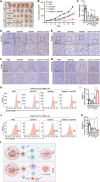
The combination of lobeline and PD1 antibody shows a stronger antitumor effect. C57BL/6 mice were subcutaneously inoculated with 1 × 106 MC38 colon cancer cells, and treated with anti‐PD1 (5  mg kg−1 i.p. once every three days) and/or lobeline (25  mg kg−1 i.p. every day) when the tumors grew to 100 mm3. All results presented are of day 13 post‐inoculation. A) Representative images of tumors. B) Mean tumor volumes of different groups. C) Mean tumor weights of different groups. N = 6 mice in each group in (A–C). D–G) IHC for SLURP1, PCNA, Foxp3, and Granzyme B in tumor sections. H–K) Histograms showing the number of M1‐like and M2‐like macrophages detected by flow cytometry in mice tumors. N = 3 in (D–K). L) Graphic illustration of the antitumor mechanism of lobeline. In the tumor microenvironment, MAPK14 translocates to the cancer cell nucleus and phosphorylates p53, which activates the negative transcriptional regulation of Slurp1 and inhibits its protein expression and secretion. Low levels of SLURP1 inhibit M1‐like macrophage polarization and promote M2‐like macrophage polarization, which leads to tumor growth. After administration of lobeline, lobeline binds to MAPK14 in cancer cells, inhibiting MAPK14 entry into the nucleus and reducing phosphorylated p53 levels, thereby inhibiting p53's negative transcriptional regulation of Slurp1 and promoting Slurp1 transcription and protein secretion. The secreted SLURP1 acts on TAMs, promoting its polarization toward M1‐like macrophages, and inhibiting its polarization toward M2‐like macrophages. SLURP1 activates T cells, down‐regulating Foxp3 and up‐regulating Granzyme B proteins’ level, thereby inhibiting tumor growth. All data are expressed as mean ± SEM from 3 independent experiments. p‐values are determined by two‐way ANOVA and Student׳s t‐test, * p < 0.05, ** p < 0.01.

References

    1. Sung H., Ferlay J., Siegel R. L., Laversanne M., Soerjomataram I., Jemal A., Bray F., CA Cancer J Clin 2021, 71, 209. - PubMed
    1. Ciardiello F., Ciardiello D., Martini G., Napolitano S., Tabernero J., Cervantes A., CA Cancer J Clin 2022, 72, 372. - PubMed
    1. a) Ganesh K., Stadler Z. K., Cercek A., Mendelsohn R. B., Shia J., Segal N. H., L. A. Diaz Jr., Nat. Rev. Gastroenterol. Hepatol. 2019, 16, 361; - PMC - PubMed
    2. b) Ganesh K., Nat. Rev. Gastroenterol. Hepatol. 2022, 19, 93; - PMC - PubMed
    3. c) Kubli S. P., Berger T., Araujo D. V., Siu L. L., Mak T. W., Nat Rev Drug Discov 2021, 20, 899; - PubMed
    4. d) Schmitt M., Greten F. R., Nat. Rev. Immunol. 2021, 21, 653; - PubMed
    5. e) Tie Y., Tang F., Wei Y. Q., Wei X. W., J. Hematol. Oncol. 2022, 15, 61. - PMC - PubMed
    1. a) Strauss L., Mahmoud M. A. A., Weaver J. D., Tijaro‐Ovalle N. M., Christofides A., Wang Q., Pal R., Yuan M., Asara J., Patsoukis N., Boussiotis V. A., Sci Immunol 2020, 5, eaay1863; - PMC - PubMed
    2. b) Dixon K. O., Tabaka M., Schramm M. A., Xiao S., Tang R., Dionne D., Anderson A. C., Rozenblatt‐Rosen O., Regev A., Kuchroo V. K., NatureNature 2021, 595, 101; - PMC - PubMed
    3. c) Seo W. I., Lee C. H., Jung S. J., Lee D. S., Park H. Y., Jeong D. H., Kim W., Chung J. I., Choi I., Cancer Immunol. Immunother. 2021, 70, 3113; - PMC - PubMed
    4. d) Veglia F., Sanseviero E., Gabrilovich D. I., Nat. Rev. Immunol. 2021, 21, 485. - PMC - PubMed
    1. Mantovani A., Marchesi F., Jaillon S., Garlanda C., Allavena P., Cell Mol. Immunol. 2021, 18, 566. - PMC - PubMed

MeSH terms

Substances

LinkOut - more resources