. 2025 Jan;14(1):e270032.
doi: 10.1002/jev2.70032.

Uncovering a new family of conserved virulence factors that promote the production of host-damaging outer membrane vesicles in gram-negative bacteria

Affiliations

Uncovering a new family of conserved virulence factors that promote the production of host-damaging outer membrane vesicles in gram-negative bacteria

Audrey Goman et al. J Extracell Vesicles. 2025 Jan.

Abstract

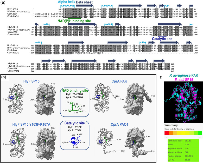
CprA is a short-chain dehydrogenase/reductase (SDR) that contributes to resistance against colistin and antimicrobial peptides. The cprA gene is conserved across Pseudomonas aeruginosa clades and its expression is directly regulated by the two-component system PmrAB. We have shown that cprA expression leads to the production of outer membrane vesicles (OMVs) that block autophagic flux and have a greater capacity to activate the non-canonical inflammasome pathway. In a murine model of sepsis, a P. aeruginosa strain deleted for cprA was less virulent than the wild-type (WT) strain. These results demonstrate the important role of CprA in the pathogenicity of P. aeruginosa. It is worth noting that CprA is also a functional ortholog of hemolysin F (HlyF), which is encoded by virulence plasmids of Escherichia coli. We have shown that other cryptic SDRs encoded by mammalian and plant pathogens, such as Yersinia pestis and Ralstonia solanacearum are functional orthologs of CprA and HlyF. These SDRs also induce the production of OMVs which block autophagic flux. This study uncovers a new family of virulence determinants in Gram-negative bacteria, offering potential for innovative therapeutic interventions and deeper insights into bacterial pathogenesis.

Keywords: OMVs; PmrAB; Pseudomonas aeruginosa; autophagy; inflammasome; pathogenicity; polymyxins.


Conflict of interest statement

The authors have declared that no competing interests exist.

Figures

FIGURE 1
FIGURE 1
Amino acid homology and structural prediction of CprA and HlyF. (a) Multiple sequence alignment of HlyF and CprA orthologous proteins from E. coli strain SP15 and P. aeruginosa strains PAK and PAO1 was performed using Clustal Omega. The mutant of HlyF was constructed by site‐directed mutagenesis of the predicted catalytic site leading to the two substitutions Y163F and K167A. The CprA protein from P. aeruginosa strain PAO1 is truncated due to a deletion of the cytosine at position 670 of the gene. This results in a frameshift and a premature stop codon at position 245. The residues' identity and similarity are highlighted in dark grey and light grey, respectively. (b) Alphafold 2 protein structure prediction was used to show the three‐dimensional structure of the HlyF and CprA proteins from E. coli and P. aeruginosa. In both panels, the NAD(P)H binding sites and catalytic sites are depicted in green and blue, respectively. (c) The iPBA web server was used to compare the secondary structure of CprA (in blue) and HlyF (in pink). (https://www.dsimb.inserm.fr/dsimb_tools/ipba/index.php). HlyF, hemolysin F, CprA, cationic peptide resistance A.
FIGURE 2
FIGURE 2
OMVs derived from CprA or HlyF‐producing E. coli and OMVs from P. aeruginosa that produce a full‐length CprA trigger autophagosomes in HeLa cells and inhibit the autophagy flux of HeLa cells. (a) This basal autophagy flux model describes the following five stages: (1) elongation and formation of a double‐membrane LC3‐II‐dependent vesicle containing cytoplasmic components; (2) newly formed autophagosome; (3) fusion of the autophagosome with the lysosome; (4) degradation and (5) recycling of autophagolysosome contents. Created with BioRender.com. (b, c) Fluorescent microscopy images of GFP‐LC3 (green) in HeLa cells expressing green fluorescent protein–fused with LC3. For panel b, cells were treated for 3 h with 5 µg/mL of OMVs purified from E. coli BL21 DE3 strain expressing the WT hlyF (hlyF WT) from E. coli strain SP15 (pAGO‐15), the mutant in the catalytic site (hlyF Y163F, K167A) (pAGO‐16) or cprA from P. aeruginosa strain PAK (pAGO‐32) or strain PAO1 (pAGO‐30). For panel c, cells treated for 5 h with 50 µg/mL of OMVs purified from P. aeruginosa strains PAK, PAO1 and PAO1 + pJN cprAPAK (pAGO‐38) or PAO1+ pJN cprAPAO1(pAGO‐37). In b and c, the scale bar in the images represents 25 µm. These images are representative of three independent experiments. (d, e) Western blot analysis of LC3 (forms I and II, as autophagy marker) and actin (as loading control) in HeLa cells. For panel d, cells were treated as in panel b. For panel e, cells were treated for 5 h with 50 µg/mL of OMVs purified from P. aeruginosa strains PAK, PAK ∆cprA, PAO1 and PAO1 + pJN cprA PAO1 (pAGO‐37) or for 1.5 h with 50 µg/mL of OMVs purified from P. aeruginosa strains PAK ∆cprA attB::PRhacprA and PAO1 + pJN cprA PAK (pAGO‐38). Each western‐blot is representative of three independent experiments. In all panels, NT were used as a control. (f) Schema for visualizing basal or blocked autophagy flux in the HeLa‐Difluo hLC3 cell line. The cells express a LC3 protein fused with RFP, which is resistant to acidic conditions and GFP, which is sensitive to acidity. In the case of basal autophagic flux, the GFP‐LC3 signal decreases due to the fusion of autophagosomes with acidic lysosomes. Subsequently, the autophagolysosome undergoes degradation and recycling, preventing the accumulation of autophagosomes. Basal autophagy is visualized through fluorescence microscopy as diffuse GFP and RFP labeling. When the autophagic flux is blocked at the lysosomal fusion step, autophagosomes accumulate with a basic pH, which is visualized as the co‐localization of GFP and/or RFP signals in foci. If the autophagic flux is blocked after the lysosomal fusion step, the autophagolysosome accumulates with an acidic pH, which is observed by a low GFP‐LC3 signal and intact RFP‐LC3 foci (Kimura et al., ; Loos et al., 2014). Created with BioRender.com (g) Confocal images of DiFluo HeLa cells were captured under different conditions: NT, deprived by incubation for 5 h in HBSS, treated for 5 h with 50 µM chloroquine or treated with 50 µg/mL of OMVs from P. aeruginosa strains PAK, PAO1 and PAO1 + pJN cprA PAK (pAGO‐38) and PAO1 + pJN cprA PAO1 (pAGO‐37). NT were used as a control. The scale bar in the images represents 10 µm. These images are representative of three independent experiments. GFP, green fluorescent protein; NT, non‐treated cells; OMV, outer membrane vesicles; PE, phosphoethanolamine; RFP, red fluorescent protein; WT, wild‐type.
FIGURE 3
FIGURE 3
OMVs from P. aeruginosa producing the full‐length CprA enhance IL‐1β secretion and cell death in human monocytes. (a) Schema of the simplified canonical pathway of NLRP3 inflammasome and non‐canonical inflammasome after activation with pathogen or danger‐associated patterns (PAMPs or DAMPs), Gram‐negative bacteria or OMVs. The assembly of the NLRP3 canonical inflammasome relies on the sensing of cytoplasmic stress signals such as K+ efflux, in response to prior detection of extracellular PAMP and DAMPs and leads to the activation of CASP1. Assembly of the non‐canonical inflammasome occurs after sensing of cytoplasmic LPS (from bacteria or OMVs) by the CARD domain of CASP4, leading to the activation of its own autocatalytic subunit. Pro‐IL‐1β was cleaved to its mature form (IL‐1β) by the activated CASP1. Activated CASP1 and CASP4 cleave gasdermin‐D (GSDMD), leading to pore formation at the plasma membrane, which triggers cell death by pyroptosis and release of the cytosolic contents such as of LDH, IL‐1β and K+, acting as a stress signal to the surrounding cells. K+ efflux induces the assembly of the NLRP3 canonical inflammasome and the maturation of pro‐IL‐1β via CASP1 (Li et al., ; Zito et al., ; Man & Kanneganti, 2015). Here, we used human monocytic THP‐1 WT cells or THP‐1 cells knocked out for NRLP3, CASP4 or GSDMD. In NLRP3 KO, there is no more secretion of mature IL‐1β, but LDH secretion, through CASP4‐mediated pyroptosis, still occurs. In CASP4 KO and in GSDMD KO, the cell death is abrogated and the mature IL‐1β is severely impaired when the non‐canonical inflammasome is activated by OMVs. Created with BioRender.com. (b) Release of LDH and (c) IL1‐β from primed WT, NLRP3 KO, CASP4 KO THP‐1 and GSDMD KO THP‐1 cells treated overnight with 12.5 µg/mL of OMVs from P. aeruginosa strains PAK, PAK ∆cprA and PAK ∆cprA attB::PRhacprA. For b and c, the graphs show the mean and the standard deviation of three independent experiments for each condition, each point represents the value obtained in one experiment. Significance was determined by a 2‐way ANOVA, with Tuckey's multiple comparisons test. ****p < 0.0001, **p < 0.005, *p < 0.05. CASP 1, caspase‐1; CASP 4, caspase‐4; OMV, outer membrane vesicles; WT, wild‐type.
FIGURE 4
FIGURE 4
CprA activity is dependent on the two‐component system PmrA/B. Western blot analysis of LC3 and actin in HeLa cells treated for 1 h with 50 µg/mL of OMVs from P. aeruginosa strain PA14, the ∆pmrAB mutant carrying the empty vector (pME6012), the mutant strain complemented with a native pmrAB allele (pABWT) or complemented with a gain‐of‐function pmrAB allele (pAB16.2). Bacteria were cultivated in M63 supplemented with two different concentrations of MgCl2: one (0.1 mM) described to activate the PmrAB two‐component regulatory system (Mcphee et al., 2006) and one (2 mM) described to inactivate the PmrAB two‐component regulatory system. NT was used as a control. Blots are representatives of three independent experiments. NT, non‐treated cells; OMV, outer membrane vesicles.
FIGURE 5
FIGURE 5
CprA increases pathogenicity during sepsis. C57BL/6 mice were infected intraperitoneally with WT P. aeruginosa PA14 strain, cprA isogenic mutant PA14 ΔcprA or with PA14 ∆cprA strain complemented with cprA (PA14 ∆cprA attB::PRhacprA). (a) Clinical score according to Table S3 at 4 and 8 hours post‐injection. The results shown are pooled from three independent experiments (n = 20–25). Differences between the experimental groups were evaluated by two‐way ANOVA followed by Tuckey's results: *p < 0.05; **p < 0.01; ***p < 0.001; ****p < 0.0001. Mean values ± SEM are shown. (b) The time to humane euthanasia (when the clinical score reached a predefined threshold) was monitored to build the survival curve. Results are pooled from two independent experiments and the total number of animals is shown (n = 10–15). The difference between the experimental groups was evaluated by the log‐rank test (Mantel‐Cox): **p < 0.01. (c) Protein levels of the cytokines IL1‐β from mouse spleen 8 h post‐infection were determined in proteins extracted from tissues using ELISA. The graphs show the mean and the standard deviation from an independent experiment (n = 10). Significance was determined by a 1‐way ANOVA, **p < 0.005, *p < 0.05. ANOVA, analysis of variance; SEM, standard error of the mean; WT, wild‐type.
FIGURE 6
FIGURE 6
OMVs from E. coli expressing various CprA orthologs trigger the accumulation of autophagosomes in HeLa cells. (a) Maximum likelihood phylogenetic tree of HlyF/CprA orthologs present in various bacterial species, rooted on the most distant and non‐functional protein found in Porphyromonas gingivalis, here the only representative of the Bacteroidia class. This protein shares the amino acids present in the NAD(P)H binding site and in the catalytic site with CprA, but lacks the highly conserved C‐terminus part of the protein with D‐F/Y‐K residues. Other bacterial species belong to two main classes: the Gammaproteobacteria and the Betaproteobacteria. The colors indicate the taxonomic order: black for the Enterobacterales, blue for the Pseudomonadales and red for the Burkholderales. Orthologs activity from the strains marked with an asterisk (*) are tested in panel b. The scale bar represents the number of substitutions per site. (b) Western blot analysis of LC3 and actin in HeLa cells treated for 3 h with 5 µg of OMVs purified from E. coli BL21 (DE3) strain expressing hlyF from E. coli SP15 (pAGO‐15) or the orthologs from E. cloacae ATCC13047 (pAGO‐17), K. pneumoniae SB4496 (pAGO‐19), K. aerogenes ATCC13048 (pAGO‐21), S. marcescens SM39 (pAGO‐23), P. aeruginosa PAK (pAGO‐32), R. solanacearum GMI1000 (pAGO‐29), P. gingivalis ATCC33277 (pAGO‐42) or with 20 µg of OMVs purified from E. coli BL21 (DE3) strain expressing the orthologs from Y. pestis KIM6+ (pAGO‐53) and Y. pseudotuberculosis IP32953 (pAGO‐55). NT was used as a control. The blot shown is representative of three independent experiments. NT, non‐treated cells; OMV, outer membrane vesicles.
FIGURE 7
FIGURE 7
Lipids from CprA producing P. aeruginosa trigger autophagosomes accumulation in HeLa cells. Western blot analysis of LC3 and actin in HeLa cells treated (a) for 1 h with 50 µg/mL of OMVs and (b) for 6 hours with an equivalent amount of lipids extracted from 3.33 µg of OMVs from P. aeruginosa PAK or PAK ∆cprA. In both panels, bacteria were cultivated in M63 supplemented with 0.1 mM MgCl2 (described to activate the PmrAB two‐component regulatory system). NT was used as a control. NT, non‐treated cells.

References

    1. Bauman, S. J. , & Kuehn, M. J. (2006). Purification of outer membrane vesicles from Pseudomonas aeruginosa and their activation of an IL‐8 response. Microbes and Infection, 8, 2400–2408. - PMC - PubMed
    1. Berni, B. , Soscia, C. , Djermoun, S. , Ize, B. , & Bleves, S. (2019). A type VI secretion system trans‐kingdom effector is required for the delivery of a novel antibacterial toxin in Pseudomonas aeruginosa. Frontiers in Microbiology, 10, 1218. - PMC - PubMed
    1. Bligh, E. G. , & Dyer, W. J. (1959). A rapid method of total lipid extraction and purification. Canadian Journal of Biochemistry and Physiology, 37(8), 911–917. - PubMed
    1. Bolard, A. , Schniederjans, M. , Haüssler, S. , Triponney, P. , Valot, B. , Plésiat, P. , & Jeannot, K. (2019). Production of norspermidine contributes to aminoglycoside resistance in pmrAB mutants of Pseudomonas aeruginosa. Antimicrobial Agents and Chemotherapy, 63, e01044–e01019. - PMC - PubMed
    1. Bricio‐Moreno, L. , Sheridan, V. H. , Goodhead, I. , Armstrong, S. , Wong, J. K. L. , Waters, E. M. , Sarsby, J. , Panagiotou, S. , Dunn, J. , Chakraborty, A. , Fang, Y. , Griswold, K. E. , Winstanley, C. , Fothergill, J. L. , Kadioglu, A. , & Neill, D. R. (2018). Evolutionary trade‐offs associated with loss of PmrB function in host‐adapted Pseudomonas aeruginosa. Nature Communications, 9, 2635. - PMC - PubMed

MeSH terms

LinkOut - more resources