. 2025 Feb 25;99(2):e0200324.
doi: 10.1128/jvi.02003-24. Epub 2025 Jan 22.

Autophagy-mediated TET2 degradation by ALV-J Env protein suppresses innate immune activation to promote viral replication

Affiliations

Autophagy-mediated TET2 degradation by ALV-J Env protein suppresses innate immune activation to promote viral replication

Shihao Chen et al. J Virol. .

Abstract

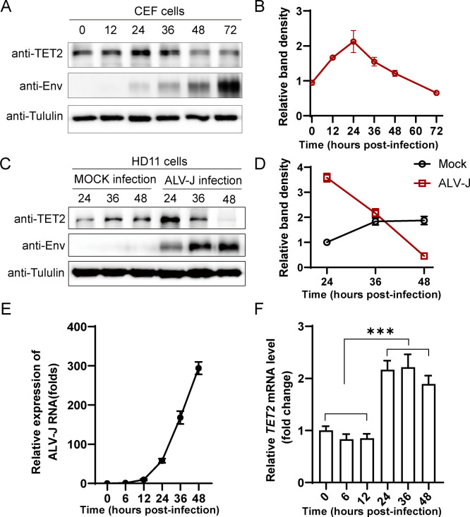
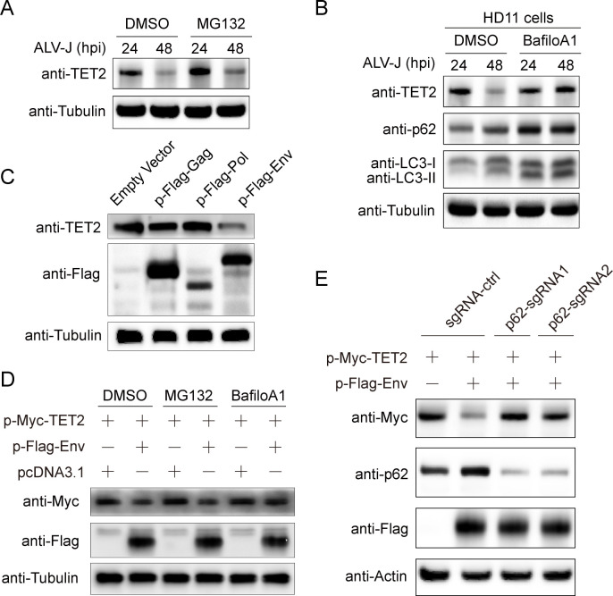
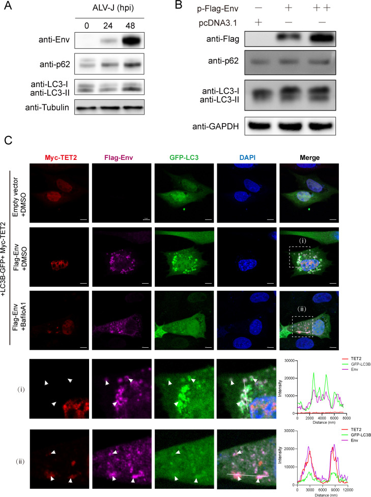
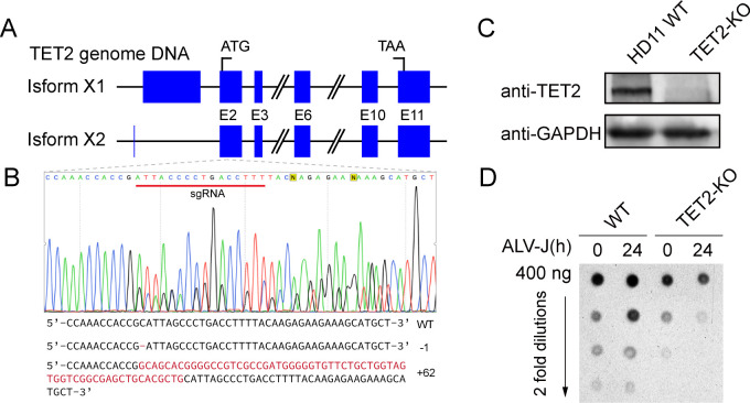
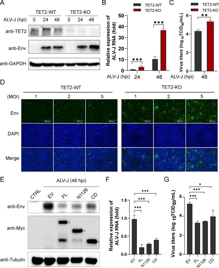
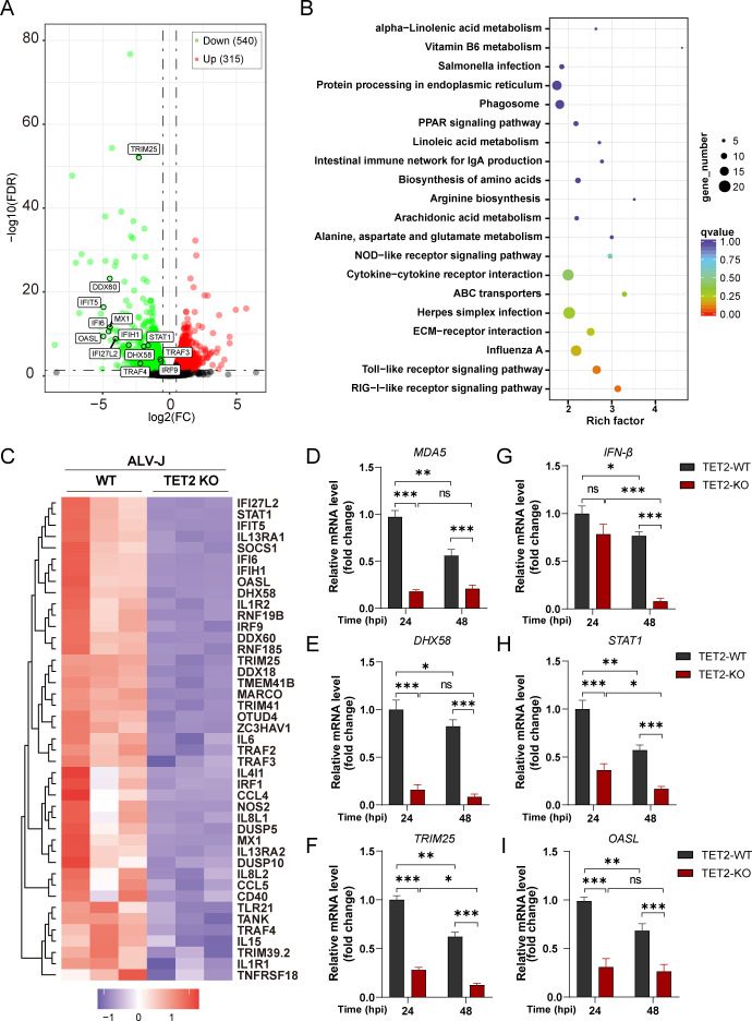
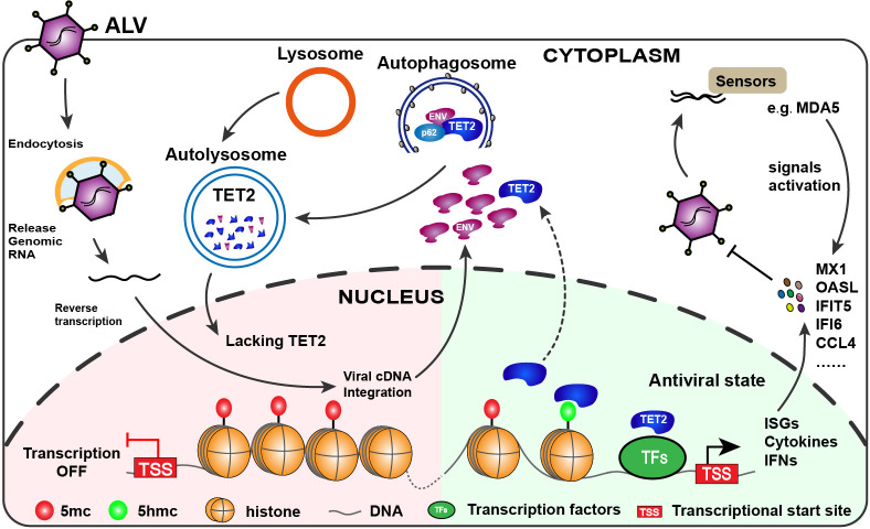
Avian leukosis virus subgroup J (ALV-J) poses a significant threat to the poultry industry; yet, our understanding of its replication and pathogenic mechanisms is limited. The Ten-Eleven Translocation 2 (TET2) is an indispensable regulatory factor in active DNA demethylation and immune response regulation. This study reports a significant and time-dependent decrease in TET2 levels following ALV-J infection and shows that the reduction of TET2 protein is mediated by the autophagy pathway. Mechanistically, we found that the accumulation of the Env protein at the late stages of ALV-J infection triggers autophagy, which, in turn, causes the TET2 protein to be exported from the nucleus and subsequently degraded in the cytoplasm. Using CRISPR-Cas9 technology, we generated TET2-deficient chicken macrophages that exhibited increased susceptibility to ALV-J replication, which was reversed by TET2 overexpression. In addition, transcriptome analysis revealed that the absence of TET2 in chicken macrophages impairs the expression of numerous cytokines and innate immune-related genes, particularly those downregulated genes enriched in the RIG-I and Toll-like signaling pathways, leading to enhanced replication of ALV-J in vitro. In summary, our research identifies TET2 as an ALV-J restriction factor. However, ALV-J exploits the autophagy machinery to promote the degradation of TET2 protein, thereby disrupting the host's innate immune responses for viral replication.

Importance: ALV-J is a carcinogenic retrovirus that plays a critical role in avian leukosis, primarily affecting chickens. Infection with ALV-J leads to decreased production performance, compromised immune function, and the development of tumors, such as myelocytoma. Currently, there are no effective treatments for ALV-J, making the control of outbreaks a significant challenge with severe economic consequences for the poultry industry. The Env protein of ALV-J has been implicated in the virus's pathogenicity. Our study shows that ALV-J infection induces autophagy in host cells through its Env protein, leading to the autophagic degradation of TET2, a key epigenetic regulator. The loss of TET2 in macrophages results in the downregulation of innate immune-related gene expression, thereby promoting viral replication. This is the first report to elucidate the role of the ALV-J Env protein in immune suppression via TET2 autophagic degradation during ALV-J infection, providing new insights into the mechanisms of viral immune evasion.

Keywords: Env; TET2; autophagic degradation; avian leukosis virus subgroup J; immunosuppression.


Conflict of interest statement

The authors declare no conflict of interest.

Figures

Fig 1
Fig 1
Time-dependent suppression of TET2 expression by ALV-J. (A) CEF cells were infected with ALV-J at an MOI of 1 and harvested at 0, 12, 24, 36, 48, and 72 hpi. TET2, viral Env, and Tubulin protein levels were analyzed via Western blot. (B) Quantification of TET2 to Tubulin protein levels is shown in the graph. (C) HD11 cells were infected with ALV-J at an MOI of 1, and samples were collected at 24, 36, and 48 hpi. Protein levels of TET2, viral Env, and Tubulin were examined by Western blot. (D) The graph shows the quantification of TET2 to Tubulin protein levels. (E and F) HD11 cells were infected with ALV-J at an MOI of 1. The fold changes in ALV-J Env (E) and TET2 (F) mRNA levels were detected by RT-qPCR at 6, 12, 24, 36, and 48 hpi, using Actin mRNA levels as an internal reference. Error bars represent the mean ± SD of three experiments. Statistical analysis was performed using Student’s t-test. ***P < 0.001.
Fig 2
Fig 2
ALV-J Env mediates the degradation of TET2 via the autophagy-lysosomal pathway. (A) HD11 cells were infected at an MOI of 1 for 24 h and 48 h, then treated with DMSO or MG132 (10 µM) for the last 6 h. Immunoblots were used to detect the expression of TET2, with Tubulin as a loading control. (B) HD11 cells were infected at an MOI of 1 for 24 h and 48 h, then treated with DMSO or bafilomycin A1 (BafioA1, 0.5 µM) for the final 4 h. Immunoblots were used to detect the expression of TET2, p62, and LC3B, with Tubulin as a loading control. (C) Endogenous TET2 expression levels were detected in HD11 cells transfected with empty vector, Gag, Pol, and Env. (D) DF-1 cells were transfected with Myc-TET2 and Flag-Env, and then treated with MG132 (10 µM) or bafilomycin A1 (BafioA1, 0.5 µM) for the last 4 h, and treated with DMSO as control. Cells were harvested for immunoblot analysis to determine TET2 and Env expression, with Tubulin as a loading control. (E) DF-1 cells were transfected with p62-targeting sgRNA or control sgRNA vectors, and GFP-positive cells were sorted by flow cytometry. These cells were plated and transfected with Myc-TET2 and Flag-Env for 24 h before harvest for Western blot detection of TET2, Env, and SQSTM1/p62, with Actin as a loading control.
Fig 3
Fig 3
ALV-J Env promotes autophagy and facilitates TET2 autophagic degradation. (A) HD11 cells were infected at an MOI of 1 for 24 and 48 h, with uninfected cells as a negative control. Western blot was utilized to assess the levels of Env, SQSTM1/p62, LC3B, and Tubulin as a loading control. (B) HD11 cells were transfected with either 1 µg or 2 µg of the Flag-Env expression vector or an empty vector for 36 h. Western blot was utilized to assess the levels of Env, SQSTM1/p62, and LC3B, with GAPDH as a loading control. (C) Confocal analysis was performed to assess the colocalization of TET2, Env, and LC3B. DF-1 cells were co-transfected with Myc-TET2 and LC3B-GFP plasmids, or with Myc-TET2, LC3B-GFP, and Flag-Env plasmids, followed by treatment with DMSO or bafilomycin A1 (0.5 µM) for 4 h as indicated, and then subjected to laser confocal analysis. The fluorescence channels for TET2 (excitation/emission at 594 nm), Env(647 nm), GFP, and DAPI were captured, followed by the overlay of images. (I) and (ii) represent the typical images of colocalization that were captured, with magnified views of the zoomed areas. The corresponding fluorescence intensity profiles of TET2 (red line), Env (pink line), and LC3B (green line) were obtained using Zeiss ZEN 3.10 analysis software. The scale bar is 5 µm.
Fig 4
Fig 4
ALV-J Env interacts with TET2 catalytic domain (CD). (A) Illustration of the complete structure of chicken TET2 and its truncated variants. (B) Interaction between Env protein and TET2. HEK293T cells were transfected with Myc-TET2 alone or co-transfected with Myc-TET2 and Flag-Env to evaluate the interaction between ALV-J Env and TET2. Cell lysates were immunoprecipitated with Myc-tag antibody, followed by Western blot analysis. (C) TET2 CD domain interacts with Env. HEK293T cells were co-transfected as indicated for 24 h, and immunoprecipitation with IgG or Myc-tag antibodies and Western blot analysis were conducted on cell lysates. (D) Env protein facilitates the degradation of the TET2 CD domain. DF-1 cells were co-transfected as indicated for 24 h and then analyzed by Western blot using cell lysates. (E) DF-1 cells were transfected with Myc-TET2-CD, EGFP-SQSTM1/p62, and Flag-Env for 24 h and were treated with bafilomycin A1 (0.5 µM) 4 h prior to cell collection. Immunoprecipitation with a GFP antibody was performed on cell lysates, followed by Western blot analysis.
Fig 5
Fig 5
TET2 gene knockout alters genome-wide levels of 5-hydroxymethylcytosine (5hmC). (A) A diagram illustrating the chicken TET2 gene locus. (B) Genomic sequencing illustrates the sgRNA-directed mutation at the chicken TET2 gene locus. (C) Western blot analysis validates the knockout of the TET2 gene in HD11 cells. (D) The global level of 5hmC was measured by a dot blot assay. Total genomic DNA was collected from the indicated cells, and the 5hmC levels was determined.
Fig 6
Fig 6
The expression of TET2 negatively regulates the replication of ALV-J. (A–D) Deletion of TET2 enhances the replication of ALV-J. (A) HD11 cells and TET2-KO cells were infected with ALV-J for 24 and 48 h, respectively, and Western blot analysis was performed to evaluate the expression of TET2 and the Env protein. (B) Subsequent to the treatments described in (A), qPCR analysis was conducted to measure the levels of ALV-J Env mRNA. (C) The viral titer in the supernatant of HD11 and TET2 KO cells at 48 hpi with ALV-J was determined using the TCID50 method. (D) After infection with indicated viral titers for 48 h, an Indirect immunofluorescence (IFA) assay was used to assess the ALV-J Env protein in HD11 and TET2 KO cells. (E–G) Overexpression of TET2 inhibits the replication of ALV-J. (E) DF-1 cells were infected with ALV-J (MOI = 1) for 24 h and then transfected with Myc-tagged TET2, deletion mutants Myc-N1126 and Myc-CD, or an empty vector for an additional 24 h. Western blot analysis was conducted to detect the expression of TET2 and ENV proteins. (F) Following the treatments described in (E), qPCR was performed to detect the expression levels of the ALV-J Env protein. (G) After the treatments described in (E), cell supernatants were collected and the viral titer was determined using the TCID50 method. Data are presented as mean ± SD. A two-tailed Student’s t-test is used for statistical analysis: *P < 0.05, **P < 0.05 and ***P < 0.001.
Fig 7
Fig 7
Deletion of TET2 significantly reduces the expression of host innate immune-related genes. (A) A volcano plot depicting the genes that are downregulated and upregulated in TET2 knockout HD11 cells as compared to the wild-type cells post 24 h ALV-J infection. (B) The genes identified as downregulated in (A) are presented according to the KEGG database, with rankings based on Q-values among the top 20 enriched pathways. (C) A heatmap illustrating the expression of innate immune-related genes in TET2 knockout HD11 cells as compared to the wild-type cells post 24 h ALV-J infection. (D–I) qPCR analysis of the expression changes in innate immune-related genes following TET2 knockout in HD11 cells that were infected for 24 and 48 h. The expression levels for each group were normalized to those of TET2-WT cells infected for 24 h. Data are presented as mean ± SD. A two-tailed Student’s t-test is used for statistical analysis: ns, no significant difference; *P < 0.05, **P < 0.01, ***P < 0.001.
Fig 8
Fig 8
Proposed model depicting the evasion of host immune defenses by ALV-J through the autophagic degradation of TET2. TET2 is essential for maintaining the expression of a series of cytokines and immune-related genes that are key to inhibiting the proliferation of ALV-J. Conversely, the Env protein of ALV-J induces autophagy, which facilitates the translocation of TET2 from the nucleus to the cytoplasm. In the cytoplasm, the interaction between the Env protein and TET2 leads to the selective autophagic degradation of TET2, preventing it from effectively activating the innate immune response and thus promoting viral replication.

Similar articles

Cited by

References

    1. Payne LN, Nair V. 2012. The long view: 40 years of avian leukosis research. Avian Pathol 41:11–19. doi:10.1080/03079457.2011.646237 - DOI - PubMed
    1. Gao YL, Qin LT, Pan W, Wang YQ, Le Qi X, Gao HL, Wang XM. 2010. Avian leukosis virus subgroup J in layer chickens, China. Emerg Infect Dis 16:1637–1638. doi:10.3201/eid1610.100780 - DOI - PMC - PubMed
    1. Chai N, Bates P. 2006. Na+/H+ exchanger type 1 is a receptor for pathogenic subgroup J avian leukosis virus. Proc Natl Acad Sci U S A 103:5531–5536. doi:10.1073/pnas.0509785103 - DOI - PMC - PubMed
    1. Yu M, Zhang Y, Zhang L, Wang S, Liu Y, Xu Z, Liu P, Chen Y, Guo R, Meng L, Zhang T, Fan W, Qi X, Gao L, Zhang Y, Cui H, Gao Y. 2024. N123I mutation in the ALV-J receptor-binding domain region enhances viral replication ability by increasing the binding affinity with chNHE1. PLoS Pathog 20:e1011928. doi:10.1371/journal.ppat.1011928 - DOI - PMC - PubMed
    1. Li T, Yao X, Li C, Zhang J, Xie Q, Wang W, Lu H, Fu H, Li L, Xie J, Shao H, Gao W, Qin A, Ye J. 2020. Gp37 regulates the pathogenesis of avian leukosis virus subgroup J via its C terminus. J Virol 94:e02180-19. doi:10.1128/JVI.02180-19 - DOI - PMC - PubMed

MeSH terms

LinkOut - more resources