Case Reports
. 2024 Nov 22;14(4):e24.00235.
doi: 10.2106/JBJS.CC.24.00235. eCollection 2024 Oct 1.

Intradural Disk Herniation at the Conus Medullaris: A Case Report With Emphasis on Patient Positioning and Neuromonitoring

Affiliations
Case Reports

Intradural Disk Herniation at the Conus Medullaris: A Case Report With Emphasis on Patient Positioning and Neuromonitoring

Michael J Kelly et al. JBJS Case Connect. .

Erratum in

Abstract

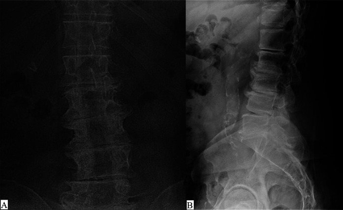
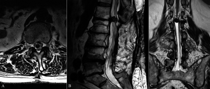
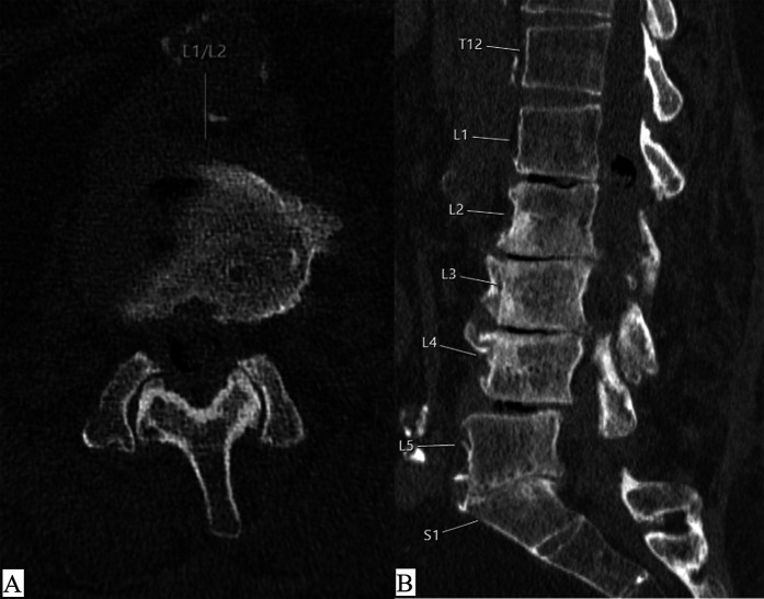
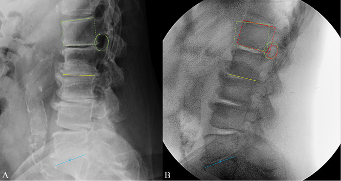
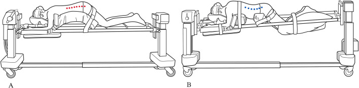
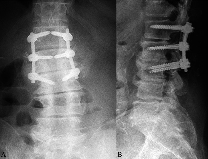
Case: A 73-year old man who underwent previous L2-S1 decompression presenting with new right radicular leg pain. Imaging suggests a large central disk herniation at L1-2 with possible intrathecal extension requiring surgical decompression. When positioned prone on a Jackson frame, neuromonitoring motor signals became diminished, and thus, the case was aborted. On returning to the operating room 2 days later, careful positioning in a more neutral/flexed position facilitated normal neuromonitoring signals, allowing for an uneventful intradural approach and discectomy.

Conclusion: With conus-level intrathecal disk herniation, consider using prepositional neuromonitoring and avoid hyperextension with positioning to ensure neurological safety.


Conflict of interest statement

Disclosure: The Disclosure of Potential Conflicts of Interest forms are provided with the online version of the article (http://links.lww.com/JBJSCC/C486).

Figures

Fig. 1
Fig. 1
Anteroposterior (Fig. 1-A) and lateral (Fig. 1-B) lumbar spine radiographs demonstrate multilevel spondylotic changes with evidence of prior L2-S1 decompression, flattening of the lumbar lordosis, and vacuum disk phenomenon most notable at the L1-2 level.
Fig. 2
Fig. 2
Axial (Fig. 2-A), sagittal (Fig. 2-B), and coronal (Fig. 2-C) MRI images of the lumbar spine (T2 image, TR/TE 4300/100 m without contrast) demonstrate postoperative findings consistent with prior L2-S1 decompression, along with a new disk extrusion at the L1-2 level extending centrally into the spinal canal, resulting in severe canal stenosis and peripheral displacement of the nerve roots. MRI = magnetic resonance imaging.
Fig. 3
Fig. 3
Axial (Fig. 3-A) and sagittal (Fig. 3-B) CT scan images of the lumbar spine demonstrate diffuse spondylotic changes in the lumbar spine, with diffuse vacuum phenomenon from L1-L5 and evidence of pneumorrhachis (intraspinal air) at the L1-2 level. CT = computed tomography.
Fig. 4
Fig. 4
Lumbar lordosis and segmental alignment relationship on the preoperative lateral radiograph (Fig. 4-A) vs. the lateral fluoroscopic image (Fig. 4-B) obtained with the patient positioned prone on the Jackson frame. Blue lines = sacral endplates with the preoperative radiograph paralleled and scaled to equal the intraoperative fluoroscopic image. Yellow lines = lower endplate of L2. Green lines = preoperative upper endplate of L1. Green dotted lines = preoperative position of L1 vertebral body and L1-2 neuroforamen. Red dotted lines = intraoperative position of L1 vertebral body and L1-2 neuroforamen. An overall increased lumbar lordosis is evident on the fluoroscopic image, along with a decrease in segmental kyphosis at L1-2, with lordosis increasing to neutral with the patient extended on the Jackson frame. Smaller neural foramina are evident on fluoroscopy. Here, lumbar spine extension has likely decreased the central canal area causing an exacerbation of neural compression between the IDH ventrally and ligamentum flavum dorsally. IDH = intradural disk herniation.
Fig. 5
Fig. 5
An example of patient positioning on the Wilson frame (Fig. 5-A), which allows for relative flexion through the thoracolumbar spine (red dots), compared with the extension moment (blue dots) precipitated by positioning on the Jackson frame (Fig. 5-B).
Fig. 6
Fig. 6
Intraoperative sterile ultrasound of the thecal sac after open exposure and laminectomy confirms the presence of an IDH (asterisk, middle of image) with dorsal displacement of the lumbosacral nerve roots (X, top of image). IDH = intradural disk herniation.
Fig. 7
Fig. 7
Fig. 7-A A midline dorsal durotomy was created, and retraction sutures were used for visualization throughout the operation. Fig. 7-B Careful mobilization of the nerve roots provided visualization of the IDH. Fig. 7-C The IDH is carefully isolated from the overlying neurological structures and dissected from the ventral dura, onto which it had partially adhered. A ventral dural defect was encountered, pointed out by the closed forceps here. IDH = intradural disk herniation.
Fig. 8
Fig. 8
Pathological analysis at 1× magnification (Fig. 8-A) and 20× magnification (Fig. 8-B), ultimately confirmed the specimen to be an IDH. IDH = intradural disk herniation.
Fig. 9
Fig. 9
Anteroposterior (Fig. 9-A) and lateral (Fig. 9-B) spine radiographs obtained at 3 months postoperative follow-up appointment.

Similar articles

Cited by

References

    1. Deyo RA, Mirza SK. CLINICAL PRACTICE. herniated lumbar intervertebral disk. N Engl J Med. 2016;374(18):1763-72. - PubMed
    1. Pojskic M, Bisson E, Oertel J, Takami T, Zygourakis C, Costa F. Lumbar disc herniation: epidemiology, clinical and radiologic diagnosis WFNS spine committee recommendations. World Neurosurg X. 2024;22:100279. - PMC - PubMed
    1. Dandy WE. Serious complications of ruptured intervertebral disks. JAMA. 1942;119(6):474-7.
    1. Hu X, Chang W. An unusual case report of intradural disc herniation mimicking the spinal intradural tumor. Asian J Surg. 2023;46(12):5546-7. - PubMed
    1. Ihejirika RC, Tong Y, Patel K, Protopsaltis T. Intradural lumbar disc herniation: illustrative case. J Neurosurg Case Lessons. 2021;2(18):CASE21336. - PMC - PubMed

Publication types