

Coronary Computed Tomography Angiography in Heart Transplant Patients: Current Insights and Future Directions
- PMID: 39841094
- PMCID: PMC12091219
- DOI: 10.1097/TP.0000000000005266
Coronary Computed Tomography Angiography in Heart Transplant Patients: Current Insights and Future Directions
Abstract
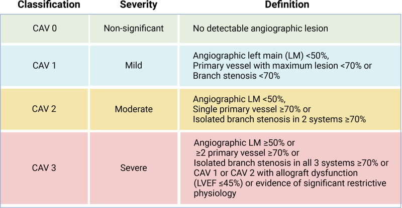
Cardiac allograft vasculopathy (CAV) remains a significant challenge after heart transplantation, necessitating effective surveillance methods. This review centers around the role of coronary computed tomography angiography (CCTA) in CAV surveillance, given its unique capabilities to visualize and quantify CAV in comparison with other imaging modalities, including invasive coronary angiography and intravascular ultrasound. CCTA has shown good diagnostic performance for detecting and monitoring CAV, exemplified by a higher sensitivity and negative predictive value compared with invasive coronary angiography. Additionally, CCTA can provide valuable functional insights with fractional flow reserve integration. An additional, considerable benefit of CCTA is that it allows for the opportunity to assess other imaging markers of cardiometabolic and general health, including coronary artery calcium score, epicardial fat volume, liver fat, vertebral bone density, and lung density, which allows for a comprehensive assessment of the overall health of the patient.
Copyright © 2024 The Author(s). Published by Wolters Kluwer Health, Inc.
Figures


References
-
- Nikolova AP, Kobashigawa JA. Cardiac allograft vasculopathy: the enduring enemy of cardiac transplantation. Transplantation. 2019;103:1338–1348. - PubMed
-
- Velleca A, Shullo MA, Dhital K, et al. The International Society for Heart and Lung Transplantation (ISHLT) guidelines for the care of heart transplant recipients. J Heart Lung Transplant. 2023;42:e1–e141. - PubMed
-
- Chih S, Chong AY, Mielniczuk LM, et al. Allograft vasculopathy: the Achilles’ heel of heart transplantation. J Am Coll Cardiol. 2016;68:80–91. - PubMed
-
- Khush KK, Cherikh WS, Chambers DC, et al. ; International Society for Heart and Lung Transplantation. The International Thoracic Organ Transplant Registry of the International Society for Heart and Lung Transplantation: Thirty-sixth adult heart transplantation report—2019; focus theme: donor and recipient size match. J Heart Lung Transplant. 2019;38:1056–1066. - PMC - PubMed
-
- Laks JA, Dipchand AI. Cardiac allograft vasculopathy: a review. Pediatr Transplant. 2022;26:e14218. - PubMed
Publication types
MeSH terms
LinkOut - more resources
Full Text Sources
Medical
