. 2025 Jan 28;122(4):e2423114122.
doi: 10.1073/pnas.2423114122. Epub 2025 Jan 22.

Itaconate mechanism of action and dissimilation in Mycobacterium tuberculosis

Affiliations

Itaconate mechanism of action and dissimilation in Mycobacterium tuberculosis

Manisha Priya et al. Proc Natl Acad Sci U S A. .

Abstract

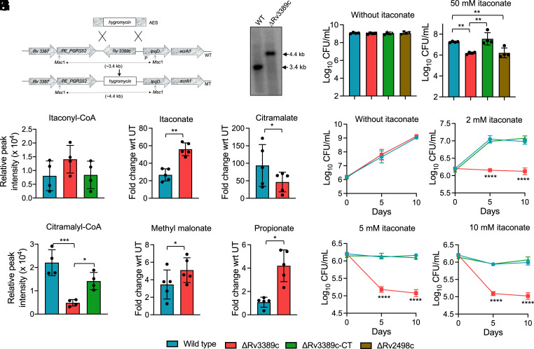
Itaconate, an abundant metabolite produced by macrophages upon interferon-γ stimulation, possesses both antibacterial and immunomodulatory properties. Despite its crucial role in immunity and antimicrobial control, its mechanism of action and dissimilation are poorly understood. Here, we demonstrate that infection of mice with Mycobacterium tuberculosis increases itaconate levels in lung tissues. We also show that exposure to itaconate inhibits M. tuberculosis growth in vitro, in macrophages, and mice. We report that exposure to sodium itaconate (ITA) interferes with the central carbon metabolism of M. tuberculosis. In addition to the inhibition of isocitrate lyase (ICL), we demonstrate that itaconate inhibits aldolase and inosine monophosphate (IMP) dehydrogenase in a concentration-dependent manner. Previous studies have shown that Rv2498c from M. tuberculosis is the bona fide (S)-citramalyl-CoA lyase, but the remaining components of the pathway remain elusive. Here, we report that Rv2503c and Rv3272 possess itaconate:succinyl-CoA transferase activity, and Rv2499c and Rv3389c possess itaconyl-CoA hydratase activity. Relative to the parental and complemented strains, the ΔRv3389c strain of M. tuberculosis was attenuated for growth in itaconate-containing medium, in macrophages, mice, and guinea pigs. The attenuated phenotype of ΔRv3389c strain of M. tuberculosis is associated with a defect in the itaconate dissimilation and propionyl-CoA detoxification pathway. This study thus reveals that multiple metabolic enzymes are targeted by itaconate in M. tuberculosis. Furthermore, we have assigned the two remaining enzymes responsible for the degradation of itaconic acid into pyruvate and acetyl-CoA. Finally, we also demonstrate the importance of enzymes involved in the itaconate dissimilation pathway for M. tuberculosis pathogenesis.

Keywords: Mycobacterium tuberculosis; dissimilation; itaconate; pathogenesis.


Conflict of interest statement

Competing interests statement:The authors declare no competing interest.

Figures

Fig. 1.
Fig. 1.
Itaconate restricts the growth of Mycobacterium tuberculosis in vitro and in vivo. (A and B) The relative levels of Irg1 (A) and itaconate (B) in the lung tissues of mice infected with M. tuberculosis at 2- and 4-wk postinfection were calculated as described in Materials and Methods. The data shown in panels (A) and (B) are obtained from three or four mice, respectively. (C) The growth of M. tuberculosis in MB7H9 medium was determined after exposure to various concentrations of sodium itaconate (ITA) or dimethyl itaconate (DI). (D) THP-1 macrophages were infected with M. tuberculosis strain at 1:10 MOI and treated with different concentrations of DI or ITA for 4 d. The data shown in panels C and D are the mean ± SD of log10 CFU obtained from two independent experiments performed in either duplicates or triplicates. (E and F) 6 to 8 wk old female BALB/c mice were infected with M. tuberculosis via aerosol route. The animals were administered intraperitoneally with 50 mg/kg DI for either 7 or 21 d. The data shown in these panels are mean ± SD of log10 CFU of lungs (E) and splenic (F) bacillary loads obtained from five animals per group. (GK) The intracellular levels of IL-1β (G), TNF-α (H), IFN–γ (I), IL-10 (J), and IL-6 (K) were determined in naïve, untreated, and DI treated groups at day 21 posttreatment by ELISA. The data shown in these panels are mean ± SD obtained from five animals. The data were statistically analyzed using one-way ANOVA (A, G, H, I, J, and K) or a two-tailed “paired” t test (B, E, and F). *P < 0.05, **P < 0.01, ***P < 0.001 and ****P < 0.0001.
Fig. 2.
Fig. 2.
(AK) The effect of itaconate exposure on the metabolite profiles of M. tuberculosis. (AC) Principal component analysis of the metabolite profiles obtained from either untreated or from early log phase cultures of M. tuberculosis exposed to either 10 mM (A), 20 mM (B), or 50 mM (C) ITA for either 3 or 6 d. (DK) This panel depicts the relative abundance of itaconate (D), citramalate (E), acetyl-CoA (F), glucose-6-phosphate (G6P, G), fructose-6-phosphate (F6P, H), dihydroxyacetone phosphate (DHAP, I), inosine monophosphate (IMP, J), and xanthosine monophosphate (XMP, K) in untreated and ITA-treated cultures. RPI represents relative peak intensity. The data were statistically analyzed using one-way ANOVA. *P < 0.05, **P < 0.01, ***P < 0.001, and ****P < 0.0001. (LU) Itaconate binds and inhibits the enzymatic activity of aldolase (Ald) and IMP dehydrogenase (GuaB2). (L and M) The binding of Ald (L) and GuaB2 (M) in the presence of various concentrations of ITA was measured by tryptophan fluorescence quenching assay as described in Materials and Methods. (N and O) The enzymatic assays of Ald (N) and GuaB2 (O) were performed in the presence of different concentrations of ITA. The data shown in these panels are mean ± SD of percentage inhibition of the enzymatic activity of Ald or GuaB2 obtained from two independent experiments performed in duplicates or triplicates. (P and Q) These panels show mean ± SD of Ald (P) or GuaB2 (Q) activity in the presence of 10 mM ITA or cis-aconitic acid (CAA) or methyl succinic acid (MSA). The data shown in these panels are obtained from two independent experiments performed in duplicates. (R and S) These panels show the molecular docking of Ald with either sodium itaconate (R) or fructose-1,6 biphosphate (S). (T and U) The molecular docking of GuaB2 with sodium itaconate (T) or IMP (U) is shown in these panels.
Fig. 3.
Fig. 3.
Functional characterization of enzymes from M. tuberculosis involved in itaconate dissimilation pathway. (A) Schematic representation of itaconate dissimilation pathway in bacteria. IcT, IcH, and CcL enzymes represent itaconate:succinyl-CoA transferase, itaconyl-CoA hydratase, and citramalyl-CoA lyase, respectively. (B) This panel shows that representative LC-chromatogram of the succinyl-CoA, itaconyl-CoA, citramalyl-CoA, and acetyl-CoA extracted at their corresponding m/z. (CE) This panel shows the relative abundance of peaks corresponding to itaconyl-CoA (C), citramalyl-CoA (D), pyruvate and acetyl-CoA (E) in enzymatic assays performed using purified proteins. The data shown in these panels are the mean ± SD of data obtained from five independent experiments. (FH) This panel shows the percentage activity of purified Rv2503c (F), Rv2499c (G), or Rv3389c (H) mutant proteins relative to the wild type protein. The data shown are mean ± SD of percentage activity for various purified proteins obtained from two independent experiments performed in triplicates. The data were statistically analyzed using one-way ANOVA. ***P < 0.001, ****P < 0.0001.
Fig. 4.
Fig. 4.
Itaconate dissimilation pathway is disrupted in ΔRv3389c strain of M. tuberculosis. (A) A schematic representation of the Rv3389c genetic locus in the genome of parental and ΔRv3389c strains is shown in this panel. The replacement of Rv3389c with the hygromycin resistance gene was confirmed by Southern blot. (B) The growth patterns of parental, ΔRv3389c, Rv3389c complemented, and ΔRv2498c strains were compared in the absence or presence of ITA for 8 d. The data shown in this panel are mean ± SD of log10 CFU/ml obtained from two independent experiments performed in duplicates. (C and D) The enzymatic reactions were performed using CFPE prepared from mid-log phase cultures of either parental or ΔRv3389c or Rv3389c complemented strain. The data shown in these panels are mean ± SD of the relative peak intensity of itaconyl-CoA (C) or citramalyl-CoA (D) obtained from four independent experiments. (EH) This panel depicts the relative fold change of itaconate (E), citramalate (F), methylmalonate (G), and propionate (H) in 50 mM ITA exposed mid-log phase cultures of parental and ΔRv3389c strain for 3 d. The data shown in this panel are the mean ± SD of five independent experiments. (IL) The growth patterns of parental, ΔRv3389c, and Rv3389c complemented strains were compared in a medium containing 10 mM propionate in the absence (I) or presence of 2 mM (J), 5 mM (K), and 10 mM sodium itaconate (L). The data shown in these panels are mean ± SD of log10 CFU obtained from two independent experiments performed in duplicates. The data were statistically analyzed using one-way ANOVA (B, C, D, J, K, and L) or two-tailed “paired” t test (E, F, G, and H). *P < 0.05, **P < 0.01, ***P < 0.001, ****P < 0.0001.
Fig. 5.
Fig. 5.
Deletion of Rv3389c impairs the survival of M. tuberculosis in macrophages, mice and guinea pigs (A) The survival of parental, ΔRv3389c and Rv3389c complemented strain was compared after exposure to oxidative stress for either 6 h or 24 h or 72 h. The data shown in this panel are mean ± SD of log10 CFU obtained from two independent experiments performed in duplicates. (BD) The growth kinetics of parental, ΔRv3389c and Rv3389c complemented strain was compared in THP-1 macrophage (B) or A549 lung epithelial cell line in the absence (C) or presence of 10 mM itaconate (D) after 2, 4, and 6 d postinfection. The data shown in these panels are mean ± SD of log10 CFU obtained from two or three independent experiments performed in triplicates. (EH) The data shown in these panels are mean ± SD of log10 CFU of lungs (E and G) and splenic (F and H) bacillary loads obtained from 5 BALB/c mice infected with various strains at 4 and 8 wk postinfection. (I) The growth of parental, ΔRv3389c and Rv3389c complemented strain was compared in BMDMs isolated from wild type or Irg1−/− C57BL/6 mice after 4 d postinfection. The data shown in this panel are mean ± SD of log10 CFU obtained from two independent experiments performed in triplicates or quadruplicates. (J) The growth patterns of parental, ΔRv3389c and Rv3389c complemented, Rv3389cQ199A complemented, and Rv3389cC212A complemented strain in THP-1 macrophages after day 6-post infection. The data shown in this panel are mean ± SD of log10 CFU obtained from two independent experiments performed in triplicates. (K and L) The data shown in these panels are mean ± SD of log10 CFU obtained from lungs (K), and spleens (L) bacillary loads in BALB/c mice (n= 5) infected with either parental or ΔRv3389c or Rv3389c complemented, Rv3389cQ199A complemented, and Rv3389cC212A complemented strains at 4 wk postinfection. (M and N) The data shown in these panels are mean ± SD of log10 CFU of lungs (M) and splenic (N) bacillary loads obtained from 5 or 6 guinea pigs infected with either parental or ΔR3389c strain at 4 and 8 wk postinfection. The data obtained were statistically analyzed using one-way ANOVA (panels A, B, D, G, H, I, J, K, and L) or two-tailed “paired” t test (panels E, F, M, and N). **P < 0.01, ***P < 0.001, and ****P < 0.0001.
Fig. 6.
Fig. 6.
Proposed model for itaconate dissimilation pathway and attenuation of ΔRv3389c strain in vivo. Our findings demonstrate that itaconate production is increased in the lung tissues of mice upon M. tuberculosis infection. We also show that itaconate inhibits M. tuberculosis growth by inhibiting the enzymatic Ald or IMP dehydrogenase (GuaB2) in addition to isocitrate lyase (ICL) enzyme. The exposure of M. tuberculosis to itaconate increases the carbon flux from glycolytic to pentose phosphate pathway and purine biosynthesis in M. tuberculosis. We also unambiguously assign the enzymes from M. tuberculosis responsible for IcT and IcH activity. These enzymes collectively degrade itaconate into acetyl CoA and pyruvate. We also demonstrate that the Rv3389c (IcH) mutant strain has a defective itaconate degradation pathway, which results in itaconyl CoA accumulation in the strain. This accumulation of itaconyl CoA might inhibit MCM enzymatic activity that is involved in the propionyl CoA detoxification pathway. Further, we show that disruption of the itaconate degradation pathway leads to reduced intracellular survival of ΔRv3389c in vivo. This figure is prepared using Biorender.

References

    1. Bagcchi S., WHO’s global tuberculosis report 2022. Lancet Microbe 4, e20 (2023). - PubMed
    1. Chang D. P. S., Guan X. L., Metabolic versatility of mycobacterium tuberculosis during infection and dormancy. Metabolites 11, 88 (2021). - PMC - PubMed
    1. Hubert C. B., de Carvalho L. P. S., Convergence and divergence in the metabolic network of Mycobacterium tuberculosis. Curr. Opin. Syst. Biol. 28, 100384 (2021).
    1. Munoz-Elias E. J., Upton A. M., Cherian J., McKinney J. D., Role of the methylcitrate cycle in Mycobacterium tuberculosis metabolism, intracellular growth, and virulence. Mol. Microbiol. 60, 1109–1122 (2006). - PubMed
    1. Savvi S., et al. , Functional characterization of a vitamin B12-dependent methylmalonyl pathway in Mycobacterium tuberculosis: Implications for propionate metabolism during growth on fatty acids. J. Bacteriol. 190, 3886–3895 (2008). - PMC - PubMed

MeSH terms

LinkOut - more resources