. 2025 Jan 22;149(1):10.
doi: 10.1007/s00401-025-02848-9.

Traumatic brain injury causes early aggregation of beta-amyloid peptides and NOTCH3 reduction in vascular smooth muscle cells of leptomeningeal arteries

Affiliations

Traumatic brain injury causes early aggregation of beta-amyloid peptides and NOTCH3 reduction in vascular smooth muscle cells of leptomeningeal arteries

Ilknur Özen et al. Acta Neuropathol. .

Abstract

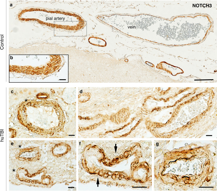
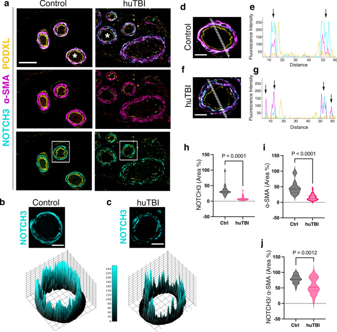
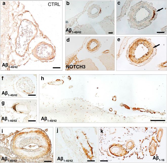
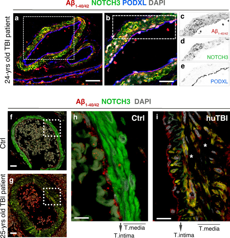
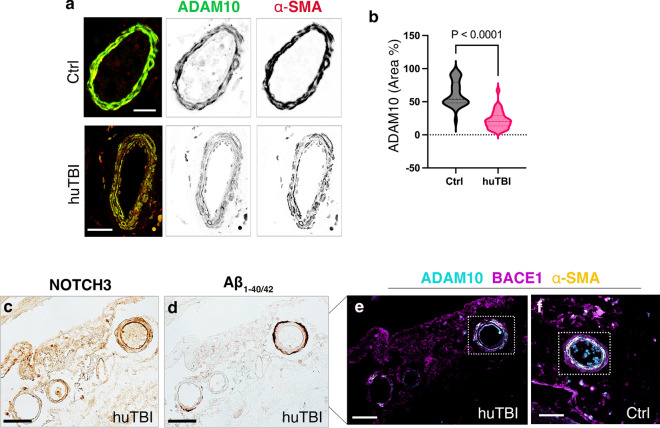
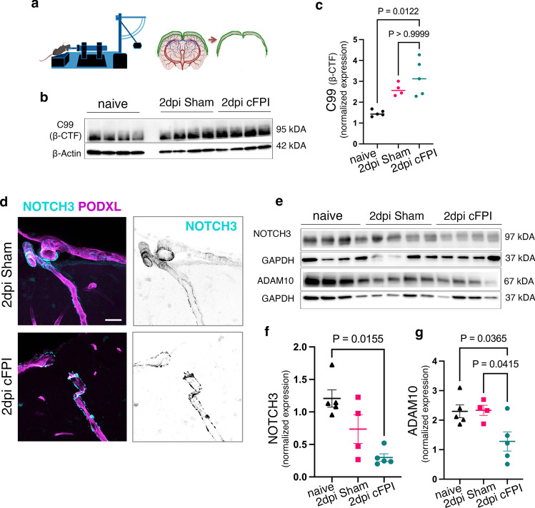
Traumatic brain injury (TBI) often leads to impaired regulation of cerebral blood flow, which may be caused by pathological changes of the vascular smooth muscle cells (VSMCs) in the arterial wall. Moreover, these cerebrovascular changes may contribute to the development of various neurodegenerative disorders such as Alzheimer's-like pathologies that include amyloid beta aggregation. Despite its importance, the pathophysiological mechanisms responsible for VSMC dysfunction after TBI have rarely been evaluated. Here, we show that acute human TBI resulted in early pathological changes in leptomeningeal arteries, closely associated with a decrease in VSMC markers such as NOTCH3 and alpha smooth muscle actin (α-SMA).These changes coincided with increased aggregation of variable-length amyloid peptides including Aβ1-40/42,1-16, and β-secretase-derived fragment (βCTF) (C99) caused by altered processing of amyloid precursor protein (APP) in VSMCs. The aggregation of Aβ1-40/42 peptides were also observed in the leptomeningeal arteries of young TBI patients. These pathological changes also included higher β-secretase (BACE1) when compared to α-secretase A Disintegrin And Metalloprotease 10 (ADAM10) expression in the leptomeningeal arteries, plausibly caused by hypoxia and oxidative stress as shown using human VSMCs in vitro. Importantly, BACE1 inhibition not only restored NOTCH3 signalling but also normalized ADAM10 levels in vitro. Furthermore, we found reduced ADAM10 activity and decreased NOTCH3, along with increased βCTF (C99) levels in mice subjected to an experimental model of TBI. This study provides evidence of early post-injury changes in VSMCs of leptomeningeal arteries that can contribute to vascular dysfunction and exacerbate secondary injury mechanisms following TBI.

Keywords: Amyloid beta; NOTCH3; Traumatic brain injury (TBI); Vascular smooth muscle cells; β-Secretase-derived fragment (βCTF) (C99).


Conflict of interest statement

Declarations. Conflict of interest: The authors declare that there is no conflict of interest.

Figures

Fig. 1
Fig. 1
Altered NOTCH3 expression in leptomeningeal arteries of human acute TBI subjects. NOTCH3 expression in the leptomeningeal arteries of a, b control subjects and cg brain samples surgically resected from severe, acute human TBI patients. a Representative bright-field images of NOTCH3 in leptomeningeal arteries of different sizes in control subjects. b The boxed area in a showing NOTCH3 staining through the full thickness of tunica media of artery walls of the control subject. c–g Transverse and longitudinal views of leptomeningeal arteries of acute human TBI patients showing a partial circumference abnormality of NOTCH3 expression in the tunica media (asterisks in c, e, and arrows in f). Scale bars = 200 μm in (a), 20 μm in (bg)
Fig. 2
Fig. 2
Acute human TBI-induced NOTCH3 changes are specific to vascular smooth muscle cells in the tunica media. a Representative confocal images of triple staining of NOTCH3 (cyan), α-SMA (magenta), and podocalyxin (yellow) in the leptomeningeal arteries of control and human TBI subjects. b, c Interactive 3D surface plots of individual leptomeningeal arteries in boxed areas in a showing spatial intensity profile of NOTCH3 expression in the tunica media. dg Higher magnification confocal images of leptomeningeal arteries (white asterisks in a) and the fluorescence intensity profiles of NOTCH3, α-SMA, and podocalyxin expression. h, i Quantification of NOTCH3 (h), α-SMA (i), NOTCH3/ α-SMA co-labelling (j) expressed as a percentage (%) of tunica media of leptomeningeal arteries of control and human TBI subjects (n = 22 arteries, control subjects, n = 46 arteries in TBI patients, Mann–Whitney test, Mean ± SEM). Scale bars = 50 μm in (a), 10 μm in (b, c), 20 μm in (d, f)
Fig. 3
Fig. 3
Amyloid beta peptides in the leptomeningeal arteries of human acute TBI subjects. ac Representative bright-field images of Aβ1-40/42 (MOAB-2) of leptomeningeal arteries in human acute TBI sections compared with control subject. Aβ1-40/42 deposits in the tunica media where NOTCH3 expression decreased (bc, arrows). Transverse and longitudinal views of leptomeningeal arteries with different sizes showing patchy Aβ1-40/42 (MOAB-2) (ac, f, g, ik) and Aβ1-16 (6E10) (k) peptides around tunica media (ac, f, g and i) or as a thin rim spanning the entire vessel wall circumference (h, j, and k). Scale bars = 50 μm in (ag, ik), 200 μm in (h)
Fig. 4
Fig. 4
Accumulation of beta-amyloid peptides is associated with changes in NOTCH3 expression in leptomeningeal arteries. a Confocal images of triple staining of Aβ1-40/42 (red), NOTCH3 (green), and podocalyxin (blue) in the leptomeningeal arteries of a 24-years old TBI patient. Scale bars = 50 μm (b) Higher magnification of boxed area in (a) showing changes in the expression of Aβ1-40/42 (red), NOTCH3 (green), and podocalyxin (blue) in vascular smooth muscle cells of tunica media. ce Confocal pictures in the boxed area (b) are inverted into greyscale. f, g Representative confocal images of double staining of NOTCH3 (green), Aβ1−40/42 (red) in medium-sized and large leptomeningeal arteries of control and human TBI subjects. Scale bars = 20 μm. hi Higher magnification confocal images of large leptomeningeal arteries in a 25-years old TBI patient (white boxed in f and g) showing a decrease in NOTCH3 (green) signalling in the vascular smooth muscle cells (VSMCs) and Aβ1-40/42 accumulation in the VSMCs. Scale bars = 10 μm in (h, i)
Fig. 5
Fig. 5
ADAM10 levels decreases in the leptomeningeal arteries following acute human TBI. a Confocal images of staining of ADAM10 (cyan) and α-SMA (yellow), which were inverted into greyscale in boxes, in the leptomeningeal arteries of control and human TBI subjects. Scale bars = 20 μm. bc Quantification of ADAM10/ α-SMA co-labelling (c) expressed as a percentage (%) of tunica media of leptomeningeal arteries of control and human TBI subjects (n = 38 arteries, control subjects, n = 41 arteries, Mann–Whitney test, Mean ± SEM) (f) A representative bright-field image showing dense Aβ deposition covering the whole wall of the medium-sized leptomeningeal arteries in the human acute TBI subject. g, h Immunofluorescence staining of ADAM10 (cyan), BACE1 (magenta), and α-SMA (yellow) in the medium-sized leptomeningeal artery (boxed in f) of (g) human TBI and (h) of control subjects. Scale bars = 50 μm
Fig. 6
Fig. 6
Human TBI increases hypoxia and proliferation in the VSMCS of leptomeningeal arteries. a, b Bright-field images of prolyl 4-hydroxylase-2 (PHD2) of leptomeningeal arteries in human acute TBI sections compared with control subject, scale 20 μm. c, d Confocal images of double staining of PHD2 (red) and PCNA (green) in leptomeningeal arteries of control and human TBI subjects, scale 20 μm. DAPI (grey) was used as counterstaining. e Higher magnification confocal image of the boxed area in large leptomeningeal arteries from human TBI subject showing co-expression of PCNA and PHD2, scale 50 μm
Fig. 7
Fig. 7
Hypoxia/oxidative stress in human VSMCs decreases NOTCH3/N3ICD and ADAM10 expression while increasing NOTCH3/ N3ECD aggregates and Aβ1-16. a The illustration shows the experimental design of oxygen and glucose deprivation /re-oxygenation (OGD/R) in vitro. b NOTCH3/N3ECD aggregates (green) on human VSMCs showing decreased NOTHC3/N3ICD levels (red) after exposure to OGD/R. c Confocal images of ADAM10 (red), Aβ1–16 levels (blue), and Phalloidin (green) staining in VSMCs exposed to OGD/R and in controls (d–e) The fluorescence intensity profiles of ADAM10 (red), Aβ1–16 levels (blue) and Phalloidin (green) in (d) control human VSMCs and (e) the ones exposed to OGD/R. Scale bars = 10 μm in (b, c)
Fig. 8
Fig. 8
BACE1 inhibition restores NOTCH3 and ADAM 10 levels in human VSMCs exposed to oxygen and glucose deprivation /re-oxygenation (OGD/R). a Confocal images of immunofluorescent staining of βCTF (C99) (magenta), BACE1 (cyan), and in Phalloidin (yellow) in VSMCs in control and OGD/R, which were inverted into greyscale Scale bar = 10 μm. bc The fluorescence intensity profiles of βCTF (C99) (magenta), BACE1 (cyan), and in Phalloidin (yellow) in VSMCs in (b) control and (c) OGD/R. d Confocal images of triple staining of NOTCH3 (green), βCTF (C99) (red), and podocalyxin (blue) in the leptomeningeal arteries of control and human TBI subjects. Scale bar = 10 μm (e) The illustration shows the experimental design of oxygen and glucose deprivation /re-oxygenation (OGD/R) in vitro and BACE1 inhibitor treatment of VSMCs. f Representative Western blots and (gh) quantification of g NOTCH3 intracellular domain (NICD3) (97 kDA) and h ADAM10, active form (67 kDA) in VSMCs exposed to OGD/R and BACE1 inhibitor. (mean ± SEM; n = 3 (Ctrl), n = 3 (OGD/R), n = 3 (OGD/R + BACE1inh); one-way ANOVA followed by Tukey post hoc test, *P < 0.05)
Fig. 9
Fig. 9
Experimental diffuse TBI causes a decrease in NOTCH3 signalling in leptomeningeal arteries accompanied by increased C99 levels and decreased ADAM10 activity. The illustration shows experimental design and the use of the central fluid percussion mouse model of TBI (cFPI; left) and isolation of sham or injured cortical consisting of leptomeningeal and penetrating leptomeningeal arteries. b Representative Western blots and (c) quantification of C99 (βCTF) band intensity in naïve, sham, and cFPI mice at 2 dpi (mean ± SEM; n = 5 (naïve), n = 4 (sham), n = 5 (cFPI); Kruskal–Wallis one-way ANOVA with Dunn’s post-test **P < 0.01). d Confocal images showing decreased NOTCH3 (magenta), expression on penetrating leptomeningeal arteries in the somatosensory cortex of sham-injured and cFPI-injured mice), NOTCH3 (magenta), and podocalyxin (cyan). Scale bar = 20 μm e Representative Western blots and fg quantification of NOTCH3 intracellular domain (NICD3) (97 kDA) (f) and ADAM10, active form (67 kDA) (g) band intensity (mean ± SEM; n = 5 (naïve), n = 4 (sham), n = 5 (cFPI); ordinary one-way ANOVA followed by Tukey post hoc test, *P < 0.05)

Similar articles

Cited by

References

    1. Abrahamson EE, Ikonomovic MD (2020) Brain injury-induced dysfunction of the blood brain barrier as a risk for dementia. Exp Neurol 328:113257. 10.1016/j.expneurol.2020.113257 - PubMed
    1. Abu Hamdeh S, Waara ER, Möller C, Söderberg L, Basun H, Alafuzoff I et al (2018) Rapid amyloid-β oligomer and protofibril accumulation in traumatic brain injury. Brain Pathol 28:451–462. 10.1111/bpa.12532 - PMC - PubMed
    1. Aldea R, Weller RO, Wilcock DM, Carare RO, Richardson G (2019) Cerebrovascular smooth muscle cells as the drivers of intramural periarterial drainage of the brain. Front Aging Neurosci 11:1–17. 10.3389/fnagi.2019.00001 - PMC - PubMed
    1. Amyot F, Kenney K, Spessert E, Moore C, Haber M, Silverman E et al (2020) Assessment of cerebrovascular dysfunction after traumatic brain injury with fMRI and fNIRS. Neuroimage Clin 25:102086. 10.1016/j.nicl.2019.102086 - PMC - PubMed
    1. Ashina H, Christensen RH, Al-Khazali HM, Iljazi A, Tolnai D, Eigenbrodt AK et al (2023) White matter hyperintensities and cerebral microbleeds in persistent post-traumatic headache attributed to mild traumatic brain injury: a magnetic resonance imaging study. J Headache Pain 24:15. 10.1186/s10194-023-01545-w - PMC - PubMed

Publication types

MeSH terms

LinkOut - more resources