. 2025 Mar 1;36(3):ar28.
doi: 10.1091/mbc.E24-07-0303. Epub 2025 Jan 22.

Roles for the canonical polarity machinery in the de novo establishment of polarity in budding yeast spores

Affiliations

Roles for the canonical polarity machinery in the de novo establishment of polarity in budding yeast spores

Benjamin Cooperman et al. Mol Biol Cell. .

Abstract

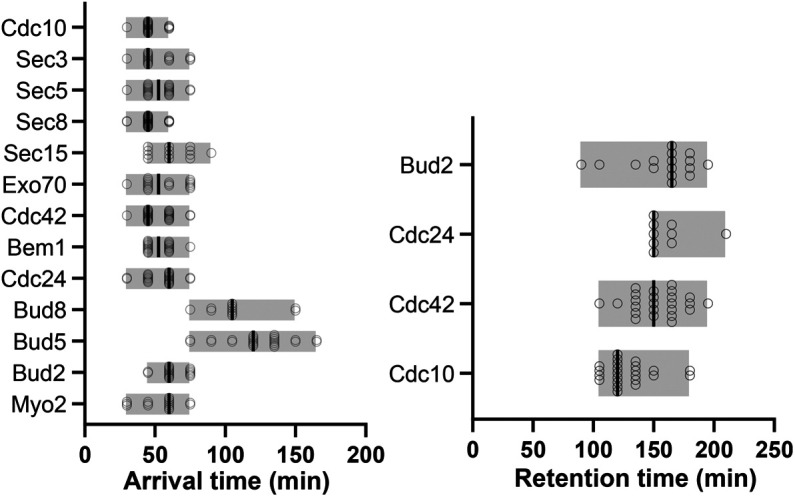
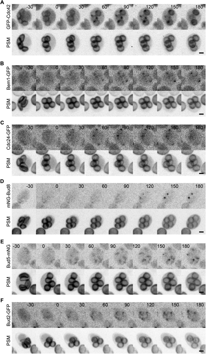
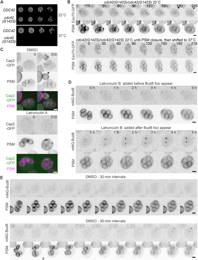
The yeast Saccharomyces cerevisiae buds at sites predetermined by cortical landmarks deposited during prior budding. During mating between haploid cells in the lab, external pheromone cues override the cortical landmarks to drive polarization and cell fusion. By contrast, in haploid gametes (called spores) produced by meiosis, a predetermined polarity site drives initial polarized morphogenesis independent of mating partner location. Spore membranes are made de novo so existing cortical landmarks were unknown, as were the mechanisms by which the spore polarity site is made and how it works. We find that the landmark canonically required for distal budding, Bud8, stably marks the spore polarity site along with Bud5, a GEF for the GTPase Rsr1 that canonically links cortical landmarks to the conserved Cdc42 polarity machinery. Cdc42 and other GTPase regulators arrive at the site during its biogenesis, after spore membrane closure but apparently at the site where membrane synthesis began, and then these factors leave, pointing to the presence of discrete phases of maturation. Filamentous actin may be required for initial establishment of the site, but thereafter Bud8 accumulates independent of actin filaments. These results suggest a distinct polarization mechanism that may provide insights into gamete polarization in other organisms.


Conflict of interest statement

Conflicts of interest: The authors declare no competing financial interests.

Figures

FIGURE 1:
FIGURE 1:
Like the septin Cdc10, Bud8, and Bud5 stably mark the future site of spore polarization upon germination. (A) Illustration of septin localization, cell polarization, and morphogenesis during the life cycle of S. cerevisiae. Gray background shading highlights events undertaken by vegetative (nonspore) cells in laboratory conditions. Ploidy is indicated by “n” values in blue nuclei. “d1” refers to the first daughter cell produced by a mother cell; “d2” is the second; “m” is the mother. The ascus wall is illustrated as a dashed line. The spore wall is shown as a thicker line. Not illustrated here are transient steps in which septins are not discretely localized. Adapted under license CC BY-NC-SA 3.0 from (Spiliotis and McMurray, 2020). (B) Fluorescence or transmitted light micrographs (or merged images thereof) of asci formed by diploid cells with one copy of the gene encoding the indicated polarity protein harboring a fluorescent protein tag. “mNG”, mNeonGreen. Strains were H07248, H07198, and H07227. (C) As in B for cells of strain H07284 coexpressing Bud5-mNeonGreen and mScarlet3-Bud8 (“mSc3-Bud8”). The dashed circles surround foci with signal for both tagged proteins. Note that in other asci not shown here, signal strength heterogeneity among foci did not obviously correlate with the presence of the other tagged protein. Scale bar, 2 µm. (D) Wild-type asci (strain H07242) were exposed to rich medium (YPD) for the indicated period of time prior to imaging of mNG-Bud8. Cartoons illustrate cell morphologies and Bud8 localization. Scale bar, 2 µm. (E) Wild-type asci expressing mNG-Bud8 (strain H07248) were imaged following 10 wk in sporulation medium. Scale bar, 2 µm. (F) A wild-type ascus expressing mNG-Bud8 (strain H07248) in sporulation medium imaged after 0, 6, and 12 h. Scale bar, 2 µm.
FIGURE 2:
FIGURE 2:
Septin and exocyst localization to a single focus per spore membrane following membrane closure. Sporulating cells were imaged at 30-min intervals, with time 0 representing the first timepoint at which closure of the prospore membrane (“PSM”, marked by an RFP-tagged fragment of Spo20 expressed from plasmid pRS425-R20) had obviously occurred. Scale bars, 2 µm. Cells coexpressed the PSM marker and (A) Cdc10-GFP, strain 132FFBB3; (B) Spr28-Envy, strain 132FFBB3; (C) Sec8-GFP, strain 132FFBB3; or (D) Exo70-GFP, strain H07283. In C and D, the boxed images at right show a different cell from the same experiment in which GFP foci were already visible at closed PSMs at the start of the time course and disappeared during the time course. The numbers in the boxed images indicate how much later, in minutes, the right image was taken relative to the left image.
FIGURE 3:
FIGURE 3:
Arrival and retention times for polarity proteins during assembly of the spore polarity site. For each of the indicated proteins, “arrival time” was approximated from the timepoint during experiments like those in Figure 2 when foci of fluorescent signal were apparent at the spore membranes within an ascus (n ≥ 11 asci per genotype), relative to the timepoint when PSM closure was first apparent. “Retention time” was calculated similarly (n ≥ 9 asci per genotype) but from the time point when foci disappeared relative to the time point when foci first appeared. Each value, shown as a circle, represents a single ascus. Lines show medians. Note that Cdc10 returns at some point after departing and, along with Bud5 and Bud8, is found stably associated with the polarity site in mature spores. Data are from strains 132FFBB3, H07191, H06741, H06742, H07182, H07237, H07201, H07202, 01ADAF2E, H07248, H07244, H07227, and H07183 all carrying pRS425-R20 except for strain H07202 which carried pRS426-R20.
FIGURE 4:
FIGURE 4:
Localization of canonical cell polarity proteins to a single focus per spore membrane following membrane closure. As in Figure 2, sporulating cells were imaged at 30-min intervals, with time 0 representing PSM closure. Scale bars, 2 µm. All strains expressed the same PSM reporter. (A) Strain H07201 with plasmid pRS425-R20, expressing GFP-Cdc42. (B) Strain H07202 with plasmid pRS426-R20, expressing Bem1-GFP. (C) Strain 01ADAF2E with plasmid pRS425-R20, expressing Cdc24-GFP. (D) Strain H07248 with plasmid pRS425-R20, expressing mNeonGreen-Bud8. (E) Strain H07244 with plasmid pRS425-R20, expressing Bud5-mNeonGreen. (F) Strain H07227 with plasmid pRS425-R20, expressing Bud2-GFP. Scale bars, 2 µm.
FIGURE 5:
FIGURE 5:
Mutating canonical polarity genes unlinks polarization during germination from the Bud8-marked site. As in Figure 1D, asci were exposed to rich medium (YPD) to induce germination for the indicated period of time prior to imaging. Cells of the indicated genotypes coexpressed mScarlet3-Bud8 and Bem1-GFP. Cartoons illustrate cell morphologies and the relevant localization of Bud8 and Bem1. Scale bars, 5 µm. (A) Wild-type ascus of strain H07280. (B) bud2∆/∆ ascus of strain H07282. (C) bud5∆/∆ ascus of strain H07281. (D) rsr1∆/∆ ascus of strain H07283.
FIGURE 6:
FIGURE 6:
Localization of the motor protein Myo2 to the spore polarity site and Bud8 localization defects upon tagging exocyst. (A) As in Figure 2, sporulating cells were imaged at 30-min intervals, with time 0 representing PSM closure. Shown is a representative ascus of strain H07183 carrying plasmid pRS425-R20. Scale bar, 2 µm. (B) Asci of strain 7FAEB7DC, in which Myo2 is tagged with GFP and Bud8 is tagged with mScarlet3 (“mSc3”), were imaged at various timepoints during sporulation. The color image shows an overlay of Bud8 and Myo2 signals. Scale bar, 2 µm. (C) A representative ascus of strain H07248 carrying plasmid pRS425-R20 (left) or C203ACF0 (right) imaged at a timepoint just before (“early”) and after PSM closure (“late”), showing Bud8 tagged with mNeonGreen (“mNG”) or mScarlet3 and the PSM reporter or GFP-tagged exocyst subunit Exo70. Scale bars, 2 µm. (D) A representative ascus of strain C203ACF0 containing mature spores, showing clear Bud8 foci at spore membranes despite earlier mislocalization throughout spore membranes shortly after PSM closure, as shown in C. Scale bar, 2 µm.
FIGURE 7:
FIGURE 7:
Following initial recruitment, polarity protein accumulation at the spore polarity site does not require actin filaments. (A) Five-fold serial dilutions of cells of strains H07237 (“CDC42”) and 0C39D4FC (“cdc42(G142S)”) were spotted on rich agar medium and incubated at the indicated temperature for 4 (top) or 3 (bottom) days prior to imaging. (B) Sporulating cells of strain 0C39D4FC carrying plasmid pRS425-R20 that coexpress the PSM reporter and a GFP-tagged version of the exocyst subunit Exo70 were imaged at 30-min intervals for 210 min. The top series shows a representative ascus maintained at 22°C throughout. The bottom series shows a representative ascus that was shifted to 37°C just after PSM closure. Scale bars, 2 µm. (C) Sporulating cells of strain A76EDCB8 carrying plasmid pRS425-R20 that coexpress the PSM reporter and a GFP-tagged version of the actin patch marker Cap2 were exposed to 1% DMSO (as a solvent control) or 200 µM Lat A dissolved in DMSO. Numbers indicate the time (in minutes) between addition of DMSO or Lat A and imaging, not counting ∼10 min required to mount the cells for imaging. A representative ascus is shown for each condition. Scale bars, 2 µm. The asterisk indicates a bud produced by an adjacent vegetative cell. (D) Sporulating cells of strain H07248 carrying plasmid pRS425-R20 that coexpress the PSM reporter and mNeonGreen-tagged Bud8 (“mNG-Bud8”) were exposed to 200 µM Lat B and imaged at 1-h intervals. As indicated, the two asci shown are representative of what was observed when Bud8 foci had not yet appeared at the time of Latrunculin addition versus when Bud8 foci were already visible. A Cap2-GFP–expressing ascus treated in the same way is shown in Supplemental Figure S2E. Scale bars, 2 µm. (E) As in D, cells of strain H07248 carrying plasmid pRS425-R20 undergoing sporulation were exposed to 1% DMSO. Two asci are shown, with the top representing what was most commonly seen, and the bottom showing a rare occasion when Bud8 foci formed. Scale bars, 2 µm.
FIGURE 8:
FIGURE 8:
Model of spore polarity site assembly in two phases. Illustrated are two phases of spore polarity site assembly, both occurring following PSM closure. During the “set up” phase (1) post-Golgi vesicles bound to Myo2 fuse at the PSM origin, which is marked by an unknown fusion target, potentially a specific phospholipid. Canonical polarity proteins (including Cdc24, Cdc42, Bem1, and Bud2) begin to accumulate at the polarity site as feedback loops develop. Three steps (2–4) comprise the “completion” phase; while they are shown as discrete steps, they likely overlap. (2) Feedback loops enforce polarity at the site, directing the fusion of newly generated vesicles which now contain Bud8. Bud8 is embedded in the membrane at the polarity site where its position is stabilized through contacts with the newly synthesized spore wall. Soluble Bud5 begins to accumulate at the polarity site through interactions with Bud8. Bud5 triggers a gradual breakdown of the polarity feedback loops, perhaps by triggering Bud2 exit (not shown). A “dilution” effect as new membrane is delivered may contribute to weakening of feedback loops; we cannot rule out contributions of other negative regulators. In step 3, the canonical polarity proteins have left the site. (4) The return of Cdc10 marks the final step of the completion phase, resulting in a spore with a mature polarity site.

Update of

Similar articles

Cited by

References

    1. Adamo JE, Moskow JJ, Gladfelter AS, Viterbo D, Lew DJ, Brennwald PJ (2001). Yeast Cdc42 functions at a late step in exocytosis, specifically during polarized growth of the emerging bud. J Cell Biol 155, 581–592. - PMC - PubMed
    1. Adams AE, Johnson DI, Longnecker RM, Sloat BF, Pringle JR (1990). CDC42 and CDC43, two additional genes involved in budding and the establishment of cell polarity in the yeast Saccharomyces cerevisiae. J Cell Biol 111, 131–142. - PMC - PubMed
    1. Ahmed SM, Nishida-Fukuda H, Li Y, McDonald WH, Gradinaru CC, Macara IG (2018). Exocyst dynamics during vesicle tethering and fusion. Nat Commun 9, 5140. - PMC - PubMed
    1. Amberg D, Burke D, Strathern J (2005). Methods in Yeast Genetics: A Cold Spring Harbor Laboratory Course Manual, 2005 Edition, Cold Spring Harbor Laboratory Press.
    1. Ayscough KR, Stryker J, Pokala N, Sanders M, Crews P, Drubin DG (1997). High rates of actin filament turnover in budding yeast and roles for actin in establishment and maintenance of cell polarity revealed using the actin inhibitor latrunculin-A. J Cell Biol 137, 399–416. - PMC - PubMed

MeSH terms

Substances

LinkOut - more resources