. 2025 May;21(5):996-1018.
doi: 10.1080/15548627.2024.2440846. Epub 2025 Jan 22.

The Epstein-Barr virus deubiquitinase BPLF1 regulates stress-induced ribosome UFMylation and reticulophagy

Affiliations

The Epstein-Barr virus deubiquitinase BPLF1 regulates stress-induced ribosome UFMylation and reticulophagy

Jiangnan Liu et al. Autophagy. 2025 May.

Abstract

The synthesis of membrane and secreted proteins is safeguarded by an endoplasmic reticulum-associated ribosome quality control (ER-RQC) that promotes the disposal of defective translation products by the proteasome or via a lysosome-dependent pathway involving the degradation of portions of the ER by macroautophagy (reticulophagy). The UFMylation of RPL26 on ER-stalled ribosomes is essential for activating the ER-RQC and reticulophagy. Here, we report that the viral deubiquitinase (vDUB) encoded in the N-terminal domain of the Epstein-Barr virus (EBV) large tegument protein BPLF1 hinders the UFMylation of RPL26 on ribosomes that stall at the ER, promotes the stabilization of ER-RQC substrates, and inhibits reticulophagy. The vDUB did not act as a de-UFMylase or interfere with the UFMylation of the ER membrane protein CYB5R3 by the UFL1 ligase. Instead, it copurified with ribosomes in sucrose gradients and abrogated a ZNF598- and LTN1-independent ubiquitination event required for RPL26 UFMylation. Physiological levels of BPLF1 impaired the UFMylation of RPL26 in productively EBV-infected cells, pointing to an important role of the enzyme in regulating the translation quality control that allows the efficient synthesis of viral proteins and the production of infectious virus.Abbreviation: BPLF1, BamH1 P fragment left open readingframe-1; CDK5RAP3, CDK5regulatory subunit associated protein 3; ChFP, mCherry fluorescent protein; DDRGK1, DDRGKdomain containing 1; EBV, Epstein-Barr virus; eGFP, enhancedGFP; ER-RQC, endoplasmicreticulum-associated ribosome quality control; LCL, EBV-carryinglymphoblastoid cell line; GFP, green fluorescent protein; RQC, ribosome quality control; SRP, signal recognition particle; UFM1, ubiquitin fold modifier 1; UFL1, UFM1 specific ligase 1.

Keywords: EBV; Macroautophagy; Ribosome; UFM1; viral DUB.


Conflict of interest statement

No potential conflict of interest was reported by the author(s).

Figures

Figure 1.
Figure 1.
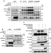
BPLF1 interacts with the UFM1 ligase UFL1 and putative UFL1 substrates. (A) curated STRING network diagram of the BPLF1 interacting proteins identified by co-immunoprecipitation and mass spectrometry. The functional annotation of the interacting proteins is color-coded: green, ribosome subunits; yellow, subunits of the EIF2 translation pre-initiation complex; turquoise, subunits of the EIF3 translation pre-initiation complex; blue, translation initiation complex; orange, translation elongation factors; pink, signal recognition particle (SRP) and SRP receptor (SRPRB) involved in the recognition and targeting of signal-sequence-tagged proteins the ER; lilac, ER translocon complex that mediates forward and retrograde transport across the ER; lime, translocon-associated N-oligosaccharyltransferase (OST) complex that links high mannose sugars to the asn-X-Ser/Thr consensus motif of ER-translocated polypeptides; light pink, ER protein CYB5R3 involved in the regulation of reticulophagy, light orange, UFM1 and ZNF598 ligases. Waved borders indicate putative UFMylation substrates. The thickness of the red connecting lanes indicates Log2 fold change: 4 points ≥ 10: 3 points ≥ 7–10; 2 points ≥ 5–7; 1 point ≥ 3–5; dotted lines ≥ 1.5–3. (B) reciprocal co-immunoprecipitation assays illustrate the interaction of BPLF1 with the UFL1 ligase. Lysates of HEK293T cells transfected with plasmids expressing FLAG-ev, BPLF1 or BPLF1C61A were immunoprecipitated with either anti-FLAG coated beads or antibodies to UFL1 followed by capture with GammaBindTM plus SepharoseTM (cytiva 17,088,601). An isotype-matched immunoglobulin control (ig) was included in the UFL1 immunoprecipitation to verify specificity. Immunoblots were probed with the indicated antibodies. Blots from one representative experiment out of three are shown in the figure. (C) affinity isolation assay illustrating the interaction of BPLF1 with the UFL1 ligase. Equimolar concentrations of purified bacterially expressed His-BPLF1 and MBP-UFL1 were mixed, and affinity isolations of the HIS and MBP tags were performed from equal aliquots. Western blots from one out of two independent experiments are shown in the figure. The involvement of contaminating nucleic acids or nonspecific interaction mediated by the MBP tag was not formally excluded. (D) Representative western bots that illustrate the interaction of BPLF1 with the putative UFMylation substrates identified in (A). FLAG immunoprecipitation was performed from extracts of HEK293T cells transfected with FLAG-ev, -BPLF1 or -BPLF1C61A and immunoblots were probed with the indicated antibodies. Each interaction was validated in at least two independent co-immunoprecipitation experiments.
Figure 2.
Figure 2.
BPLF1 inhibits the UFMylation of RPL26 on ER-associated ribosomes. (A) BPLF1 inhibits the UFMylation of RPL26 in ANS-treated cells. Control HeLa cells and cells transfected with RPL26-MYC together with FLAG-ev, -BPLF1 or -BPLF1C61A were treated with 50 ng/ml ANS for 1 h, followed by lysis under denaturing conditions to destroy non-covalent interactions and immunoprecipitation with anti-MYC coupled beads. Immunoblots from one representative experiment out of four are shown in the figure. (B) BPLF1 inhibits the ANS-induced UFMylation of RPL26 on ER-associated ribosomes. Total cell lysates (W), cytosolic (C), and ER membrane fractions (E) of ANS-treated cells cotransfected with RPL26-MYC and FLAG-ev, -BPLF1 or -BPLF1C61A were probed with the indicated antibodies. Untreated FLAG-ev transfected cells were included to assess the abundance and subcellular localization of endogenous UFM1 adducts. UFMylated species of RPL26-MYC and endogenous RPL26 are indicated by red and black arrows, respectively. Immunoblots from one representative experiment out of three are shown in the figure.
Figure 3.
Figure 3.
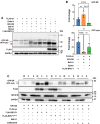
BPLF1 inhibits the UFMylation of endogenous RPL26. (A) FLAG-ev, -BPLF1 or -BPLF1C61A transfected HeLa cells were cultured for 24 h and then treated with 50 ng/ml ANS for 1 h before lysis in buffer containing NEM and iodoacetamide to inhibit DUB activity. High molecular weight species corresponding to the size of mono- di- and tri-UFMylated RPL26 were detected in immunoblots probed with antibodies specific for RPL26, and bands of the same size were detected after stripping and re-probing the blot with the UFM1 antibody. Representative blots from one out of four independent experiments are shown in the figure. (B) densitometry quantification of the intensity of the UFMylated species. A red dotted box indicates the area included in the densitometry scan. The intensity was normalized to the intensity of the RPL26 band in short exposure of the same blots. The mean ± SD fold increase in ANS treated relative to untreated cells in four independent experiments is shown. Significance was calculated by unpaired two-tailed Student’s t-test. (C) schematic illustration of the modified ER-RQC reporter. The reporter expresses in-frame an N-terminal ER-targeting signal sequence followed by an N-glycosylation site, GFP, and a stretch of lys residues encoded by AAA codons (K20) and RFP. Stalling of the ribosome at the poly(A) sequence traps the nascent ER-inserted polypeptide in the translocon. (D) BPLF1 inhibits the UFMylation of endogenous RPL26 induced by the ribosome stall-inducing reporter. HEK293T cells were cotransfected with FLAG-ev, -BPLF1 or -BPLF1C61A, and the modified ER-K20 reporter and immunoblots of cells harvested after 24 h were probed with the indicated antibodies. Immunoblots from one representative experiment out of four are shown in the figure. (E) densitometry quantification of the UFMylated proteins in four independent experiments. A red dotted box indicates the area included in the densitometry scan. Fold change was calculated relative to vector-transfected cells after normalization to the intensity of the RPL26 band in short exposure of the same blots. Statistical analysis was performed using an unpaired two-tailed Student’s t-test.
Figure 4.
Figure 4.
BPLF1 does not have deUfmylase activity. (A) Representative immunoblots of an in vitro UFMylation reaction performed in the presence or absence of BPLF1 or UFSP2. UFMylation reactions were performed in the presence of purified components of conjugation cascade: 0.25 μM recombinant His-UBA5 (E1), 5 μM GST-UFC1 (E2), 1 μM His-UFL1/Strep-DDRGK1 (E3), 0.5 μM of H3/H4 complex (substrate), and 10 μM His-UFM1 in the absence (lane 1) or presence of 5 mM ATP (lanes 2–9). Where indicated increasing concentrations of recombinant BPLF1 (lanes 3–5; 0.3, 0.75, and 1.5 μM, respectively), His-BPLF1C61A (CM, lane 6; 1.5 μM) or His-UFSP2 (lane 7–9; 0.03, 0.075, and 0.15 μM, respectively) were included in the reaction. The DUB activity of recombinant BPLF1 was confirmed by labeling 1.5 μM His-BPLF1 with 1 μM of the functional probe HA-Ubiquitin-vinyl-sulphone (Ub-Vs) that forms covalent adducts with the catalytic cys residue (lane 10–11). All reactions were incubated at 37°C for 90 min and analyzed in immunoblots using the indicated antibodies. UFMylated H3/H4, UFL1, and DDRGK1 were detected when the reaction was performed in the presence of ATP (lane 2). The addition of increasing amounts of BPLF1 or BPLF1C61A had no effect (lanes 3–6), while efficient de-UFMylation was induced by minute amounts of UFSP2 (lanes 7–9). The experiment was repeated twice with comparable results. UFM1 dimers were detected by the UFM1 antibody as a strong band of approximately 20 kDa. (B) recombinant BPLF1 does not cleave a UFM1-GFP reporter. Increasing amounts of purified recombinant BPLF1 (0.3, 0.75, 1.5 µM) were mixed with the UFM1-GFP or Ub-GFP reporters (0.3 µM) in reaction buffer and incubated for 1 h at 37°C followed by immunoblot analysis. One representative experiment out of three is shown in the figure. (C) Representative immunoblots illustrating the failure of BPLF1 to cleave a UFM1-GFP reporter in cells. U2OS-UFSP2-knockout cells were cotransfected with a UFM1-GFP fusion protein-expressing plasmids and plasmids expressing BPLF1 or the cellular deUfmylase UFSP2. The production of free GFP and UFM1 was monitored in immunoblots probed with specific antibodies. The reporter was cleaved only in cells expressing UFSP2. One representative experiment out of two is shown.
Figure 5.
Figure 5.
BPLF1 does not affect the assembly and activity of the UFL1 ligase complex. (A) reciprocal co-immunoprecipitation assays illustrate the failure of BPLF1 to affect the interaction of UFL1 with DDRGK1 and CDK5RAP3 in untreated cells. Lysates of HEK293T transfected with FLAG-ev, -BPLF1 or -BPLF1C61A were divided into equal aliquots, and co-immunoprecipitation was carried out with the indicated antibodies. Comparable levels of catalytic active and mutant BPLF1 were detected by the FLAG antibody in the UFL1, DDRGK1, and CDK5RAP3 immunoprecipitates, but their presence did not affect the co-immunoprecipitation of the ligase components. One representative experiment out of three is shown in the figure. (B) BPLF1 does not affect the assembly of the ligase in ANS-treated cells. HEK293T were transfected with FLAG-ev or -BPLF1, and one aliquot of the FLAG-BPLF1 transfected cells was treated with 50 ng/ml ANS for 30 min before harvesting. Immunoprecipitates of endogenous DDRGK1 were probed with the indicated antibodies. Immunoblots from one out of two independent experiments are shown. (C) BPLF1 does not affect the activity of the UFL1 ligase. The UFMylation of CYB5R3 was induced in HEK293-UFSP2-knockout cells by transfection of ha-tagged UFL1 and DDRGK1 and MYC-tagged active UFM1 (UFM1ΔC2) with or without co-transfection of FLAG-BPLF1 or -UFSP2. Representative immunoblots from one of two independent experiments are shown.
Figure 6.
Figure 6.
BPLF1 counteracts a ZNF598- and LTN1-independent ubiquitination event required for RPL26 UFMylation. (A) the activity of the ZNF598 ligase is not required for UFMylation of RPL26 in ANS-treated cells. Control (WT) and HEK293T-ZNF598-KO cells were treated with 50 ng/ml ANS for the indicated times before analysis of RPL26 UFMylation. Immunoblots from one representative experiment out of three are shown. (B) the activity of the LTN1 ligase is not required for UFMylation of RPL26 in ANS-treated cells. HEK293T cells were transfected with a control scrambled siRNA or a previously characterized LTN1 siRNA for 72 h before treatment with 50 ng/ml ANS for 30 min and analysis of RPL26 UFMylation. Immunoblots from one representative experiment out of two are shown. (C) the inhibition of de novo ubiquitination by pretreatment with a ubiquitin E1 small molecule inhibitor prevents RPL26 UFMylation in ANS-treated cells. HEK293T cells were pretreated with the indicated concentrations of TAK243 or the SUMO E1 inhibitor ML-792 before ANS treatment. UFC1*UFM1 thioesters were detected by running the same lysates without reducing agents. Immunoblots from one representative experiment out of three are shown. (D) cleared lysates of HEK293T transfected with FLAG-ev or -BPLF1 and treated with 50 ng/ml ANS for 20 min were fractionated by centrifugation over a 5–50% sucrose gradient. Fractions enriched in 40S, 60S, and 80S ribosomes and the first polysome fraction were identified (Figure S5) and analyzed by immunoblotting using the indicated antibodies. Immunoblots from one of two independent experiments showing comparable results are shown.
Figure 7.
Figure 7.
BPLF1 rescues ER-RQC substrates in the cytosol and the ER. (A) BPLF1 stabilizes an ER-RQC substrate. HEK293T cotransfected with the ER-K20 reporter and FLAG-ev, -BPLF1 or -BPLF1C61A were cultured for 24 h. Aliquots of FLAG-ev and ER-K20 transfected cells were treated with 100 nM Baf A1 or 100 nM epoxomicin (epoxo) overnight before harvesting. The de-glycosylated GFP-ER product is indicated by a red arrow. Immunoblots from one representative experiment out of three are shown in the figure. A nonspecific band detected by the anti-GFP antibody is indicated by an asterisk (*). (B) Densitometry quantification of the GFP bands. Fold increase was calculated as the ratio between the band’s intensity in treated cells versus cells transfected with the ER-K20 reporter alone. The average fold increase in three independent experiments is shown. Statistical analysis was performed using an unpaired two-tailed Student’s t-test. (C) BPLF1 stabilizes the ER-RQC substrate in the cytosol and ER. HEK293T cells were transfected and treated as described in (A), except that carfilzomib was used instead of epoxomicin to inhibit proteasome activity. Unfractionated (W), cytosolic (C), and ER-membrane (E) fractions were produced as described in the methods section. Immunoblots from one out of three independent experiments are shown in the figure.
Figure 8.
Figure 8.
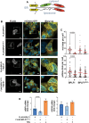
BPLF1 inhibits reticulophagy and triggers ER-stress responses. (A) schematic illustration of the ER-Autophagy Tandem Reporter (EATR). The reporter expresses in-frame the coding sequence of the SERP1 subunit of the ER translocon complex followed by the coding sequences of eGFP and ChFP. Upon ER insertion of the SERP1 domain, eGFP and ChFP face the cytosol and emit equal fluorescence, whereas, due to the selective loss of eGFP fluorescence at low pH, ER-loaded autophagosomes appear as distinct red fluorescent dots. (B) Representative confocal images that illustrate the failure to accumulate red fluorescent dots or yellow fluorescent dots in cells expressing active BPLF1 starved in the absence or presence of Baf A1, respectively. Stable HCT116-EATR cells were transfected with plasmids expressing FLAG-BPLF1 or -BPLF1C61A and then starved overnight in EBSS medium before visualizing the formation of ER-loaded autophagosomes by confocal microscopy. Scale bar: 10 μm. (C) quantification of the number of red (upper panel) and yellow (lower panel) fluorescent dots in BPLF1- or BPLF1C61A-positive and -negative cells from the same transfection experiments. The cumulative data from two independent experiments where approximately 50 vDUB-positive and -negative cells scored from the same slide are shown. Significance was calculated using an unpaired two-tailed Student t-test. (D) BPLF1 promotes the accumulation of the ER stress markers spliced XBP1 mRNA and the transcriptional upregulation of HSPA5. FLAG-ev transfected cells treated with thapsigargin during the last 2 h before harvest were included as a reference of ER-stress induction. Quantification of sXBP1 and HSPA5 mRNAs in four independent experiments. Fold induction was calculated relative to untreated FLAG-ev transfected cells after normalization to the GAPDH housekeeping gene. Significance was calculated using an unpaired two-tailed Student t-test.
Figure 9.
Figure 9.
Physiological levels of BPLF1 inhibit RPL26 UFMylation in productively EBV-infected cells. (A) the productive virus cycle was induced in LCLs immortalized with recombinant EBV expressing wild-type (LCL-WT) or catalytic mutant (LCL-CM) BPLF1. After 72 h, equal aliquots of cells were treated with 50 ng/ml ANS for 30 min, followed by the analysis of RPL26 UFMylation by immunoblots. Blots from one representative experiment out of three are shown. (B) densitometry quantification of the UFMylated species in three independent experiments. A red dotted box indicates the area included in the densitometry scan. Data are shown as fold induction of UFMylated RLP26 in untreated versus ANS-treated cells after normalization to the TUBA loading control and total RPL26. The mean ± SD fold change in induced relative to uninduced cells is shown. Significance was calculated using an unpaired two-tailed Student t-test.

Similar articles

References

    1. Jayaraj GG, Hipp MS, Hartl FU.. Functional modules of the proteostasis network. Cold Spring Harb Perspect Biol. 2020;12(1):a033951. doi: 10.1101/cshperspect.a033951 - DOI - PMC - PubMed
    1. Brandman O, Hegde RS. Ribosome-associated protein quality control. Nat Struct Mol Biol. 2016;23(1):7–15. doi: 10.1038/nsmb.3147 - DOI - PMC - PubMed
    1. Joazeiro CAP. Mechanisms and functions of ribosome-associated protein quality control. Nat Rev Mol Cell Biol. 2019;20(6):368–383. doi: 10.1038/s41580-019-0118-2 - DOI - PMC - PubMed
    1. Sitron CS, Brandman O. Detection and degradation of stalled nascent chains via ribosome-associated quality control. Annu Rev Biochem. 2020;89(1):417–442. doi: 10.1146/annurev-biochem-013118-110729 - DOI - PMC - PubMed
    1. Vind AC, Genzor AV, Bekker-Jensen S. Ribosomal stress-surveillance: three pathways is a magic number. Nucleic Acids Res. 2020;48(19):10648–10661. doi: 10.1093/nar/gkaa757 - DOI - PMC - PubMed

Publication types

Substances

LinkOut - more resources