. 2025 Apr;18(2):441-452.
doi: 10.1016/j.mucimm.2025.01.002. Epub 2025 Jan 20.

Breast milk delivery of an engineered dimeric IgA protects neonates against rotavirus

Affiliations

Breast milk delivery of an engineered dimeric IgA protects neonates against rotavirus

Stephanie N Langel et al. Mucosal Immunol. 2025 Apr.

Abstract

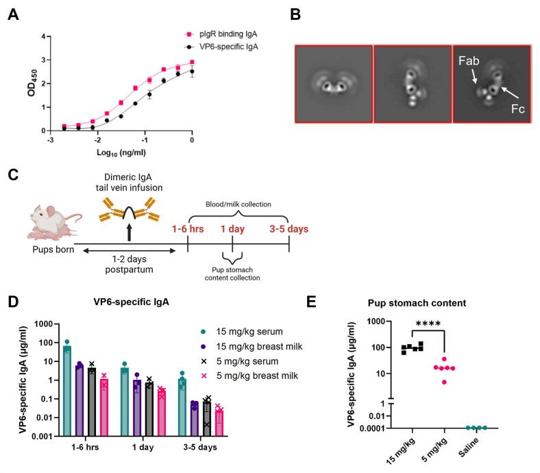
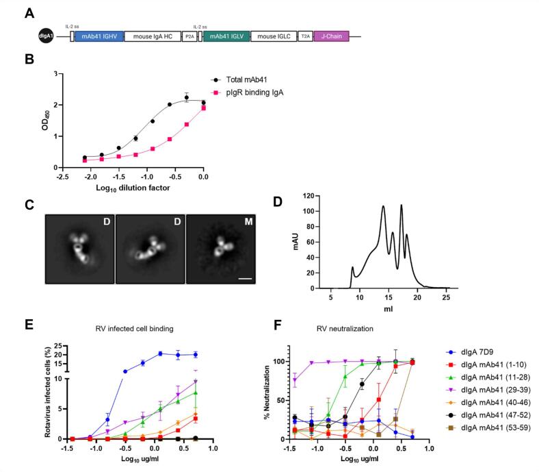
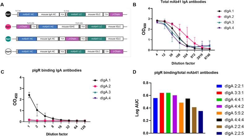
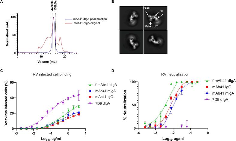
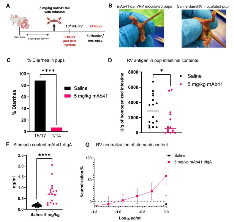
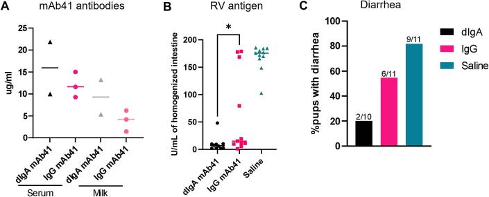
Dimeric IgA (dIgA) is the dominant antibody in many mucosal tissues. It is actively transported onto mucosal surfaces as secretory IgA (sIgA) which plays an integral role in protection against enteric pathogens, particularly in young children. Therapeutic strategies that deliver engineered, potently neutralizing antibodies directly into the infant intestine through breast milk could provide enhanced antimicrobial protection for neonates. Here, we developed a murine model of maternal protective transfer against human rotavirus (RV) using systemic administration of a dimeric IgA monoclonal antibody (mAb). First, we showed that systemically administered dIgA passively transferred into breast milk and the stomach of suckling pups in a dose-dependent manner. Next, we optimized the recombinant production of a potently RV-neutralizing, VP4-specific dIgA (mAb41) antibody. We then demonstrated that systemic administration of dIgA and IgG mAb41 in lactating dams conferred protection from RV-induced diarrhea in suckling pups, with dIgA resulting in lower diarrhea incidence from IgG. Systemic delivery of engineered antimicrobial dIgA mAbs should be considered as an effective strategy for sIgA delivery to the infant gastrointestinal tract via breast milk to increase protection against enteric pathogens.

Keywords: Breast milk; Dimeric IgA; Maternal immunity; Neonatal immunity; Passive transfer; Rotavirus.


Conflict of interest statement

Declaration of competing interest The authors declare the following financial interests/personal relationships which may be considered as potential competing interests: [J.E.C. has served as a consultant for Luna Biologics, is a member of the Scientific Advisory Board of Meissa Vaccines and is Founder of IDBiologics. The Crowe laboratory at Vanderbilt University Medical Center has received unrelated sponsored research agreements from Takeda Vaccines, IDBiologics and AstraZeneca. S.R.P. provides individual consulting services to Moderna, Merck, Dynavax, GSK, and Pfizer on CMV vaccines. Merck Vaccines and Moderna have provided grants and contracts for S.R.P. sponsored programs.].

Figures

Fig. 1
Fig. 1
Dimeric IgA antibodies administered systemically to lactating mice are transferred to milk and the stomach contents of suckling pups. (A) The rotavirus (RV) VP6-specific, non-neutralizing 7D9 hybridoma derived antibodies bound to RV VP6 (black circles) and polymeric immunoglobulin receptor (pIgR; magenta squares) via ELISA. Data are plotted as mean ± SD of two technical replicates. (B) Negative stain electron microscopy representative images of the hybridoma-purified dimeric IgA (dIgA) 7D9 antibodies. The Fab and Fc regions are indicated. Scale bar represents 10 nm. (C) Schematic of tail vein injections of BALB/c lactating dams given 5 mg/kg or 15 mg/kg dIgA 7D9 at 1 to 2 days postpartum. Blood and milk were collected from dams at 1–6 hrs, 1-, and 3–5-days post injection. A subset of pups (n = 6) per treatment group were sacrificed at 1-day post injection to collect their stomach content. (D) 7D9 antibodies were detected in blood and milk of injected dams via a RV VP6-specific IgA antibody ELISA. The 5 mg/kg (x) and 15 mg/kg (circle) treatment groups are indicated for serum and milk. Data are plotted as mean ± SD of two technical replicates and represent individual mice. (E) 7D9 antibodies were detected in the stomach content of suckling pups via a RV VP6-specific IgA antibody ELISA (15 mg/kg = black squares, 5 mg/kg = pink circles; saline = teal circles). Data are plotted as mean of two technical replicates per individual pup. A significant difference between the compared groups (****p < 0.0001) was determined using an ANOVA. (For interpretation of the references to colour in this figure legend, the reader is referred to the web version of this article.)
Fig. 2
Fig. 2
Recombinant production and characterization of an RV-neutralizing mouse-human chimeric dimeric IgA antibody. (A) Schematic of the plasmid used to produce the mouse-human chimeric mAb41 dimeric IgA (dIgA). The mAb41 recombined immunoglobulin heavy chain variable region genes (HC), recombined immunoglobulin light chain variable region genes (LC), and the joining chain (J-chain) gene (purple box), are indicated in that order. Schematic created with BioRender. (B) J-chain containing IgA (magenta squares) and total IgA (black circles) antibodies were detected via ELISA. Data are plotted as mean ± SD of two technical replicates. (C) Representative negative stain electron microscopy images of purified dimeric (D) and monomeric (M) mAb41 IgA antibodies. Size exclusion chromatography using a Superose 6 10/300 GL revealed multiple different peaks (Fig. S4). Each of the peaks were fractionated (corresponding fractions 1–10, 11–28, 29–39, 40–46, 47–52 and 52–59) and functionally characterized by rotavirus (RV)-infected cell binding (D) and neutralization assays (E). The non-neutralizing dIgA 7D9 (blue line) was used as a positive control for RV infected cell binding and a negative control for RV neutralization. The IgA isotype control was used as a negative control for both RV infected cell binding and RV neutralization. Data are plotted as mean ± SD of two technical replicates. (For interpretation of the references to colour in this figure legend, the reader is referred to the web version of this article.)
Fig. 3
Fig. 3
Antibody chains position and ratio impacts the recombinant production of RV-neutralizing mouse-human chimeric mAb41 dimeric IgA. (A) Schematic of the different constructs generated to determine if the position of the J-chain gene in the plasmid cassette impacts dimeric IgA (dIgA) production. Illustration created with BioRender. (B) Total IgA antibodies as determined by ELISA. Data are plotted as mean ± SD of experimental duplicates. (C) Polymeric immunoglobulin receptor (pIgR) binding IgA antibodies determined via ELISA. Data are plotted as mean ± SD of experimental duplicates. (D) pIgR IgA antibodies and total mAb41 IgA antibodies were detected via ELISA. Log area under the curve (AUC) was calculated for each co-transfection and graphed as a ratio of pIgR binding IgA antibodies over total mAb41 IgA antibodies.
Fig. 4
Fig. 4
Recombinant mAb41 dimeric IgA antibodies demonstrate greater rotavirus (RV) binding and neutralization potency than mAb41 monomeric IgA or IgG antibodies. (A) Size exclusion chromatography by Superose 6 Increase 10/300 GL column in 1XPBS of mAb41 dimeric IgA (dIgA) antibodies at a flow rate of 0.75 ml/min. Molecular weight markers are listed above the dashed lines at 440 kDa and 158 kDa, respectively. (B) Negative stain electron microscopy of the purified dimeric antibodies. Fab and Fc regions are indicated respectively by white arrows. (C) RV infected cell binding assay. Data are plotted as mean ± SD of two technical replicates. (D) RV neutralization assay. Data are plotted as mean ± SD of two technical replicates.
Fig. 5
Fig. 5
Passive maternal immunization with systemic mAb41 dIgA protects against rotavirus (RV)-induced diarrhea in suckling pups. (A) Schematic of tail vein injections of BALB/c lactating dams with 5 mg/kg f-dIgA mAb41 antibodies at 5 days postpartum. Pups were orally inoculated with 1 × 106 FFU of RV (Wa strain) at 1–2 hrs post dam injection and euthanized 24 hrs later. Schematic created with BioRender. (B) Representative image of RV inoculated suckling pups from f-dIgA mAb41 infused dams, which excreted urine or hard stool upon abdomen gentle palpation. (C) Representative image of RV inoculated suckling pups from saline infused dams, which excreted yellow, liquid and/or stick stool after gentle abdomen palpation. (D) Diarrhea was reported as % of animals with clinical symptoms upon gentle abdomen palpation in each treatment group (5 mg/kg = blue; saline = red). The number of animals with diarrhea out of the total number of animals are reported at the top of each bar graph. (E) RV antigen in homogenized intestinal tissue was detected via a commercial RV antigen binding ELISA. Significant differences between the compared groups were determined using a Mann-Whitney U test (**p < 0.05). (F) mAb41 antibodies were detected in stomach content of suckling pups using an mAb41 anti-idiotypic IgA antibody ELISA. Significant differences between the compared groups were determined using a Mann-Whitney U test (***p < 0.001). (G) Stomach content was assessed for RV neutralization at different dilutions and plotted as neutralization % in n = 6 pups per treatment group (5 mg/kg = blue; saline = red). Data from C-F are from 3 litters per treatment group (for litter distribution see Supplementary Table 1 and Fig. S6). Data are plotted as the mean ± SD of two technical replicates. (For interpretation of the references to colour in this figure legend, the reader is referred to the web version of this article.)
Fig. 6
Fig. 6
Passive immunization with dimeric IgA mAb41 decreases intestinal rotavirus antigen and diarrhea scores in suckling pups. (A) Concentrations of dIgA and IgG mAb41 in dams’ serum and milk were determined by mAb41 anti-idiotypic antibody ELISA. (B) RV antigen in pups homogenized intestinal tissue was detected via a commercial RV antigen binding ELISA. Significant differences between the compared groups were determined using a one-way ANOVA (*p < 0.05). (C) Diarrhea was reported as the percentage of animals with diarrhea at day 1 post-challenge in each treatment group. The number of animals with diarrhea out of the total number of animals are reported at the top of each bar graph. Data from B-C are from 3 L per treatment group (for litter distribution see Supplementary Table 1 and Fig. S6). Data are plotted as the mean ± SD of two technical replicates.

Similar articles

References

    1. Chodirker W.B., Tomasi T.B., Jr. Gamma-globulins: quantitative relationships in human serum and nonvascular fluids. Science. 1963;142(3595):1080–1081. - PubMed
    1. Bemark M., Angeletti D. Know your enemy or find your friend?-Induction of IgA at mucosal surfaces. Immunol Rev. 2021;303(1):83–102. - PMC - PubMed
    1. Muramatsu M., Yoshida R., Yokoyama A., et al. Comparison of antiviral activity between IgA and IgG specific to influenza virus hemagglutinin: increased potential of IgA for heterosubtypic immunity. PLoS One. 2014;9(1) - PMC - PubMed
    1. Okuya K., Yoshida R., Manzoor R., et al. Potential role of nonneutralizing IgA antibodies in cross-protective immunity against Influenza A viruses of multiple hemagglutinin subtypes. J Virol. 2020;94(12) - PMC - PubMed
    1. Renegar K.B., Small P.A., Jr. Passive transfer of local immunity to influenza virus infection by IgA antibody. J Immunol. 1991;146(6):1972–1978. - PubMed

MeSH terms