. 2025 Jan 22;16(1):957.
doi: 10.1038/s41467-025-56336-5.

Chemical tools to define and manipulate interferon-inducible Ubl protease USP18

Affiliations

Chemical tools to define and manipulate interferon-inducible Ubl protease USP18

Griffin J Davis et al. Nat Commun. .

Abstract

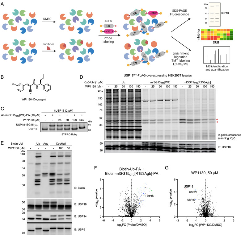
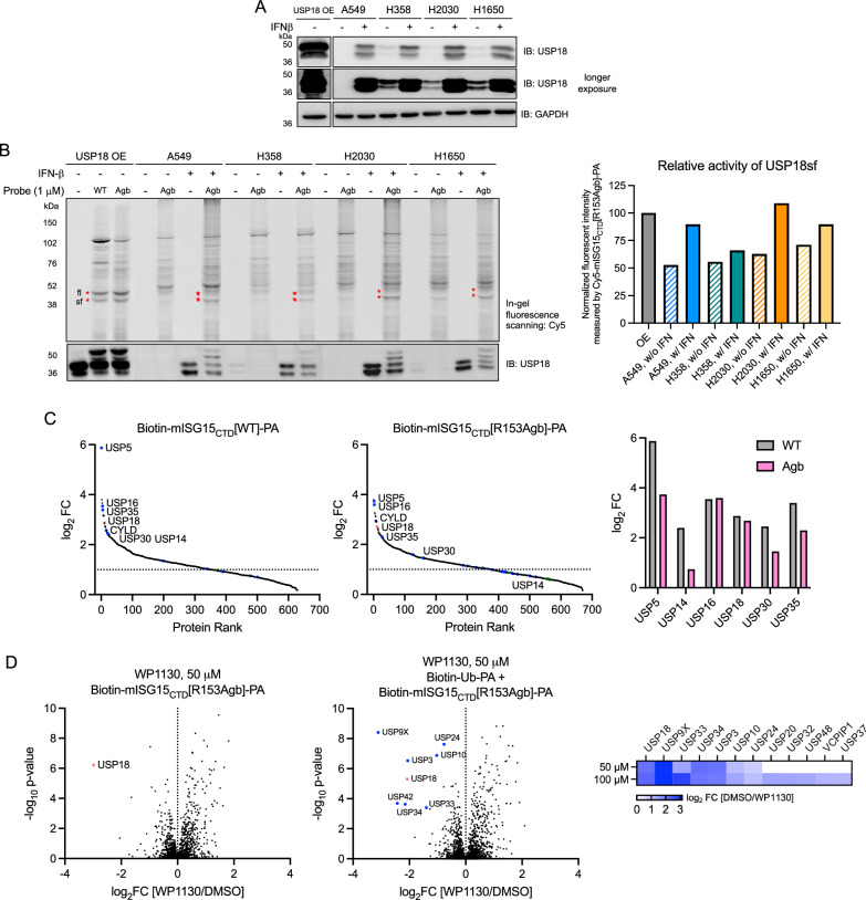
Ubiquitin-specific protease 18 (USP18) is a multifunctional cysteine protease primarily responsible for deconjugating the interferon-inducible ubiquitin-like modifier ISG15 from protein substrates. Here, we report the design and synthesis of activity-based probes (ABPs) that incorporate unnatural amino acids into the C-terminal tail of ISG15, enabling the selective detection of USP18 activity over other ISG15 cross-reactive deubiquitinases (DUBs) such as USP5 and USP14. Combined with a ubiquitin-based DUB ABP, the USP18 ABP is employed in a chemoproteomics screening platform to identify and assess inhibitors of DUBs including USP18. We further demonstrate that USP18 ABPs can be utilized to profile differential activities of USP18 in lung cancer cell lines, providing a strategy that will help define the activity-related landscape of USP18 in different disease states and unravel important (de)ISGylation-dependent biological processes.


Conflict of interest statement

Competing interests: The authors declare no competing interests.

Figures

Fig. 1
Fig. 1. Fluorogenic substrates for USP18.
A Catalytic triad of USP18. The isopeptide bond between the C-terminal Gly residue of ISG15 and the Lys residue of the substrate protein is hydrolyzed by the USP18 protease activity. B Structure of USP18 complexed with ISG15 (PDB: 5CHV). Active site residues of USP18 that interact with conserved C-terminal LRLRGG of ISG15 are highlighted in comparison to other ISG15 reactive DUBs. C Structure of ISG15-based ACC-labeled substrate. The C-terminal LRGG motif is highlighted. D Schematic overview of HyCoSuL screening created in BioRender. Yoo, E. (2025) https://BioRender.com/z61a243. E Substrate profiles presented as heatmaps indicating the percentage of relative cleavage measured by a fluorescent signal produced upon USP18 binding with two sub-libraries, Ac-Mix-P3-Gly-Gly-ACC and Ac-P4-Mix-Gly-Gly-ACC (where Mix represents an equimolar mixture of natural amino acids). Source data are provided as a Source Data file. F Structures (left panel) and rate of hydrolysis (right panel) of selected tetrapeptide fluorogenic substrates. mUSP18 (10 μM), USP2 (2 μM), and USP5 (1 μM) were incubated with 100 μM of Ac-LXGG-ACC substrates for 1 h at RT and initial release of fluorescent ACC (V0, RFU/s) by enzyme was measured at Ex: 360 nm / Em: 460 nm. Agb 2-amino-4-guanidino-butyric acid, hArg homoarginine, Phe(guan) guanidino-phenylalanine. Data represent mean values (n = 2 independent replicates). Source data are provided as a Source Data file.
Fig. 2
Fig. 2. Preparation of mISG15CTD-based probes.
A Synthesis of C-terminal domain of mouse ISG15 and ISG15 variants. Leu80~Gly154 sequence was assembled using SPPS and the N-terminal end was modified by either an acetyl group, fluorescent cyanine dye, or biotin affinity handle. After the cleavage from the solid support, the C-terminal end was functionalized with propargylamine (PA) replacing Gly155. Arg153 was varied with unnatural amino acids such as Agb (2-amino-4-guanidino-butyric acid), hArg (homoarginine), Phe(guan)(guanidino-phenylalanine). B LC-MS analysis of synthetic Ac-mISG15CTD-PA and Cy5-mISG15CTD-PA probes.
Fig. 3
Fig. 3. Reactivity and selectivity of mISG15CTD-based probes.
A, B Recombinant mouse and human USP18 (2 μM) or USP5 (1 μM) were incubated with Ac-mISG15CTD-PA probes (10 μM) for 3 h at RT. Protein samples were analyzed by SDS-PAGE and SYPRO Ruby staining. The result is a representative of three experiments (n = 3 independent replicates). Source data are provided as a Source Data file. C WT or C64/65A mutant of USP18-FLAG overexpressing HEK293T cell lysates were incubated with Cy5-mISG15CTD-PA probes at indicated concentrations for 3 h at RT. Protein samples were analyzed by SDS-PAGE and in-gel fluorescence scanning for Cy5 signal. Red marks indicate the appearance of new protein bands only in USP18WT overexpressing cell lysates after labeling by probes corresponding to the expected molecular weight of USP18⎯probe conjugate. Expression of USP18 was confirmed by western blotting. The result is a representative of three experiments (n = 3 independent replicates). Source data are provided as a Source Data file. D Volcano (left) and protein rank (right) plots of quantitative proteomic analysis of streptavidin beads pulldowns after labeling of USP18-FLAG overexpressing HEK293T cell lysates by Biotin-mISG15CTD[R153Agb]-PA probe (5 μM, 3 h, RT) showing significantly enriched proteins (log2 ratio >1, p-value ≤ 0.05). USP18 is marked red and other DUBs are colored based on subfamilies (blue: USP, red: UCH, green: OTU, and cyan: JAMM). Data represent mean values (n = 3 independent replicates). p-values were calculated by two-tailed t-test. Source data are provided with this paper (Supplementary Data 2).
Fig. 4
Fig. 4. USP18 inhibitor assay by competitive activity-based protein profiling.
A Schematic workflow for quantitative activity-based protein profiling created in BioRender. Yoo, E. (2025) https://BioRender.com/k51r701. Lysates were treated with DMSO or compound, followed by treatment with an activity-based DUB probe. Labeled proteins were enriched using biotin-streptavidin pulldown, digested, and analyzed by LC-MS/MS. B Chemical structure of WP1130. C Recombinant human USP18 (2 μM) was pretreated with WP1130 for 30 min and incubated with Ac-mISG15CTD[WT]-PA probe (10 μM) for 2 h at RT. Protein samples were analyzed by SDS-PAGE and SYPRO Ruby staining. The result is a representative of two experiments (n = 2 independent replicates). Source data are provided as a Source Data file. D USP18WT-FLAG overexpressing HEK293T cell lysates were preincubated with WP1130 for 1 h followed by labeling with 1 μM of Cy5-Ubl-PA for 2 h at RT. Protein samples were analyzed by SDS-PAGE, in-gel fluorescence scanning for Cy5 signal, and immunoblotting for USP18. The result is a representative of two experiments (n = 2 independent replicates). Source data are provided as a Source Data file. EG USP18WT-FLAG overexpressing HEK293T cell lysates were preincubated with WP1130 for 2 h followed by labeling with 1 μM of Biotin-Ub-PA or 5 μM of Biotin-mISG15CTD[R153Agb]-PA or a cocktail of probes (1 μM of Biotin-Ub-PA + 5 μM of Biotin-mISG15CTD[R153Agb]-PA) for 3 h at RT. Protein samples were analyzed by SDS-PAGE and immunoblotting (E). The result is a representative of two experiments (n = 2 independent replicates). Volcano plots of quantitative proteomic analysis of streptavidin beads pulldowns after pretreating cell lysates with either DMSO (F) or 50 μM of WP1130 (G) and labeling by a cocktail of probes. Significantly enriched proteins are shown (log2 ratio >1, p-value ≤ 0.05). USP18 is marked red and other DUBs are colored based on subfamilies (blue: USP, red: UCH, green: OTU, and cyan: JAMM). Data represent mean values (n = 3 independent replicates). p-values were calculated by two-tailed t-test. Source data are provided with this paper (Supplementary Data 2).
Fig. 5
Fig. 5. USP18 expression and activity profiles in lung cancer cell lines.
A Constitutive and induced USP18 expression levels upon IFN-β stimulation (50 ng/mL, 48 h) across lung cancer cell lines. Source data are provided as a Source Data file. The result is a representative of three experiments (n = 3 independent replicates). B Each of the cell lysates were incubated with 1 μM of Cy5-mISG15CTD[R153Agb]-PA probe (3 h, RT). Left, protein samples were analyzed by SDS-PAGE and in-gel fluorescence scanning for Cy5 signal. Red marks indicate the appearance of USP18⎯probe conjugates. Expression of USP18 was confirmed by western blotting. Right, the normalized intensity of the fluorescent protein band corresponding to USP18sf⎯probe conjugate to the fluorescent intensity of USP18sf⎯probe conjugate measured from USP18 overexpressing conditions. The result is a representative of three experiments (n = 3 independent replicates). Source data are provided as a Source Data file. C Protein rank plots of quantitative proteomic analysis of streptavidin bead pulldowns after labeling of lysates of A549 cells stimulated with IFN-β by 5 μM of either Biotin-mISG15CTD[WT]-PA (left) or Biotin-mISG15CTD[R153Agb]-PA probe (middle), showing significantly enriched proteins (log2 ratio >1, p-value ≤ 0.05). USP18 is marked and other DUBs are colored based on subfamilies (blue: USP, red: UCH, green: OTU, and cyan: JAMM). Enrichment ratios (log2 [Probe/DMSO]) of most highly enriched DUBs are compared (right). Data represent mean values (n = 3 independent replicates). p-values were calculated by two-tailed t-test. Source data are provided as a Source Data file and with this paper (Supplementary Data 4). D Lysates of A549 cells stimulated with IFN-β were preincubated with 50 μM of WP1130 for 2 h followed by labeling with either 5 μM of Biotin-mISG15CTD[R153Agb]-PA (left) or a cocktail of probes (1 μM of Biotin-Ub-PA + 5 μM of Biotin-mISG15CTD[R153Agb]-PA) (middle). Volcano plots of quantitative proteomic analysis comparing samples treated with WP1130 to DMSO are shown with DUBs inhibited by the compound marked. USP18 is marked and other DUBs are colored based on subfamilies (blue: USP, red: UCH, green: OTU, and cyan: JAMM). Heat map analysis of competition ratios (log2 [DMSO/WP1130]) comparing samples pretreated with 50 μM or 100 μM of WP1130 followed by labeling with a cocktail of probes indicates several DUBs inhibited by the compound (right). Data represent mean values (n = 3 independent replicates). p-values were calculated by two-tailed t-test. Source data are provided as a Source Data file and with this paper (Supplementary Data 5).
Fig. 6
Fig. 6. Functional effect of WP1130 on protein ISGylation.
A Lysates of A549 cells stimulated with IFN-β (48 h) were incubated with WP1130 (50 μM) for 2 h in the absence or presence of recombinant human USP18 (5 μM). Protein samples were analyzed by SDS-PAGE and immunoblotted for protein ISGylation. The result is a representative of three experiments (n = 3 independent replicates). Source data are provided as a Source Data file. B A549 cells were stimulated with IFN-β (48 h) and incubated with the indicated concentration of WP1130 for 2 h. Lysates were separated into soluble and insoluble fractions and probed for protein ISGylation. The result is a representative of three experiments (n = 3 independent replicates). Source data are provided as a Source Data file.
Fig. 7
Fig. 7. Reactivity with USP16.
A Top, recombinant USP16 (0.45 μM) was incubated with Ac-Ubl-PA probes for 3 h at RT. Protein samples were analyzed by SDS-PAGE and SYPRO Ruby staining. Bottom, mean density of labeled protein band corresponding to the USP16⎯Ubl probe conjugate. The result is a representative of two experiments (n = 2 independent replicates). Source data are provided as a Source Data file. B Left, recombinant USP16 (0.25 μM) was incubated with Cy5-Ubl-PA probes (3 h, RT). Protein samples were analyzed by SDS-PAGE, in-gel fluorescence scanning for Cy5, and SYPRO Ruby staining. Right, mean density of labeled protein band corresponding to the USP16⎯Ubl probe conjugate. The result is a representative of two experiments (n = 2 independent replicates). Source data are provided as a Source Data file. C Left, USP16-FLAG overexpressing HEK293T cell lysates were incubated with Biotin-Ubl-PA probes (3 h, RT). Protein samples were analyzed by SDS-PAGE and immunoblotting. Right, mean density of labeled protein band corresponding to the USP16⎯Ubl probe conjugate. The result is a representative of two experiments (n = 2 independent replicates). Source data are provided as a Source Data file.

Update of

Similar articles

References

    1. Komander, D., Clague, M. J. & Urbe, S. Breaking the chains: structure and function of the deubiquitinases. Nat. Rev. Mol. Cell Biol.10, 550–563 (2009). - PubMed
    1. Sacco, J. J., Coulson, J. M., Clague, M. J. & Urbe, S. Emerging roles of deubiquitinases in cancer-associated pathways. IUBMB Life62, 140–157 (2010). - PMC - PubMed
    1. Malakhov, M. P., Malakhova, O. A., Kim, K. I., Ritchie, K. J. & Zhang, D. E. UBP43 (USP18) specifically removes ISG15 from conjugated proteins. J. Biol. Chem.277, 9976–9981 (2002). - PubMed
    1. Narasimhan, J., Potter, J. L. & Haas, A. L. Conjugation of the 15-kDa interferon-induced ubiquitin homolog is distinct from that of ubiquitin. J. Biol. Chem.271, 324–330 (1996). - PubMed
    1. Wong, J. J. Y., Pung, Y. F., Sze, N. S. K. & Chin, K. C. HERC5 is an IFN-induced HECT-type E3 protein ligase that mediates type I IFN-induced ISGylation of protein targets. Proc. Natl Acad. Sci. USA103, 10735–10740 (2006). - PMC - PubMed

MeSH terms