. 2025 Jan 22;16(1):39.
doi: 10.1038/s41419-025-07357-x.

RNAi-based ALOX15B silencing augments keratinocyte inflammation in vitro via EGFR/STAT1/JAK1 signalling

Affiliations

RNAi-based ALOX15B silencing augments keratinocyte inflammation in vitro via EGFR/STAT1/JAK1 signalling

Megan A Palmer et al. Cell Death Dis. .

Abstract

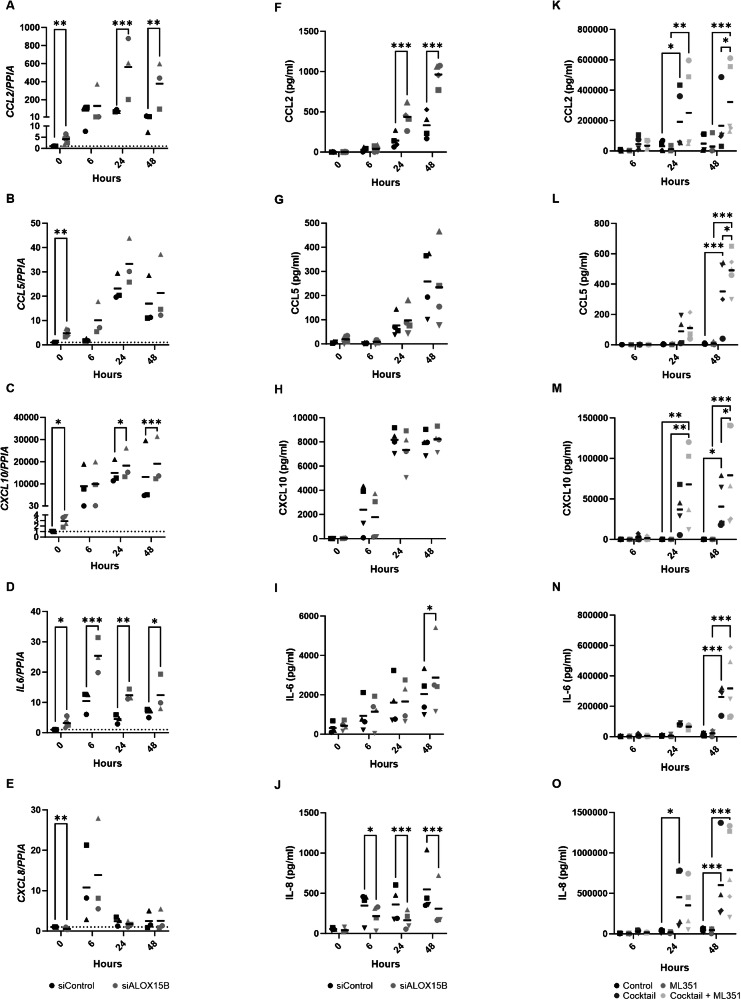
Arachidonate 15-lipoxygenase type B (ALOX15B) peroxidises polyunsaturated fatty acids to their corresponding fatty acid hydroperoxides, which are subsequently reduced into hydroxy-fatty acids. A dysregulated abundance of these biological lipid mediators has been reported in the skin and blood of psoriatic compared to healthy individuals. RNAscope and immunohistochemistry revealed increased ALOX15B expression in lesional psoriasis samples. Using a cytokine cocktail containing IL-17A, interferon-gamma and tumour necrosis factor-alpha to produce a psoriasis-like phenotype, a role for ALOX15B in human epidermal keratinocyte inflammation was investigated. siRNA-mediated silencing of ALOX15B increased CCL2 expression and secretion. In addition to CCL2, secretion of CCL5 and CXCL10 were elevated in skin equivalents treated with lipoxygenase inhibitor ML351. Inhibition of the JAK1/STAT1 pathway reversed the enhanced CCL2 expression found with ALOX15B silencing. Previous studies have linked epidermal growth factor receptor (EGFR) inhibition with the upregulation of cytokines including CCL2, CCL5 and CXCL10. ALOX15B silencing reduced EGFR expression and inhibition of EGFR signalling potentiated the effect of ALOX15B silencing on increased CCL2, CCL5 and CXCL10 expression. Confirming previous findings, gene expression of cholesterol biosynthesis genes was reduced via reduced ERK phosphorylation. Reduced ERK phosphorylation was dependant on EGFR and NRF2 activation. Furthermore, plasma membrane lipids were investigated via confocal microscopy, revealing reduced cholesterol and lipid rafts. This study suggests a role for ALOX15B in keratinocyte inflammation through modulation of lipid peroxidation and the EGFR/JAK1/STAT1 signalling axis.


Conflict of interest statement

Competing interests: The authors declare no competing interests. Ethics statement: The study was approved by the ethics committee of the Clinic of the Goethe-University (116/11); written, informed consent was obtained from all patients and control participants. The Declaration of Helsinki protocols were followed.

Figures

Fig. 1
Fig. 1. ALOX15B is upregulated in Psoriasis.
A Representative images from dual immunofluorescence and RNAscope in human skin for ALOX15B (yellow, protein; magenta, RNA) and CD68 (cyan, macrophages). Scale bar 100 µm, white boxes represent zoomed-in regions, zoomed in region scale bar 5 µm. Analysis of (B) RNAscope and (C) immunofluorescence staining of ALOX15B, data represented as mean dots/intensity per mm2 of the epidermis (N = 3). Gene expression (D) or Western (E) analysis of ALOX15B in keratinocytes treated with control or ALOX15B siRNA for 72 h, followed by cytokine cocktail of IL-17A, IFNγ and TNFα for 6, 24, 48 or 72 h (Western blot 72 h). Gene expression is relative to control siRNA and normalised to PPIA (N = 3, or 4 for 72 h). Western analysis is relative to control siRNA and normalised to loading control (N = 3). Analysis of 15-LOX specific oxylipins (F, N = 7) and confocal microscopy of lipid peroxidation sensor BODIPY C11 (G, N = 5) in keratinocytes treated with control or ALOX15B siRNA for 72 h. Scale bar 50 µm, image analysis (H) of mean pixel intensity per cell of oxidised BODIPY C11. Individual biological replicates represented by different symbol sets, line is mean, dotted line represents normalisation to siControl at 1. t tests (tissue) and two-way ANOVA (cells) were performed; significance denoted by *P < 0.05, **P < 0.01, ***P < 0.001.
Fig. 2
Fig. 2. ALOX15B silencing augments keratinocyte inflammation.
Gene expression (AE: N = 6 for 0 h and N = 3 for 6–48 h) and cytokine secretion (FJ: N = 4) in keratinocytes transfected with control or ALOX15B siRNA for 72 h, followed by a cocktail of 50 ng/ml IL-17A, TNFα and 10 ng/ml IFNγ for 6, 24 or 48 h. Gene expression is relative to control siRNA (represented by dotted line) and normalised to PPIA. Cytokine secretion from human skin equivalents (KO) treated with cytokine cocktail with or without 10 µM ML351 for 6, 24 or 48 h (N = 5). Individual biological replicates represented by different symbol sets, line is mean. Two-way ANOVA performed; significance denoted by *P < 0.05, **P < 0.01, ***P < 0.001.
Fig. 3
Fig. 3. JAK1/STAT1 pathway is altered with ALOX15B silencing.
AC Keratinocytes transfected with control or ALOX15B siRNA for 72 h, (B, C) followed by 30-min pre-treatment with 1 µM JAK inhibitor I, then a cocktail of 50 ng/ml IL-17A, TNFα and 10 ng/ml IFNγ or IFNγ alone for 6 h. D, E Keratinocytes transfected with control, ALOX15B or STAT1 siRNA for 72 h, followed by cocktail treatment. Gene expression (C, E) is relative to control siRNA and normalised to PPIA (C: N = 4, E: N = 5). Western analysis (A: N = 6, B: N = 4, D: N = 4 or 3 for cocktail treated) of total and phospho STAT1 (Y701) relative to control siRNA. Individual biological replicates represented by different symbol sets, line is mean, dotted line represents normalisation to siControl at 1. Two-way ANOVA or one sample t test (A) performed; significance denoted by *P < 0.05, **P < 0.01, ***P < 0.001.
Fig. 4
Fig. 4. ALOX15B silencing reduces EGFR expression.
Keratinocytes transfected with ALOX15B siRNA for 72 h alone (AD) or followed by 30 min pre-treatment with 2 µM PD168393 or vehicle (DMSO), then combined with cocktail treatment of 50 ng/ml IL-17A, TNFα and 10 ng/ml IFNγ for 30 min (E, F) or IFNγ alone or 6 h (F). Gene expression analysis is relative to control siRNA and normalised to PPIA (A: N = 9, F: N = 4). Western analysis of EGFR normalised to loading control (B, N = 10). Immunocytochemistry (C) of lysosomal marker LAMP1 (Magenta) and EGFR (Green). Scale bar 20 µm, zoomed-in region scale bar 2 µm. White boxes represent zoomed-in regions, and white overlays represent lysosomal regions. Mander’s M1 and M2 co-localisation analysis of LAMP1 and EGFR, data are mean per cell per biological replicate (N = 6). Western analysis (E) of total and phospho EGFR (Y1068) relative to control siRNA (N = 5). Individual biological replicates represented by different symbol sets, line is mean, dotted line represents normalisation to siControl at 1. Two-way ANOVA (DF), one-sample t-test (A), one-sample Wilcoxon test (B); significance denoted by *P < 0.05, **P < 0.01, ***P < 0.001.
Fig. 5
Fig. 5. ALOX15B modulates cholesterol homoeostasis through EGFR/ERK.
Gene expression analysis of cholesterol biosynthesis pathway enzymes in keratinocytes transfected with ALOX15B siRNA for 72 h (A) or treated with 2 µM 5 PD168393, 10 µM PD98059 or vehicle (DMSO) for 24 h. (D). Normalised to PPIA and relative to control siRNA (represented by dotted line), one sample t test performed (N = 7–9). SREBP2 (magenta) immunocytochemistry staining in keratinocytes transfected with ALOX15B siRNA for 72 h, counterstained with DAPI (green) (B). Scale bar 20 µm, image analysis of mean pixel intensity per nucleus. Western analysis (C) of total and phospho ERK (Thr202/Tyr204) relative to control siRNA (N = 5). Two-way ANOVA (C), one-sample t test (A, D); significance denoted by *P < 0.05, **P < 0.01, ***P < 0.001.
Fig. 6
Fig. 6. ALOX15B silencing reduces NRF2 transcription.
Gene expression analysis of NRF2 target genes in keratinocytes transfected with ALOX15B siRNA for 72 h compared to siControl (A). Western analysis of NRF2 (B), EGFR (D) compared to total protein, phospho ERK compared to total ERK (E), and gene expression of EGFR (C), NQO1 (F) and HMGCR (G) in keratinocytes transfected with control or ALOX15B siRNA for 72 h followed by 24 h treatment with 100 nm CDDO-Imidazole (CDDO-Im). Individual biological replicates represented by different symbol sets, line is mean (N = 5 for gene expression and 6 for Western). t test (B, H) or two-way ANOVA performed (DF, I); significance denoted by *P < 0.05, **P < 0.01, ***P < 0.001. For all images, scale bar 20 µm, white boxes represent zoomed-in regions.
Fig. 7
Fig. 7. ALOX15B silencing alters membrane lipids.
Analysis of membrane lipids via confocal microscopy in keratinocytes transfected with control or ALOX15B siRNA for 72 h. Ratiometric images of NR12A staining (A) and analysis (B, N = 7) of the ratio of liquid order to liquid disorder. Immunocytochemistry staining (C) of endoplasmic reticulum marker Calnexin (magenta) in combination with plasma membrane marker MemGlow 488 (yellow) and cholesterol staining by filipin (Cyan). Mean pixel intensity of filipin staining at the plasma membrane and total per cell (D), Mander’s M1 and M2 co-localisation analysis of MemGlow 488 (E) or Calnexin (F) and filipin, data are mean per cell per biological replicate (N = 6). Lipid raft staining (G) via Alexa Fluor 488 conjugated cholera toxin subunit B (CTX-AF488) and analysis (H) of mean pixel intensity per cell (N = 6). Median fluorescence intensity of propidium iodide (I) following 1 h streptolysin O pore formation measured by flow cytometry (N = 5). Individual biological replicates represented by different symbol sets, line is mean. t test (B, H) or two-way ANOVA performed (DF, I); significance denoted by *P < 0.05, **P < 0.01, ***P < 0.001. For all images, scale bar 20 µm, white boxes represent zoomed-in regions.
Fig. 8
Fig. 8. Mechanism of ALOX15B in regulation of cholesterol biosynthesis and STAT1 inflammation.
Silencing of ALOX15B reduces polyunsaturated fatty acid (PUFA) derived lipid hydroperoxides (LOOH), thus reducing lipid peroxidation. Lipid peroxidation induces NRF2 activation and transcription of EGFR. ERK is phosphorylated via EGFR activation, and reduced ERK phosphorylation attenuates in the transcription of genes involved in the cholesterol biosynthesis pathway. Inhibition of EGFR activation increased STAT1 phosphorylation and in turn increased transcription and secretion of CCL2, CCL5 but reduced CXCL8. Created with BioRender.com.

Similar articles

Cited by

References

    1. Nicolaou A. Eicosanoids in skin inflammation. Prostaglandins, Leukotrienes Ess. Fat. Acids 2013;88:131–8. - PubMed
    1. Schweiger D, Fürstenberger G, Krieg P. Inducible expression of 15-lipoxygenase-2 and 8-lipoxygenase inhibits cell growth via common signaling pathways. J. Lipid Res. 2007;48:553–64. - PubMed
    1. Damiani G, Bragazzi NL, Karimkhani Aksut C, Wu D, Alicandro G, McGonagle D, et al. The global, regional, and national burden of psoriasis: results and insights from the global burden of disease 2019 study. Front Med (Lausanne). 2021;8:743180 10.3389/fmed.2021.743180 - PMC - PubMed
    1. Guo J, Zhang H, Lin W, Lu L, Su J, Chen X. Signaling pathways and targeted therapies for psoriasis. Signal Transduct. Target Ther. 2023;8:437 10.1038/s41392-023-01655-6 - PMC - PubMed
    1. Simard M, Morin S, Ridha Z, Pouliot R Current knowledge of the implication of lipid mediators in psoriasis. Front Immunol 2022. 10.3389/fimmu.2022.961107. - PMC - PubMed

MeSH terms

LinkOut - more resources