. 2025 Jan 22;15(1):2793.
doi: 10.1038/s41598-025-87184-4.

S100A8-CAMKK2-AMPK axis confers the protective effects of mild hypothermia against cerebral ischemia-reperfusion injury in rats

Affiliations

S100A8-CAMKK2-AMPK axis confers the protective effects of mild hypothermia against cerebral ischemia-reperfusion injury in rats

Dandan Zhang et al. Sci Rep. .

Abstract

To investigate the neuroprotective mechanism of mild hypothermia (MH) in ameliorating cerebral ischemia reperfusion (IR) injury. The Pulsinelli's four-vessel ligation method was utilized to establish a rat model of global cerebral IR injury. To investigate the role of S100A8 in MH treatment of cerebral IR injury, hippocampus-specific S100A8 loss or gain of function was achieved using an adeno-associated virus system. We examined the effect of S100A8 over-expression or knock-down on the function of the SH-SY5Y cell line subjected to oxygen-glucose deprivation reoxygenation (OGDR) injury under MH treatment and delved into the underlying mechanisms. MH significantly ameliorates IR-induced neurological injury in the brain. Similarly to MH, knock-down of S100A8 significantly reduced neuronal oxidative stress, attenuated mitochondrial damage, inhibited apoptosis, and improved cognitive function in IR rats. Conversely, over-expression of S100A8 attenuated MH's protective effect and aggravated brain IR injury. In vitro, low expression of S100A8 significantly inhibited the decline in mitochondrial membrane potential induced by OGDR, reduced oxidative stress response, and decreased cell apoptosis, acting as a protective agent nearly equivalent to MH in SH-SY5Y cells. However, over-expression of S100A8 significantly inhibited these protective effects of MH. Mechanistically, MH down-regulated S100A8 expression, enhancing mitochondrial function via activation of the CAMKK2/AMPK signaling pathway. Moreover, with MH treatment, the administration of CAMKK2 and AMPK inhibitors STO-609 and Dorsomorphin significantly increased oxidative stress, mitochondrial damage, and cell apoptosis, thereby diminishing MH's neuroprotective effect against cerebral IR injury. Our study identified S100A8 as a master regulator that enables MH to ameliorate neurological injury during the early stage of cerebral IR injury by enhancing mitochondrial function. By targeting the S100A8-initiated CAMKK2/AMPK signaling pathway, we may unlock a novel therapeutic intervention or develop a refined MH therapeutic strategy against cerebral IR injury.

Keywords: Apoptosis; CAMKK2; Cerebral ischemia reperfusion; Mild hypothermia; S100A8.


Conflict of interest statement

Declarations. Competing interests: The authors declare no competing interests. Ethics approval and consent to participate: Male Wistar Rats (body weight: 250–300 g, age: 6–8 weeks) were obtained from Jinan Pengyue Laboratory Animal Breeding Company (Jinan, China). The animal experiments were reviewed and approved by the Qingdao University Medical Ethics Committee (QDU-AEC-2024473). Consent for publication: Not applicable. Statement on ARRIVE guidelines: We declared that this study was carried out in compliance with the ARRIVE guidelines.

Figures
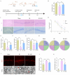
Fig. 1
Fig. 1
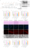
Protective effects of MH on hippocampal neurological damage, mitochondrial oxidative stress, and cognitive dysfunction after cerebral IR injury. (A) Schematic diagram of the animal experiment. (B) Neurodeficit scores for each group (n = 10, per group). (C) HE and Nissl staining of the CA1 region in the hippocampus (Scale bar: 50 μm) (n = 5, per group). (D) The time that rats spent to find the platform during 4 training days. (EG) Mean crossing times, and the time and distance rats spent in the target quadrant in the probe test (n = 10, per group). (H) Routes taken by rats in the probe test. (I) Representative images of DHE and TEM of neurons in the CA1 region of the hippocampus (Scale bars: DHE-50 μm, TEM-500 nm). (J) Intensity of DHE fluorescence (n = 5, per group). (K) Scores assessing mitochondrial injury (n = 5, per group). Sham: Sham-operation group; IR: Group subjected to cerebral IR injury followed by normothermia (37 °C); IR + MH: Group subjected to cerebral IR injury followed by 4 h of MH (33 ± 0.5 °C). Results are presented as the mean formula image standard deviation (SD). *p < 0.05, **p < 0.01.
Fig. 2
Fig. 2
Impact of MH on S100A8 expression after cerebral IR injury. (A) The volcano plot from proteomic analysis, proteins significantly altered are highlighted, with S100A8 indicated in red (n = 7, per group). (B) Western blot showing temporal expression of S100A8 protein at various time points after reperfusion. (C) Quantitative analysis of Western blots from (B) (n = 3, per group). (D) Western blot of S100A8 expression cross three different groups. (E) Quantitative analysis of Western blots from (D) (n = 3, per group). (F, G) Immunofluorescence staining of S100A8 in the CA1 region of the hippocampus (Scale bar: 50 μm) and its expression quantification (n = 5, per group). Sham: Sham-operation group; IR: Group subjected to cerebral IR injury followed by normothermia (37 °C); IR + MH: Group subjected to cerebral IR injury followed by 4 h of MH (33 ± 0.5 °C). Results are presented as the mean formula image SD. *p < 0.05, **p < 0.01.
Fig. 3
Fig. 3
In vivo, MH ameliorates IR-induced neuronal injury by decreasing S100A8 expression. (A) Schematic diagram of the animal experiment. (B) Western blot showing the expression of S100A8, bax, and caspase3 protein, with β-actin serving as the loading control. (CE) Quantification of the Western blots from (B). Levels of S100A8, bax and cleaved-caspase3/pro-caspase3 normalized to the corresponding loading control across three independent trials (n = 3, per group). (F) Representative images of HE, TUNEL, DHE staining and TEM in the CA1 region of the hippocampus (Scale bars: HE-50 μm, TUNEL-50 μm, DHE-50 μm, TEM-500 nm). (GI) Quantitative analysis of cell apoptosis ratio, DHE fluorescence intensity, and mitochondrial injury score (n = 5, per group). Sham + AAV9.NC: bilateral lateral ventricle injected with AAV9.NC 28 days before sham-operation; IR + AAV9.NC: bilateral lateral ventricle injected with AAV9.NC 28 days before cerebral IR injury; IR + AAV9.ShS100A8: bilateral lateral ventricle injected with AAV9.ShS100A8 28 days before cerebral IR injury; IR + MH + AAV9.NC: bilateral lateral ventricle injected with AAV9.NC 28 days before cerebral IR injury followed by 4 h of MH (33 ± 0.5 °C). IR + MH + AAV9.S100A8: bilateral lateral ventricle injected with AAV9.S100A8 28 days before cerebral IR injury followed by 4 h of MH (33 ± 0.5 °C). Results are presented as the mean formula image SD. *p < 0.05, **p < 0.01.
Fig. 4
Fig. 4
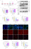
In vitro, MH ameliorates OGDR injury by decreasing the expression of S100A8 in SH-SY5Y cells. (A) Schematic diagram of the cell experiment. (B) Western blot showing the expression of S100A8, bax, and caspase3 protein, with β-actin was serving as the loading control. (CE) Quantification analysis of the Western blots from (B). Levels of S100A8, bax and cleaved-caspase3/pro-caspase3 normalized to the corresponding loading control across three independent trials (n = 3, per group). (F) Representative images of TUNEL, DHE and TMRE staining in SH-SY5Y cells (Scale bar: 50 μm). (GI) Quantitative analysis of apoptosis ratio, DHE, and TMRE fluorescence intensity (n = 5, per group). Control: Control group; OGDR: oxygen-glucose deprivation 2 h and reoxygenation 24 h; OGDR + Si-S100A8: SH-SY5Y were infected S100A8 siRNA using siRNA transfection kit for knocking down the expression level of S100A8 48 h before OGDR injury; OGDR + MH: OGDR injury followed by 4 h of MH (33 ± 0.5 °C). OGDR + MH + pcDNA3.1-S100A8: SH-SY5Y were infected with pcDNA3.1-S100A8 using Lipofectamine 2000 for overexpressing the expression level of S100A8 before OGDR injury followed by 4 h of MH (33 ± 0.5 °C). Results are presented as the mean formula image SD. *p < 0.05, **p < 0.01.
Fig. 5
Fig. 5
MH activates the CAMKK2/AMPK pathway by downregulating S100A8 expression. (A) Volcano plot from proteomic analysis, highlighting potential signaling pathways, with markers for S100A8 and CAMKK2 (n = 7, per group). (B) Western blot analysis displaying in vivo expression of S100A8, CAMKK2, p-AMPK, and AMPK, with β-actin serving as the loading control. (CE) Quantification analysis of the Western blots from (B) (n = 3, per group). (F) Western blot analysis showing in vitro expression of S100A8, CAMKK2, p-AMPK, and AMPK, with β-actin serving as the loading control. (GI) Quantification analysis of the Western blots from (F). Levels of S100A8, CAMKK2, p-AMPK, and AMPK normalized to corresponding loading control are summarized across three independent trials (n = 3, per group). Sham + AAV9.NC: bilateral lateral ventricle injected with AAV9.NC 28 days before sham-operation; IR + AAV9.NC: bilateral lateral ventricle injected with AAV9.NC 28 days before cerebral IR injury; IR + MH + AAV9.NC: bilateral lateral ventricle injected with AAV9.NC 28 days before cerebral IR injury followed by 4 h of MH (33 ± 0.5 °C). IR + MH + AAV9.S100A8: bilateral lateral ventricle injected with AAV9.S100A8 28 days before cerebral IR injury followed by 4 h of MH (33 ± 0.5 °C); Control: Control group; OGDR: oxygen-glucose deprivation 4 h and reoxygenation 24 h; OGDR + MH: OGDR injury followed by 4 h of MH (33 ± 0.5 °C). OGDR + MH + pcDNA3.1-S100A8: SH-SY5Y were infected with pcDNA3.1-S100A8 using Lipofectamine 2000 for over-expressing S100A8 before OGDR injury followed by 4 h of MH (33 ± 0.5 °C). Results are presented as the mean formula image SD. *p < 0.05, **p < 0.01.
Fig. 6
Fig. 6
Inhibition of the CAMKK2/AMPK pathway reduces the protective effect of MH on neuronal apoptosis, oxidative stress, and mitochondrial injury in IR rats. (A) Schematic diagram of the animal experiment. (B) Western blot showing in vivo expression of S100A8, CAMKK2, p-AMPK, AMPK, bax and caspase 3, with β-actin serving as the loading control. (CG) Quantification of the Western blots of (B) (n = 3, per group). (H) Representative images of TUNEL, DHE staining and TEM in the CA1 region of the hippocampus (Scale bars: TUNEL-50 μm, DHE-50 μm, TEM-500 nm). (IK) Quantitative analysis of cell apoptosis ratio, DHE fluorescence intensity, and mitochondrial injury score (n = 5, per group). Sham: Sham-operation group; IR: Group subjected to cerebral IR injury followed by normothermia (37 °C); IR + MH: Group subjected to cerebral IR injury followed by 4 h of MH (33 ± 0.5 °C); IR + MH + STO-609: STO-609 microinjected bilaterally into the lateral ventricles 30 min before cerebral IR injury followed by 4 h of MH (33 ± 0.5 °C); IR + MH + Dor: Dor microinjected bilaterally into the lateral ventricles 30 min before cerebral IR injury followed by 4 h of MH (33 ± 0.5 °C). Results are presented as the mean formula image SD. *p < 0.05, **p < 0.01.
Fig. 7
Fig. 7
Inhibition of the CAMKK2/AMPK pathway reduces the protective effect of MH on apoptosis and oxidative stress in SH-SY5Y cells after OGDR injury. (A) Schematic diagram of the cell experiment; (B) Western blot analysis shows the expression of S100A8, CAMKK2, p-AMPK, AMPK, bax and caspase3 protein expression, with β-actin serving as the loading control; (CG) Quantification of the Western blots from (B). Normalized S100A8, CAMKK2, p-AMPK/AMPK, bax and cleaved-caspase3/pro-caspase3 to corresponding loading control are summarized across three independent trials (n = 3, per group). (H) Representative images of TUNEL, DHE and TMRE staining (Scale bar: 50 μm) of SH-SY5Y cells; (IK) Quantitative analysis of apoptosis ratio, DHE and TMRE fluorescence intensity (n = 5, per group). Control: Control group; OGDR: oxygen-glucose deprivation 2 h and reoxygenation 24 h; OGDR + MH: OGDR injury followed by 4 h of MH (33 ± 0.5 °C); OGDR + MH + STO-609: SH-SY5Y were treated with STO-609 for inhibiting the activation of CAMKK2 30 min before OGDR injury followed by 4 h of MH (33 ± 0.5 °C); OGDR + MH + Dor: SH-SY5Y were treated with Dor for inhibiting the activation of AMPK 30 min before OGDR injury followed by 4 h of MH (33 ± 0.5 °C). Results are presented as the mean formula image SD. *p < 0.05, **p < 0.01.
Fig. 8
Fig. 8
Mechanism diagram of MH regulating S100A8-CAMKK2-AMPK axis against cerebral IR injury. Cerebral IR injury leads to an increase in the expression of S100A8, which inhibits the activity of the CAMKK2/AMPK pathway. This inhibition leads to mitochondrial dysfunction, including oxidative damage, decreased membrane potential, and loss of mitochondrial criste structure, along with an increase in apoptosis-related proteins such as bax and cleaved-caspase3. Consequently, this results in neuronal apoptosis. By contrast, MH down-regulates the high expression of S100A8, thereby enhancing mitochondrial function via activation of the CAMKK2/AMPK signaling pathway. As a result, this inhibits neuronal apoptosis and ameliorates neurological injury during cerebral IR in rats.

Similar articles

References

    1. Fanning, J. P. et al. Perioperative stroke. Nat. Rev. Dis. Primers10, 3. 10.1038/s41572-023-00487-6 (2024). - PubMed
    1. Xu, Y. et al. Mild hypothermia therapy attenuates early BBB leakage in acute ischaemic stroke. J. Cerebral Blood Flow Metab.1, 2. 10.1177/0271678x241275761 (2024). - PMC - PubMed
    1. Li, W. et al. Near-infrared-II imaging revealed hypothermia regulates neuroinflammation following brain injury by increasing the glymphatic influx. ACS Nano18, 13836–13848. 10.1021/acsnano.4c02652 (2024). - PubMed
    1. Polderman, K. H. Mechanisms of action, physiological effects, and complications of hypothermia. Crit. Care Med.37, S186-202. 10.1097/CCM.0b013e3181aa5241 (2009). - PubMed
    1. Wang, J. et al. Proteome profiling of hippocampus reveals the neuroprotective effect of mild hypothermia on global cerebral ischemia-reperfusion injury in rats. Sci. Rep.13, 14450. 10.1038/s41598-023-41766-2 (2023). - PMC - PubMed

Publication types

MeSH terms

Substances

LinkOut - more resources