. 2024 Sep 2;2(1):22.
doi: 10.1038/s44259-024-00039-2.

Stalled ribosome rescue factors exert different roles depending on types of antibiotics in Escherichia coli

Affiliations

Stalled ribosome rescue factors exert different roles depending on types of antibiotics in Escherichia coli

Mayu Mikami et al. NPJ Antimicrob Resist. .

Abstract

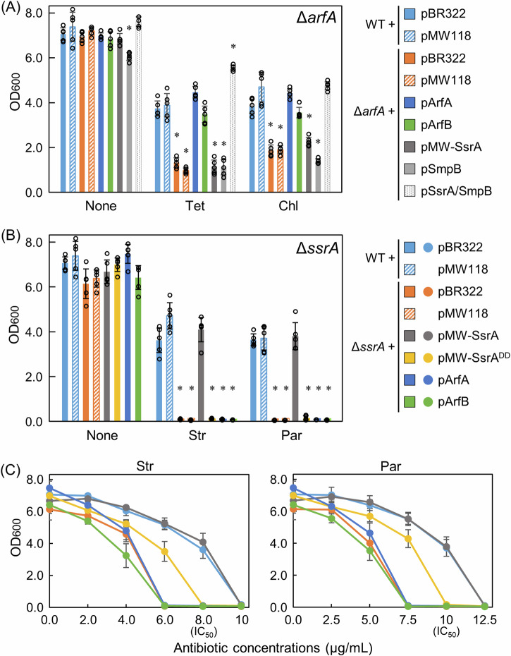
Escherichia coli possesses three stalled-ribosome rescue factors, tmRNA·SmpB (primary factor), ArfA (alternative factor to tmRNA·SmpB), and ArfB. Here, we examined the susceptibility of rescue factor-deficient strains from E. coli SE15 to various ribosome-targeting antibiotics. Aminoglycosides specifically decreased the growth of the ΔssrA (tmRNA gene) strain, in which the levels of reactive oxygen species were elevated. The decrease in growth of ΔssrA could not be complemented by plasmid-borne expression of arfA, arfB, or ssrAAA to DD mutant gene possessing a proteolysis-resistant tag sequence. These results highlight the significance of tmRNA·SmpB-mediated proteolysis during growth under aminoglycoside stress. In contrast, tetracyclines or amphenicols decreased the growth of the ΔarfA strain despite the presence of tmRNA·SmpB. Quantitative RT-PCR revealed that tetracyclines and amphenicols, but not aminoglycosides, considerably induced mRNA expression of arfA. These findings indicate that tmRNA·SmpB, and ArfA exert differing functions during stalled-ribosome rescue depending on the type of ribosome-targeting antibiotic.


Conflict of interest statement

Competing interests: The authors declare no competing interests.

Figures

Fig. 1
Fig. 1. Comparison of inhibitory effects of ribosome-targeting antibiotics on growth among the wild-type and the three ribosome rescue factor-deficient strains.
A Growth of the strains in the presence of various antibiotics. At the starting point, each strain was inoculated into 25 ml of LB medium at 37 °C in a 50 ml tube with a filter screw cap at a starting OD600 of 0.001. The LB medium contains each antibiotic at the IC50 concentration. After 5 h of growth in a shaking incubator, the OD600 of each culture was measured. It should be noted that the OD600 values of the wild-type strain after 5-h growth in the presence of each antibiotic at the IC50 concentration (particularly, tetracyclines and amphenicols) were not half of those in its absence, as the IC50 values were determined based on data after 8-h growth (Supplementary Fig. 2). Data are presented as the mean ± standard deviation of five independent experiments. Asterisks indicate significant differences compared to wild-type (Student’s t test, *p < 0.01; **p < 0.001). B Antibiotic concentration-dependent inhibition of growth of the wild-type and the three ribosome rescue factor-deficient strains. Each culture medium contained an antibiotic at the indicated concentration, and the OD600 of each medium was measured after 8 h of growth.
Fig. 2
Fig. 2. Suppression of the antibiotic susceptibility phenotypes of ΔarfA and ΔssrA by a plasmid-borne ribosome rescue factor.
A ΔarfA was transformed with a derivative of plasmid pBR322 harboring arfA (pArfA), arfB (pArfB), smpB (pSmpB), or both ssrA and smpB (pSsrA/SmpB) or that of plasmid pMW118 harboring ssrA (pMW-SsrA). As the controls, the wild-type and ΔarfA strains were transformed with an empty plasmid pBR322 or pMW118. In all experiments, the LB medium contains not only each indicted antibiotic at the IC50 concentration but also ampicillin for plasmid maintenance (pBR322, 50 µg/ml; pMW118, 12.5 µg/ml). The OD600 value of each medium was measured after 8 h of growth. Data are presented as the mean ± standard deviation of five independent experiments. Asterisks indicate significant differences compared to wild-type (Student’s t test, *p < 0.001). B ΔssrA was transformed with pMW-SsrA, pMW-SsrADD, pArfA, or pArfB. As the controls, the wild-type and ΔssrA strains were transformed with an empty plasmid pBR322 or pMW118. C Str- and Par-concentration-dependent inhibition of growth of the transformant used in (B). The symbol legend is indicted in (B). For the control, the data for pMW118 are not shown in the graphs, as they were virtually identical to those for pBR322. Data are presented as the mean ± standard deviation of five independent experiments.
Fig. 3
Fig. 3. Significant ROS generation induced by the ribosome-targeting antibiotics in ΔssrA.
A Percentages of fluorescent-positive cells of antibiotic-treated wild-type, ΔssrA, ΔarfA, and ΔarfB strains. After the strains were grown for 2.5 h, Str at 1.5-fold the IC50 concentrations (12.2 µg/ml), Par (14.6 µg/ml), or Tet (0.48 µg/ml) was added to the media, and they were further incubated for 3 h. The strain samples were stained with a photo-oxidation resistant derivative of DCFH-DA, and were adjusted to OD600 values of 2.0. All images were obtained by a fluorescence microscope (magnification, ×40; scale bar: 20 µm). Three fields per sample were analyzed, and each time, at least 100 cells were counted. Two independent experiments were performed for the wild-type and the mutant strains (n = 6). The results were expressed as percentage of fluorescent-positive cells versus cells observed in bright-field images. Data are presented as the mean ± standard deviation of the six data. Asterisks indicate significant difference compared to wild-type (Student’s t test, *p < 0.001). Representative merged bright-field and fluorescent microscopy images are presented in Supplementary Fig. 6A. B Representative merged bright-field and fluorescent microscopy images of antibiotic-treated cells of the wild-type and ΔssrA strains transformed with an empty plasmid, pMW-SsrA, or pMW-SsrADD that were stained with the oxidant-sensing probe. Only representative images regarding non-antibiotic- and Par-treated cells are presented. The other images are presented in Supplementary Fig. 6B. C Percentages of fluorescent-positive cells of the antibiotic-treated transformants of the wild-type and ΔssrA. For all others, refer to the legend in (A). D Percentages of fluorescent-positive cells of antibiotic-treated wild-type and ΔssrA depending on the antibiotic concentrations. The variable slope sigmoidal dose-response best-fit curves generated in GraphPad Prism 9.3.1 are plotted (R2: WT [0.933 and 0.901] and ΔssrA [0.944 and 0.984] for Str and Par, respectively). The curves for the wild-type and ΔssrA are indicated by solid and dotted lines, respectively.
Fig. 4
Fig. 4. Effects of antibiotics on mRNA expression of smpB, ssrA, arfA, and arfB from total RNA extracted from the wild-type SE15 strain.
When the culture medium reached an OD600 of 0.3 ~ 0.4, the indicated antibiotic at double the IC50 concentration was added to the medium. The samples were incubated for 30 min. idnT was used as an internal reference gene (see details in the text). Relative target gene expression levels were calculated using the equation 2−ΔΔCt, where ΔΔCt = ((Ctantibiotictarget – CtantibioticidnT) – (Ctnontreatedtarget – CtnontreatedidnT)). All Ct values used in the calculations are presented in Supplementary Fig. 8. Data are presented as the mean ± standard deviation of five independent experiments. Asterisks indicate significant differences compared to wild-type (Student’s t test, *p < 0.001). Values are described above the bars marked with asterisks.
Fig. 5
Fig. 5. Schematic diagrams of roles of tmRNA·SmpB and ArfA in rescue of antibiotic-dependent stalled ribosomes in E. coli.
A The stalled-ribosome rescue process in the presence of the aminoglycosides. Upper: tmRNA·SmpB rescues stalled ribosomes caused by the aminoglycosides by adding the tag peptide to the nascent polypeptides (indicated by a wide blue arrow). Tag-dependent proteolysis of the resultant aberrant proteins rapidly occurs so that ROS generation can be suppressed. Lower: in the absence of tmRNA·SmpB, ArfA is expressed; however, it does not help to rescue ribosomes exposed to aminoglycosides. Misreading of mRNA and/or stop codon read thorough occurs, and consequently, aberrant proteins accumulate in cells (see the text). This results in an increase in intracellular ROS levels that may lead to severe growth reduction or cell death. B The stalled-ribosome rescue process in the presence of tetracyclines and amphenicols. Red and blue vertical arrows indicate increase and decrease in mRNA induced by the antibiotics, respectively. A dotted T-shaped lines indicate a weakening of inhibition. When ArfA begins to increase in cells, this causes a positive feedback mechanisms where ArfA further increases itself (indicated by a rotated arrow). Although full-length ArfA protein that is produced when arfA mRNA is not cleaved by RNase III may also be more expressed, it is unstable and degraded immediately, as it possesses an extremely hydrophobic C-terminal tail that leads to aggregation and degradation of the full-length protein. ArfA can rescue stalled ribosomes caused by the bacteriostatic antibiotics even more efficiently (a blue arrow) than can tmRNA·SmpB (a light blue arrow). Note that neither tetracyclines nor amphenicols induce ROS generation.
Fig. 6
Fig. 6. Possible models in which each antibiotic that binds to 16S rRNA inhibits the binding of specific ribosome rescue factors to the empty A site of a ribosome.
A Left: Close-up views of superimposition of 16S rRNA of the small ribosomal subunit (PDB ID: 4V4Q) (green) and that bound with Par (4V5Y) (yellow). The backbone trace of 16S rRNA is depicted in either color; only A1492 and A1493 nucleotides in helix 44 are presented. A red arrow indicates changes in the base orientation caused by the Par binding. Middle and Right: Close-up views of superimposition of 16S rRNA bound with Par (4V5Y) and that with ArfB (6YSU) (middle) and that of 16S rRNA bound with Par and that with ArfA (5H5U) (right). van der Waals surfaces are presented for the bases of A1492 and A1493. The ribbon representations of ArfB and ArfA are depicted in red. Locations of putative clashes are surrounded by dotted skeleton boxes. Additionally, positions of putative clashed amino acid residues are indicated. All PDB data used in this Figure are from E. coli with the exception of 4DR3 (Str) from Thermus thermophiles. PyMOL (PyMOL Molecular Graphics System, Version 2.0, Schrödinger, LLC) was used to visualize and analyze all structures. B Left: Close-up views of superimposition of 16S rRNA (4V4Q) (green) and that bound with of Str (4DR3) (yellow). Only A1492 and A1493 nucleotides in the Str-bound 16S rRNA are indicated by black lines. A red arrow indicates a shift of the backbone of helix 44 laterally in the direction of ribosomal protein S12 and helix 18. Middle and Right: Close-up views of superimposition of 16S rRNA bound with Str (4DR3) and that with ArfB (6YSU) (middle) and that of 16S rRNA bound with Str and that with ArfA (5H5U) (right). C Left: Close-up views of the interaction between 16S rRNA and Tet (5J5B). Tet stacks with C1054 that is involved in decoding. The gray balls indicate Mg2+. The black and purple dashed lines indicate hydrogen bonds and electrical interactions, respectively. Middle: Close-up view of the superimposed small subunits structures focusing on the Tet-binding (5J5B), the SmpB-binding (7AC7), and the tRNA/mRNA binding (4V66). tRNA and mRNA are depicted in warmpink and salmon, respectively. A van der Waals surface is presented for Tet. Right, Top: Putative clash between Tet (5J5B) and the first nucleotide of anticodon (G34) in tRNA (4V66). Right, bottom: Putative clashes between Lys134 in SmpB (7AC7) and Tet (5J5B) and between Lys134 and C1054 in 16S rRNA (5J5B). van der Waals surfaces are presented for G34 and Lys134.

Similar articles

References

    1. Keiler, K. C. Mechanisms of ribosome rescue in bacteria. Nat. Rev. Microbiol.13, 285–297 (2015). - PubMed
    1. Keiler, K. C. & Feaga, H. A. Resolving nonstop translation complexes is a matter of life or death. J. Bacteriol.196, 2123–2130 (2014). - PMC - PubMed
    1. Müller, C., Crowe-McAuliffe, C. & Wilson, D. N. Ribosome rescue pathways in bacteria. Front. Microbiol.12, 652980 (2021). - PMC - PubMed
    1. Abo, T. & Chadani, Y. The fail-safe system to rescue the stalled ribosomes in Escherichia coli. Front. Microbiol.5, 156 (2014). - PMC - PubMed
    1. Himeno, H., Nameki, N., Kurita, D., Muto, A. & Abo, T. Ribosome rescue systems in bacteria. Biochimie114, 102–112 (2015). - PubMed

LinkOut - more resources