. 2025 Jan 22;15(1):2756.
doi: 10.1038/s41598-025-86716-2.

Mechanism of engineered macrophage membrane bionic gene-carrying nanospheres for targeted drug delivery to promote wound repair in deep second-degree burns

Affiliations

Mechanism of engineered macrophage membrane bionic gene-carrying nanospheres for targeted drug delivery to promote wound repair in deep second-degree burns

Zhihan Zhu et al. Sci Rep. .

Abstract

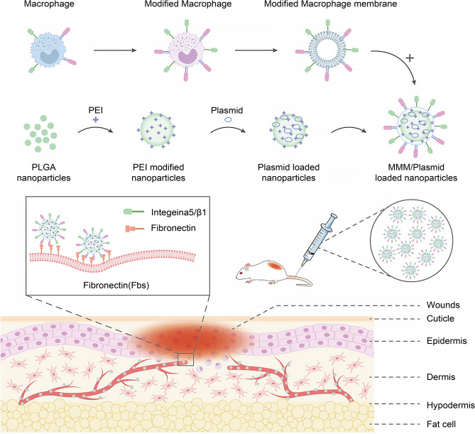
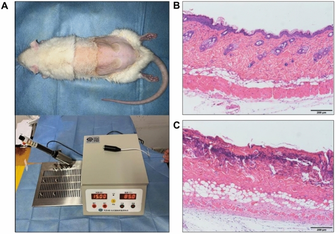
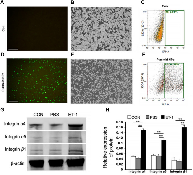
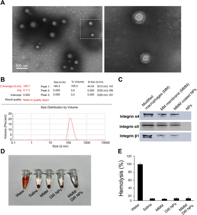
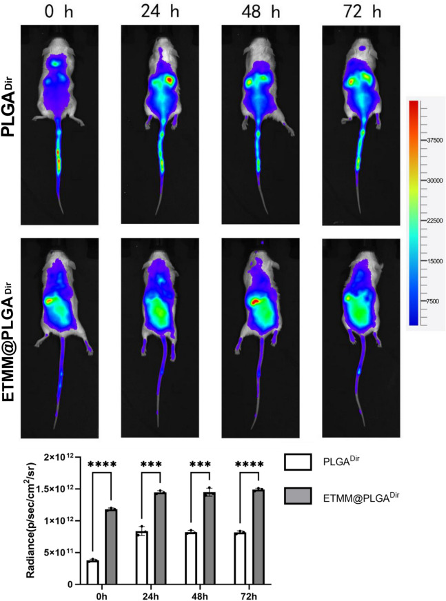
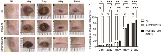
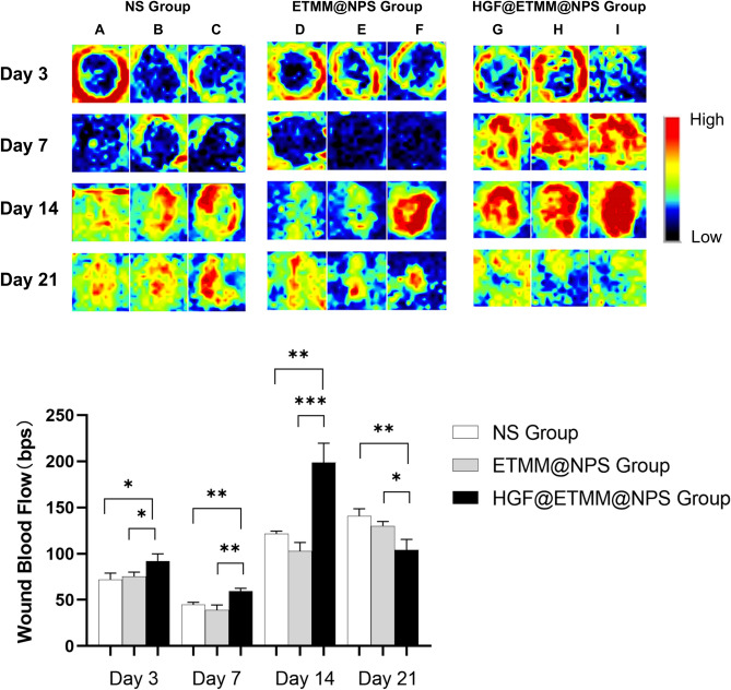
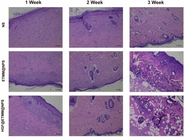
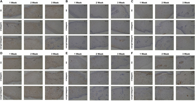
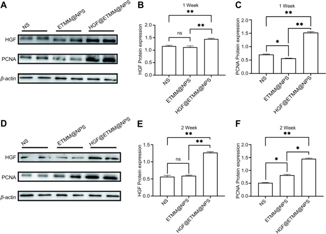
Hepatocyte growth factor (HGF) is a substance that stimulates the proliferation of hepatocytes which promote healing. We developed a macrophage membrane-encapsulated nanosphere drug delivery system containing HGF for the study of burn wound healing. Twenty-seven Sprague-Dawley rats were randomly divided into three groups: a saline control (NS) group, an engineered macrophage membrane-encapsulated nanospheres (ETMM@NPS) group, and an engineered macrophage membrane-encapsulated nanospheres treatment with HGF-loaded gene (HGF@ETMM@NPS) group.The wound tissue sections were examined histologically using hematoxylin and eosin (H&E) and Masson trichrome staining. Immunohistochemistry and Western blotting were performed to determine the expression of relevant proteins. The wound-healing, blood flow and complete epithelialization rates were significantly better in the HGF@ETMM@NPS group compared to the NS and ETMM@NPS groups. Expression of B-cell lymphoma 2-associated X-protein was significantly lower, and B-cell lymphoma 2, cluster of differentiation 31, HGF, alpha smooth muscle actin, and PCNA expression was significantly higher in the HGF@ETMM@NPS group compared with the other two groups. PCNA and HGF expression was significantly up-regulated in the HGF@ETMM@NPS group. The HGF@ETMM@NPS complex drug delivery system used in this research promoted wound healing via effective delivery of HGF to burn wounds, thereby accelerating skin cell growth and migration.

Keywords: Deep second-degree burns; Engineered macrophage membranes; PLGA nanospheres; Targeted drug delivery system; Trabecular repair.


Conflict of interest statement

Declarations. Competing interests: The authors declare no competing interests. Ethical statement: The study is reported in accordance with ARRIVE guidelines.The animal experiments of the study were reviewed and approved by the Animal Experiment Ethics Committee of Nantong University.

Figures

Fig. 1
Fig. 1
Schematic diagram of the mechanism of the engineered macrophage membrane-encapsulated gene-carrying nanosphere drug delivery system to promote skin burn repair. Abbreviations: PLGA, polylactic-co-glycolic acid; PEI, polyetherimide; MMM, modified macrophage membrane (engineered macrophage membrane).
Fig. 2
Fig. 2
Establishment of a rat model of deep second-degree burns. (A) Schematic representation of the rat model. (B) H&E micrograph of normal rat skin tissue. (C) H&E-micrograph of burnt rat skin tissue. Abbreviation: H&E, hematoxylin and eosin.
Fig. 3
Fig. 3
Relevant features of the gene-carrying nanospheres. (A) Scanning electron microscope image of gene-carrying nanospheres. (B, C) Particle size analysis graph of gene-carrying nanospheres. (D) Gel blocking assay. (E) In vitro release of gene-carrying nanospheres. Abbreviation: N/P ratio: ratio of ammonia root of polyetherimide to the phosphate group of plasmid.
Fig. 4
Fig. 4
Up-regulation of target protein expression by pEGFP-N1-loaded nanospheres. (A-F) Transfection of macrophages with pEGFP-N1-loaded nanospheres effectively transfected plasmids into macrophages. (G, H) Integrin expression in macrophages. Abbreviations: pEGFP-N1, plasmid enhanced green fluorescent protein-N1; CON, control; PBS, phosphate-buffered saline; ET-1, endothelin. **P < 0.01.
Fig. 5
Fig. 5
Membrane-coated DiR-loaded nanospheres electron micrographs and related features. (A) Transmission electron micrograph of modified macrophage membrane-coated DiR-loaded nanospheres. (B) Particle size map of membrane-coated DiR-loaded nanospheres. (C) MMM/DiR-NPS carrying integrins. (D, E) MMM/DiR-NPS have good hemocompatibility. Abbreviations: DiR, 1,1′-dioctadecyl-3,3,3′,3′-tetramethylindotricarbocyanine iodide; MMM/DiR-NPS, modified macrophage membrane-coated DiR-loaded nanospheres.
Fig. 6
Fig. 6
Modified macrophage membrane-encapsulated loaded DiR nanospheres targeting burn site.***P < 0.001, ****P < 0.0001. DiR, 1,1′-dioctadecyl-3,3,3′,3′-tetramethylindotricarbocyanine iodide; MMM/DiR-NPS, modified macrophage membrane-coated DiR-loaded nanospheres; M/DiR-NPS, membrane-coated DiR-loaded nanospheres.
Fig. 7
Fig. 7
Comparison of wound healing of deep second-grade burns in rats. Abbreviations: NS, normal saline; ETMM@NPS, engineered macrophage membrane-encapsulated nanospheres; HGF@ETMM@NPS, engineered macrophage membrane-encapsulated nanospheres with HGF-loaded gene; bps, beats per second. *P < 0.05, **P < 0.01, ***P < 0.001, ns, not significant.
Fig. 8
Fig. 8
Blood flow perfusion of burn wounds. Black areas indicate low or no blood flow and blue, yellow, green, and red areas indicate increased blood flow, in that order. A, B, and C represent the three rats in the NS group, D, E, and F represent the three rats in the ETMM@NPS group, and G, H, and I represent the three rats in the HGF@ETMM@NPS group. Abbreviations: HGF, hepatocyte growth factor; NS, normal saline; ETMM@NPS, engineered macrophage membrane-encapsulated nanospheres; HGF@ETMM@NPS, engineered macrophage membrane-encapsulated nanospheres with HGF-loaded gene; bps, beats per second. *P < 0.05, **P < 0.01, ***P < 0.001.
Fig. 9
Fig. 9
H&E staining of skin burn wounds at 7 d, 14 d, and 21 d post-injury.
Fig. 10
Fig. 10
(A) Results of immunohistochemical staining with Bax antibody. (B) Results of immunohistochemical staining with Bcl-2 antibody. (C) Results of immunohistochemical staining with CD31 antibody. (D) Results of immunohistochemical staining with HGF antibody. (E) Results of immunohistochemical staining with α-SMA antibody. (F) Results of immunohistochemical staining with PCNA antibody. Abbreviations: HGF, hepatocyte growth factor; PCNA, proliferating cell nuclear antigen; Bcl-2, B-cell lymphoma 2; Bax, Bcl-2 associated X-protein; CD31, cluster of differentiation 31; α-SMA, alpha smooth muscle actin.
Fig. 11
Fig. 11
Immunoblot analysis of HGF and PCNA protein expression in rat wounds in the three groups. (A) Western blot detection of HGF and PCNA protein expression in the NS, ETMM@NPS, and HGF@ETMM@NPS groups. (B) The expression of HGF and PCNA proteins in the NS, ETMM@NPS, and HGF@ETMM@NPS groups at 7 days after burn injury. (C) Relative expression of PCNA proteins in the NS, ETMM@NPS, and HGF@ETMM@NPS groups at 7 days after burn injury. (D) Western blot detection of the expression of HGF and PCNA proteins in the NS, ETMM@NPS, and HGF@ETMM@NPS groups at 14 days after burn injury. (E) Expression of HGF and PCNA proteins in the NS, ETMM@NPS, and HGF@ETMM@NPS groups at 14 days after burn injury. (F) Relative expression of PCNA proteins in the NS, ETMM@NPS, and HGF@ETMM@NPS groups 14 days after burn injury. Abbreviations: HGF, hepatocyte growth factor; PCNA, proliferating cell nuclear antigen; NS, normal saline; ETMM@NPS, engineered macrophage membrane-encapsulated nanospheres; HGF@ETMM@NPS, engineered macrophage membrane-encapsulated nanospheres HGF-loaded.

Similar articles

References

    1. Costa de Oliveira Souza, C. M. et al. Nanostructured cellulose–gellan–xyloglucan–lysozyme dressing seeded with mesenchymal stem cells for deep second-degree burn treatment. Int. J. Nanomed.16, 833–850. 10.2147/ijn.S289868 (2021). - PMC - PubMed
    1. Ebrahimpour, N. et al. The efficacy of a traditional medicine preparation on second-degree burn wounds in rats. J. Ethnopharmacol.10.1016/j.jep.2020.112570 (2020). - PubMed
    1. Homaeigohar, S., Li, M. & Boccaccini, A. R. Bioactive glass-based fibrous wound dressings. Burns Trauma10, tkac038. 10.1093/burnst/tkac038 (2022). - PMC - PubMed
    1. Norman, G. et al. Antiseptics for burns. Cochrane Database Syst. Rev.7, CD011821. 10.1002/14651858.CD011821.pub2 (2017). - PMC - PubMed
    1. Shao, Y. et al. Neutrophil extracellular traps contribute to myofibroblast differentiation and scar hyperplasia through the Toll-like receptor 9/Kappanuclear Kappafactor Kappa-B/interleukin-6 pathway. Burns Trauma10, tkac044. 10.1093/burnst/tkac044 (2022). - PMC - PubMed

Publication types

Substances