. 2025 Jan 22;16(1):939.
doi: 10.1038/s41467-025-56258-2.

Treadmill exercise prevents stress-induced anxiety-like behaviors via enhancing the excitatory input from the primary motor cortex to the thalamocortical circuit

Affiliations

Treadmill exercise prevents stress-induced anxiety-like behaviors via enhancing the excitatory input from the primary motor cortex to the thalamocortical circuit

Zhihua Luo et al. Nat Commun. .

Abstract

Physical exercise effectively prevents anxiety disorders caused by environmental stress. The neural circuitry mechanism, however, remains incomplete. Here, we identified a previously unrecognized pathway originating from the primary motor cortex (M1) to medial prefrontal cortex (mPFC) via the ventromedial thalamic (VM) nuclei in male mice. Besides anatomical evidence, both ex vivo and in vivo recordings showed enhanced excitability of M1-VM inputs to the prelimbic (PrL) region of mPFC upon 14-day treadmill exercise on a chronic restraint stress (CRS) mouse model. Further functional interrogations demonstrated that the activation of this neural circuit is both necessary and sufficient to direct the anxiolytic effect of exercise training in CRS mice. Our findings provide more insights into the neural circuits connecting motor and mental regions under exercise paradigm and implicate potential targets for neuromodulation in treating anxiety disorders.


Conflict of interest statement

Competing interests: The authors declare no competing interests.

Figures
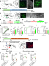
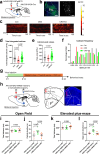
Fig. 1
Fig. 1. Treadmill exercise activates prelimbic-projecting ventromedial thalamic neurons to prevent stress-induced anxiety-like behaviors.
a Left, viral injection sites; Right, Retrograde labelling of neurons in the primary motor cortex (M1) and the ventromedial thalamus (VM). PrL, prelimbic; CPU, caudate putamen. Green, EGFP. Scale bar, 100 μm (left- and right-most) and 500 μm (middle panel). b Timelines of experimental design. c Left, viral injection sites; Right, distribution of PrL-projecting neurons in VM, and patch-clamp recording of one representative cell. Green, EGFP. Scale bar, 100 μm (left) and 20 μm (right). d Sample spikes of VM neurons giving fixed (240 pA) injection currents. e Exercise training (Ex) elevates the total number of spikes in CRS mice. Two-way ANOVA with respect to the group effect, F(2,525) = 15.40, P < 0.0001. f Left, CRS+Ex animals showed elevated resting membrane potential (RMP). One-way ANOVA, F(2,33) = 5.472, P = 0.0089. Right, CRS+Ex animals displayed lowered rheobase value. F(2,33) = 5.562, P = 0.0083. n = 12 neurons from 3 mice in each group in (e, f). g Representative trances of miniature excitatory postsynaptic currents (mEPSCs) in all groups. h Distribution of mEPSC frequency. Exercise training enhanced the frequency of mEPSC in CRS-treated animals. F(2,36) = 13.49, P < 0.0001. i Distribution of mEPSC amplitude. Exercise did not affect mEPSC amplitude. One-way ANOVA, F(2,36) = 0.1044, P = 0.9011. n = 13 neurons from 3 mice in each group in (h, i). j Timelines of experimental design. k Left, viral injection sites. Right, distribution of PrL-projecting neurons in VM. Red, mCherry. Scale bar, 100 μm. l Chemogenetic manipulation did not change overall locomotor ability in the open field. F(2,21) = 0.05169, P = 0.9497. m The inhibition of PrL-projecting VM neurons decreased time spent in the central region. F(2,21) = 6.873, P = 0.0051. n CNO infusion did not affect the total distance travelled on the elevated plus-maze. F(2,21) = 1.799, P = 0.1900. o Chemogenetic inhibition of PrL-projecting VM cells decreased the time spent in the open arm region. F(2,21) = 7.292, P = 0.0039. N = 8 mice each group in (lo). Tukey’s multiple comparison test was employed to make comparisons between two specified groups in a two-sided manner. All data were presented as mean ± sem.
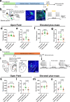
Fig. 2
Fig. 2. The activation of VM-PrL pathway is necessary for exercise-mediated anxiolytic effects.
a Schematic illustration of virus injection. b Left, fluorescent images of viral infection. Green, GCaMP. Scale bar, 300 μm. Right, the field of view (FOV) of in vivo 2-photon imaging. Scale bar, 100 μm. c Heatmaps of individual in vivo neuronal calcium activities during a 150-sec recording window. A total of 40 sampled neurons from 4 mice were plotted in each group. d Exercise training potentiated the total integrated calcium strength. Nonparametric Kruskal-Wallis test statistic=39.27, P < 0.0001. e Exercise training increased the peak value of individual calcium transient. Nonparametric Kruskal-Wallis test statistic=41.74, P < 0.0001. f Distribution of calcium transient frequency. n = 133 neurons from 4 mice in each group in (df). g Timelines of experimental design. h Left, viral injection sites. Right, distribution of VM innervating neurons in PrL. Scale bar, 250 μm. Red, mCherry. i Chemogenetic manipulation did not change overall locomotor ability in the open field. One-way ANOVA, F(2,21) = 0.5787, P = 0.5693. j The inhibition of VM innervating PrL neurons decreased time spent in the central region. F(2,21) = 5.903, P = 0.0092. k CNO infusion did not affect the total distance travelled on the elevated plus-maze. F(2,21) = 0.1739, P = 0.8416. l Chemogenetic inhibition of VM innervating PrL cells decreased the time spent in the open arm region. F(2,21) = 7.363, P = 0.0038. N = 8 mice each group in (il). The comparison between two specified groups was performed using Dunn’s multiple comparison test in a two-sided manner in (de), and Tukey’s multiple comparison test in a two-sided manner in (il). All data were presented as mean ± sem.
Fig. 3
Fig. 3. M1 neurons project to VM for improving anxiety disorders.
a Left, schematic illustration of viral injection; Right, Retrograde labelling of neurons in M1 across the anteroposterior axis. M1, primary motor cortex; M2, secondary motor cortex; DCN, deep cerebellar nuclei. Green, GFP. Scale bar, 500 μm. b Upper, timelines of experimental design. Lower left, viral injection sites; Lower right, distribution of VM-projecting neurons in PrL. S1, primary sensory cortex; Scale bar, 250 μm. c The quantification of VM-projecting cell number in M1 after cell ablation. Multiple t-test was used for the comparison in a two-sided manner. N = 6 mice per group. d The ablation of VM-projecting M1 neurons did not change overall locomotor ability in the open field. One-way ANOVA, F(2,21) = 0.4272, P = 0.6578. e The specific cell ablation decreased time spent in the central region. F(2,21) = 9.225, P = 0.0013. f Cell ablation in M1 did not affect the total distance travelled on the elevated plus-maze. F(2,21) = 0.2087, P = 0.8133. g The blockade of M1-VM pathway decreased the time spent in the open arm region. F(2,21) = 8.785, P = 0.0017. N = 8 mice each group in (dg). h Timelines of experimental design. i Left, viral injection sites. Right, distribution of VM-projecting neurons in M1. Red, mCherry. Scale bar, 100 μm. j Current-evoked action potentials in a representative VM-projecting M1 neuron labelled with hM4Di recorded before, during and after CNO perfusion (10 μM). k CNO infusion effectively repressed cellular excitability. One-way ANOVA with repeated measures, F(1.241,6.207) = 93.14, P < 0.0001. n = 6 neurons from 3 mice. l Chemogenetic manipulation did not change overall locomotor ability in the open field. F(2,24) = 0.8324, P = 0.4472. m The inhibition of VM-projecting M1 neurons decreased time spent in the central region. F(2,24) = 26.81, P < 0.0001. n CNO infusion did not affect the total distance travelled on the elevated plus-maze. F(2,24) = 0.9322, P = 0.4075. o Chemogenetic inhibition of PrL-projecting VM cells decreased the time spent in the open arm region. F(2,24) = 6.958, P = 0.0041. N = 9 mice each group in (lo). Tukey’s multiple comparison test was employed to make comparisons between two specified groups in a two-sided manner. All data were presented as mean ± sem.
Fig. 4
Fig. 4. VM nuclei is activated by M1 for relieving anxiety-like behaviors.
a Timelines of experimental design. b Left, schematic illustration of viral injection; Right, distribution of M1 innervating neurons in VM, and patch-clamp recording of one representative cell. Red, mCherry. Scale bar, 100 μm (left) and 20 μm (right). c Sample spikes of VM neurons giving fixed (240 pA) injection currents. d Exercise training (Ex) elevates the total number of spikes in CRS mice. Two-way ANOVA with respect to the group effect, F(2,495) = 34.52, P < 0.0001. e Left, CRS+Ex animals showed elevated resting membrane potential (RMP). One-way ANOVA, F(2,36) = 3.972, P = 0.0276. Right, CRS+Ex animals displayed lowered rheobase value. F(2,36) = 9.229, P = 0.0006. n = 13 neurons from 3 mice in each group in (d, e). f Representative trances of miniature excitatory postsynaptic currents (mEPSCs) in all groups. g Distribution of mEPSC frequency. Exercise training enhanced the frequency of mEPSC in CRS-treated animals. F(2,33) = 4.362, P = 0.0209. h Distribution of mEPSC amplitude. Exercise did not affect mEPSC amplitude. F(2,33) = 0.9798, P = 0.3857. n = 12 neurons from 3 mice in each group in (g, h). Tukey’s multiple comparison test was employed to make comparisons between two specified groups in a two-sided manner. All data were presented as mean ± sem.
Fig. 5
Fig. 5. Exercise training potentiates excitatory input to PrL by M1-VM pathway.
a Viral injection schemes. b Fluorescent colabelling of antero- and retro-grade labelling VM neurons. FrA, frontal association cortex; PrL, prelimbic region; MO, medial orbital cortex; LO, lateral orbital cortex; VO, ventral orbital cortex; ZI, zona incerta; VM, ventromedial thalamus; Sub, subcoeruleus nucleus; mt, medial terminal nucleus of the accessory optic tract. Scale bar, 150 μm and 25 μm (enlarged views only). c Percentage of excitatory (CaMKIIα) neurons in co-labelled VM neurons. N = 6 mice. d Left, viral injection and ex vivo optogenetic stimulation schemes. Right, fluorescent images showing viral infection in VM. Scale bar, 250 μm. e Representative traces showing EPSCs when slices were sequentially perfused with ACSF (left), TTX (1 µM, middle left), TTX + 4-AP (100 µM, middle right) and TTX + 4-AP + CNQX (20 µM, right). f Summary plots of the EPSC amplitudes in (e). One-way ANOVA with repeated measures, F(1.167,7.002) = 93.14, P = 0.0004. n = 7 neurons from 3 mice. g Viral injection schemes. h Fluorescent images showing viral infection in VM and PrL. DLO, dorsolateral orbital cortex Green, GCaMP. Scale bar, 500 μm (left) and 100 μm (right). i Heatmaps of in vivo calcium activities of axonal terminals during a 150 s recording window. A total of 20 sampled axons from 4 mice were plotted in each group. j Exercise training potentiated the total integrated calcium strength. Nonparametric Kruskal-Wallis test statistic=111.2, P = 0.0001. k Exercise training also increased the peak value of individual calcium transient. Nonparametric Kruskal-Wallis test statistic=101.0, P < 0.0001. l Distribution of calcium transient frequency. n = 116 neurons from 4 mice in each group in (jl). The comparison between two specified groups was performed using Tukey’s multiple comparison test in a two-sided manner in (f), and Dunns multiple comparison test in a two-sided manner in (j, k). All data were presented as mean ± sem.
Fig. 6
Fig. 6. The M1-VM-PrL pathway mediates exercise effect in relieving anxiety-like behaviors.
a Timelines of experimental design. b Left, viral injection sites. Right, fluorescent images showing viral infection in VM and PrL. Green, EGFP. Scale bar, 150 μm. c Sample spikes of VM neurons giving fixed (300 pA) injection currents plus optogenetic inhibition. d Light inhibition (eNpHR3) did not change overall locomotor ability in the open field. One-way ANOVA, F(2,27) = 0.6808, P = 0.5147. e Specific light inhibition of M1-VM terminus in PrL region decreased time spent in the central region. F(2,27) = 7.535, P = 0.0025. f The optogenetic manipulation did not affect the total distance travelled on the elevated plus-maze. F(2,27) = 0.9190, P = 0.4110. g The inhibition of M1-VM terminus decreased the time spent in the open arm region. F(2,27) = 5.352, P = 0.0110. N = 10 mice each group in (dg). h Timelines of experimental design. i Left, viral injection sites. Right, fluorescent images showing viral infection in VM. Green, EGFP Scale bar, 250 μm. j Current-evoked action potentials in a representative M1-innervated, and PrL-projecting VM neurons were labelled with hM4Di recorded before, during and after CNO perfusion (10 μM). k Chemogenetic inhibition (CNO group) did not change overall locomotor ability in the open field. One-way ANOVA, F(2,21) = 0.1865, P = 0.8312. l CNO infusion decreased time spent in the central region. F(2,21) = 6.876, P = 0.0050. m The chemogenetic manipulation did not affect the total distance travelled on the elevated plus-maze. F(2,21) = 0.5705, P = 0.5737. n The chemogenetic inhibition of M1-VM-PrL pathway decreased the time spent in the open arm region. F(2,21) = 4.975, P = 0.0170. N = 8 mice each group in (kn). Tukey’s multiple comparison test was employed to make comparisons between two specified groups in a two-sided manner. All data were presented as mean ± sem.
Fig. 7
Fig. 7. Activation of M1-VM-PrL pathway relieves stress-induced anxiety-like behaviors.
a Timelines of experimental design. b Left, viral injection sites. Right, M1-inneravting VM neurons and their terminus in PrL. Green, EGFP Scale bar, 100 μm. c Sample spikes of PrL neurons under light stimulation. d Light activation (ChR2) did not change overall locomotor ability in the open field. One-way ANOVA, F(3,32) = 0.7786, P = 0.9956. e Specific light activation of M1-VM terminus in PrL region increased time spent in the central region. F(3,32) = 6.508, P = 0.0015. f The optogenetic manipulation did not affect the total distance travelled on the elevated plus-maze. F(3,32) = 0.07647, P = 0.9674. g The activation of M1-VM terminus increased the time spent in the open arm region. F(3,32) = 5.067, P = 0.0055. N = 9 mice each group in (dg). h Timelines of experimental design. i Left, viral injection sites. Right, fluorescent images showing viral infection in VM. Green, EGFP Scale bar, 250 μm. j Sample traces of VM neurons giving under CNO infusion. k Chemogenetic activation (CNO group) did not change overall locomotor ability in the open field. F(2,21) = 0.09855, P = 0.9066. l CNO infusion increased time spent in the central region. F(2,21) = 7.931, P = 0.0027. m The chemogenetic manipulation did not affect the total distance travelled on the elevated plus-maze. F(2,21) = 0.2438, P = 0.7858. n The chemogenetic activation of M1-VM-PrL pathway increased the time spent in the open arm region. F(2,21) = 8.759, P = 0.0017. N = 8 mice each group in (kn). Tukey’s multiple comparison test was employed to make comparisons between two specified groups in a two-sided manner. All data were presented as mean ± sem.

Similar articles

Cited by

References

    1. Tyrer, P. & Baldwin, D. Generalised anxiety disorder. Lancet368, 2156–2166 (2006). - PubMed
    1. Villagrasa, B. et al. Prevalence of anxiety disorder among older adults in Spain: a meta-analysis. J. Affect Disord.246, 408–417 (2019). - PubMed
    1. Aune, T., Nordahl, H. M. & Beidel, D. C. Social anxiety disorder in adolescents: prevalence and subtypes in the Young-HUNT3 study. J. Anxiety Disord.87, 102546 (2022). - PubMed
    1. Delpino, F. M. et al. Prevalence of anxiety during the COVID-19 pandemic: a systematic review and meta-analysis of over 2 million people. J. Affect Disord.318, 272–282 (2022). - PMC - PubMed
    1. Bosman, R. C., et al., Prevalence and course of subthreshold anxiety disorder in the general population: A three-year follow-up study. J. Affect Disord. 247, 105–113 (2019). - PubMed

MeSH terms