. 2025 Jan 22;31(1):18.
doi: 10.1186/s10020-025-01066-z.

Tousled-like kinase loss confers PARP inhibitor resistance in BRCA1-mutated cancers by impeding non-homologous end joining repair

Affiliations

Tousled-like kinase loss confers PARP inhibitor resistance in BRCA1-mutated cancers by impeding non-homologous end joining repair

Min-Ah Kim et al. Mol Med. .

Abstract

Background: Double-strand breaks (DSBs) are primarily repaired through non-homologous end joining (NHEJ) and homologous recombination (HR). Given that DSBs are highly cytotoxic, PARP inhibitors (PARPi), a prominent class of anticancer drugs, are designed to target tumors with HR deficiency (HRD), such as those harboring BRCA mutations. However, many tumor cells acquire resistance to PARPi, often by restoring HR in HRD cells through the inactivation of NHEJ. Therefore, identifying novel regulators of NHEJ could provide valuable insights into the mechanisms underlying PARPi resistance.

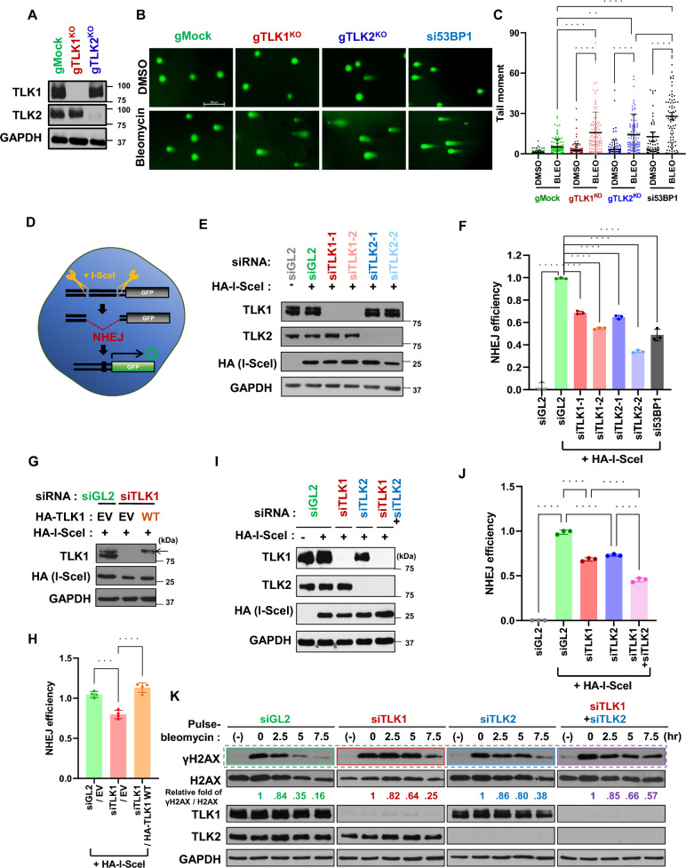
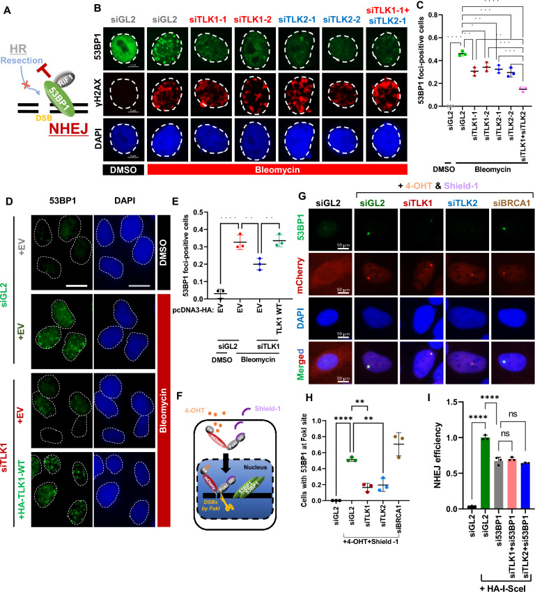
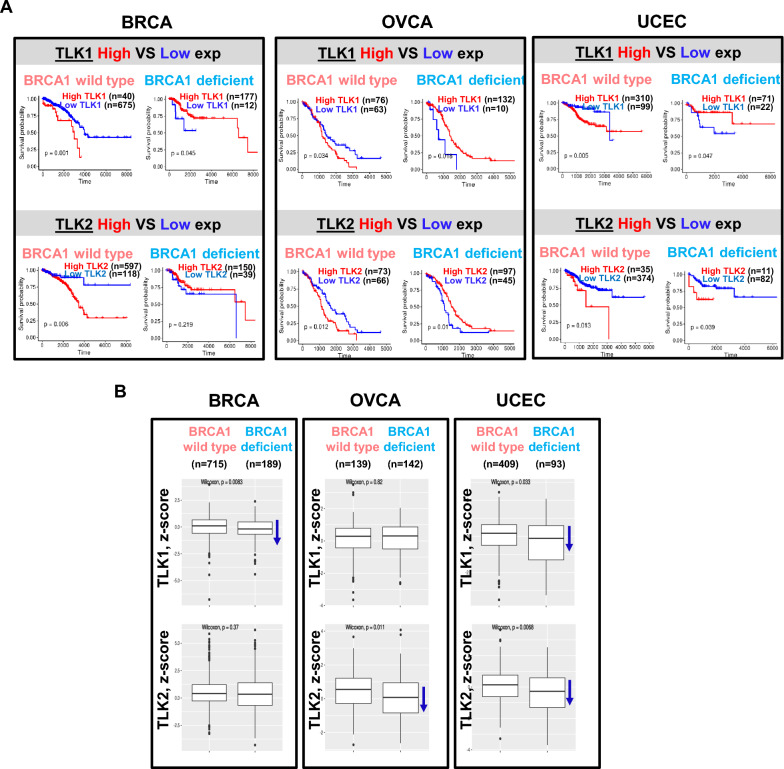
Methods: Cellular DSBs were assessed using neutral comet assays and phospho-H2AX immunoblotting. Fluorescence-based reporter assays quantified repair via NHEJ or HR. The recruitment of proteins that promote NHEJ and HR to DSBs was analyzed using immunostaining, live-cell imaging following laser-induced microirradiation, and FokI-inducible single DSB generation. Loss-of-function experiments were performed in multiple human cancer cell lines using siRNA-mediated knockdown or CRISPR-Cas9 gene knockout. Cell viability assays were conducted to evaluate resistance to PARP inhibitors. Additionally, bioinformatic analyses of public databases were performed to investigate the association between TLK expression and BRCA1 status.

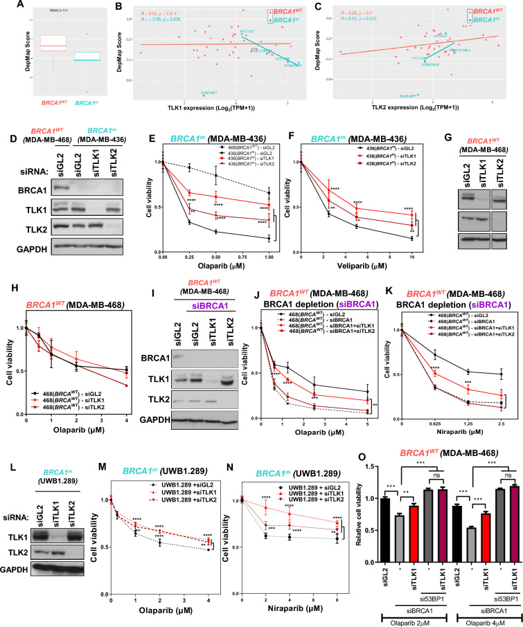
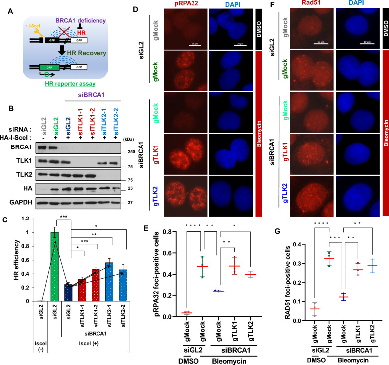
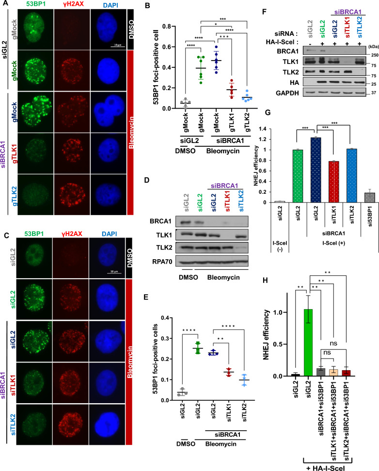
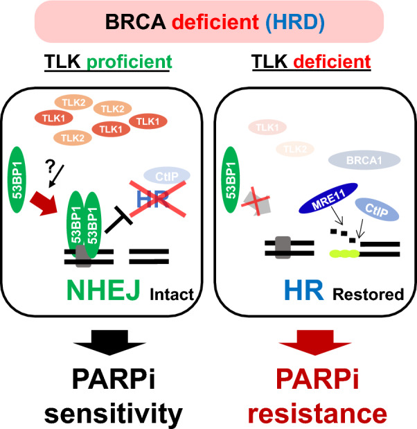
Results: We demonstrate that human tousled-like kinase (TLK) orthologs are essential for NHEJ-mediated repair of DSBs and for PARPi sensitivity in cells with BRCA1 mutation. TLK1 and TLK2 exhibit redundant roles in promoting NHEJ, and their deficiency results in a significant accumulation of DSBs. TLKs are required for the proper localization of 53BP1, a key factor in promoting the NHEJ pathway. Consequently, TLK deficiency induces PARPi resistance in triple-negative breast cancer (TNBC) and ovarian cancer (OVCA) cell lines with BRCA1 deficiency, as TLK deficiency in BRCA1-depleted cells, impairs 53BP1 recruitment to DSBs and reduces NHEJ efficiency, while restoring HR.

Conclusions: We have identified TLK proteins as novel regulators of NHEJ repair and PARPi sensitivity in BRCA1-depleted cells, suggesting that TLK repression may represent a previously unrecognized mechanism by which BRCA1 mutant cancers acquire PARPi resistance.

Keywords: BRCA1; Cancer Therapy; Cancer treatment; DNA damage repair; Double-stranded breaks (DSBs); Drug resistance; Homologous recombination (HR); Non-homologous end joining (NHEJ); PARP inhibitor; Tousled-like kinase.


Conflict of interest statement

Declarations. Ethics approval and consent to participate: Not applicable. Consent of publication: Not applicable. Competing interests: The authors declare no competing interests.

Figures

Fig. 1
Fig. 1
TLK1 and TLK2 promote NHEJ-mediated DSB repair. A to C Increased levels of DSBs following bleomycin treatment in TLK1 or TLK2 knockout (KO) cells. TLK1 knockout (gTLK1KO) and TLK2 knockout (gTLK2KO) U2OS cells were generated using the CRISPR/Cas9 system (A), and a neutral comet assay was performed on the cells in presence of DMSO or bleomycin for 12 h. Representative images (B) and quantifications (C) are shown. Each dot depicts the tail moment of individual cells, and the mean with S.D. is presented from at least 100 cells. si53BP1 used as a positive control. **P < 0.01; ****P < 0.0001. Scale bar, 200 μm. D to F Decreased NHEJ in TLK1- and TLK2-depleted cells. Schematic of the GFP reporter assay used to measure NHEJ efficiency using U2OS EJ5-GFP cells (D). Cells were transfected with two different siRNAs targeting either TLK1 or TLK2. Cell extracts were used for immunoblotting as shown in (E) and NHEJ efficiency was measured and plotted in (F). Transient expression of HA-I-SceI was observed in the immunoblot using anti-HA antibody. The ratio of HA-I-SceI to GAPDH was quantified from the immunoblots and normalized to the percentage of GFP-positive cells in each sample. NHEJ efficiency was represented as the relative ratio compared to the siGL2 + HA-I-SceI. si53BP1 used as a positive control. Mean ± S.D. of triplicates. ****P < 0.0001. G and H Restoration of NHEJ by siRNA-resistant wild type TLK1 (WT) in endogenous TLK1-depleted cells. siTLK1-1 were used, and immunoblots (G) and quantification of NHEJ efficiency (H) are shown. Mean ± S.D. of quadruplicates. EV, empty vector. ***P < 0.001; ****P < 0.0001. I and J Further decrease of NHEJ in co-depletion of TLK1 and TLK2. Cells were transfected with siRNAs against TLK1 and/or TLK2. Immunoblots (I) and NHEJ efficiency (J) are shown. Mean ± S.D. of triplicates. K Delayed disappearance of bleomycin-induced γH2AX (phospho-H2AX at Ser139) in TLK-depleted cells. After transfection with the indicated siRNA, U2OS cells were pulse-treated with 20 μg/mL bleomycin for 1 h and harvested at the indicated time points. The amount of protein in the γH2AX and H2AX immunoblots was quantified using ImageJ software, and their ratio was calculated
Fig. 2
Fig. 2
TLK loss impairs DSB localization of 53BP1. A A schematic explaining the critical role of 53BP1 localization at DSBs in promoting NHEJ by antagonizing DSB end resection. B and C Suppression of 53BP1 focus formation at DSBs in TLK-depleted cells. A549 cells transfected with the indicated siRNAs were treated with 10 μg/mL bleomycin for 1.5 h. Representative images are shown in (B), and cell lysates were subjected to immunoblotting (Fig. S2A). Cells with > 20 foci were counted as positives and plotted in (C). Hereafter, siTLK1 and siTLK2 indicate siTLK1-1 and siTLK2-1, respectively. Mean ± S.D. of triplicates. *P < 0.05; **P < 0.01; ***P < 0.001; ****P < 0.0001. D and E Restoration of 53BP1 focus formation by siRNA-resistant TLK1 in endogenous TLK1-depleted cells. A549 cells stably expressing EV or siTLK1-resistant TLK1 was subjected to siRNA transfection to deplete endogenous TLK1. Representative images (D) and quantification of 53BP1 foci-positives (E). Scale bar, 10 μm. F A schematic of U2OS ER-mCherry-LacI-FokI-DD cells to monitor the recruitment of protein-of-interest at artificial DSBs generated by the FokI endonuclease. The ER-mCherry-LacI-FokI-DD fusion proteins is stabilized and translocated into the nucleus by co-treatment with Shield-1 and 4-OHT to generate DSBs at LacO repeats. G and H Reduced localization of 53BP1 at the FokI cut site in TLK1- or TLK2-depleted cells. U2OS ER-mCherry-LacI-FokI-DD cells were transfected with the indicated siRNAs and stained with an anti-53BP1 antibody. Cells were counted when 53BP1 foci colocalized with mCherry. Representative images are shown in (G) and the counted % graphed in (H). I TLK’s NHEJ function depends on 53BP1. The NHEJ assay using U2OS EJ5-GFP cells was performed with the indicated siRNAs, as described in Fig. 1D to F. Data are presented as mean ± S.D. of triplicates. ****P < 0.0001; ns, not significant
Fig. 3
Fig. 3
TLK deficiency increases PARP inhibitor resistance in BRCA1-mutated cancers. A Dependency effect of PARP1 on BRCA1 mutation status using the DepMap breast cancer cell line dataset. DepMap scores for cells with wild type and mutated BRCA1 were compared, and statistical significance was assessed using the Wilcoxon test. The DepMap score reflects the impact of PARP1 knockout on cell survival. B and C Correlation analysis of TLK1 (B) or TLK2 (C) expression with PARP1 DepMap scores using breast cancer cell lines from the CCLE database. DepMap scores were compared between cell lines with wild type and mutated BRCA1. D to F Increase of PARP inhibitor resistance following TLK1 or TLK2 depletion in BRCA1-mutated MDA-MB-436 breast cancer cells. After siRNA transfection, cell viability was measured five days post-treatment with PARPi at indicated concentration. Immunoblots (D) and cell survival quantification following olaparib (E) and veliparib (F) treatment are shown. Data are presented as mean ± S.D. of triplicates. *P < 0.05; **P < 0.01; ****P < 0.0005. G and H Effect of TLK loss on PARP inhibitor sensitivity in BRCA1-proficient MDA-MB-468 cells. Immunoblots for the indicated siRNAs (G) and quantification of cell survival (H) are shown. I to K Enhanced PARP inhibitor resistance following TLK1 depletion in siBRCA1-transfected MDA-MB-468 cells. Immunoblots (I), cell survival quantification following olaparib (J) and niraparib (K) treatment are shown. L to N Increased PARP inhibitor resistance in depletion of TLK1 or TLK2 in BRCA1-mutated UWB1.289 HGSOC cells. Immunoblots (L), cell survival quantification following olaparib (M) and niraparib (N) treatment are shown. **P < 0.01; ****P < 0.0005. O PARPi resistance caused by 53BP1 depletion in BRCA1-deficient cells is not strengthened by TLK1 co-depletion. MDA-MB-468 cells were treated with the indicated siRNAs and concentrations of olaparib, and the cell viability of all samples was normalized to that of siGL2 treated with 2 μM olaparib. ns, not significant
Fig. 4
Fig. 4
TLK loss restores HR in BRCA1-deficient cells. A Schematic depicting the measurement of HR repair efficiency using U2OS DR-GFP cells. Cells were transfected with siBRCA1 to reduce HR efficiency, and recovery of HR was measured by GFP reporter expression via FACS analysis. B and C Restoration of HR efficiency following TLK depletion under BRCA1 deficiency. Immunoblots of cell extracts (B) and HR efficiency (C) after depletion of the indicated proteins are shown. Mean ± S.D. of triplicates. *P < 0.05; **P < 0.01; ***P < 0.005. D to G Restoration of nuclear pRPA32 and RAD51 foci following TLK1 or TLK2 knockout in BRCA1-deficient cells. BRCA1 was depleted by siRNA in the indicated cell lines. Cells were treated with DMSO or bleomycin for 2 h, and pre-extracted before fixation. Cells with more than 20 and 5 foci were counted as positive for pRPA32 and RAD51, respectively. Representative images (D and F) and quantification of foci-positive cells (E and G) for nuclear pRPA32 and Rad51 are shown. Mean ± S.D. of triplicates. *P < 0.05; **P < 0.01; ***P < 0.005; ****P < 0.0005. Scale bar, 10 μm
Fig. 5
Fig. 5
TLKs are essential for 53BP1-mediated NHEJ in BRCA1-deficient cells. A and B TLK1 or TLK2 knockout suppresses 53BP1 focus formation at DSBs in the absence of BRCA1. U2OS TLK knockout cells were transfected with si-control (siGL2) or siBRCA1 for 48 h, and treated with bleomycin before fixation. Representative images (A) and quantification of 53BP1 foci-positive cells (B) are shown. *P < 0.05; ***P < 0.005; ****P < 0.0005. Scale bar, 10 μm. C to E Reduced 53BP1 foci at DSBs following TLK1 or TLK2 depletion in BRCA1-deficient A549 cells. 53BP1 foci-positive cells are quantified after indicated siRNA transfections. Representative images (C), immunoblots (D), and quantification of 53BP1 positives (E) are shown. **, P < 0.01; ****, P < 0.0005. F and G Decrease of NHEJ efficiency by TLK depletion in BRCA1-deficient cells. NHEJ/DsRed293B cells were transfected with siBRCA1 and the indicated siRNAs. Immunoblots (F) and FACS analysis of DsRed-positive cells (G) are shown to assess NHEJ efficiency. si53BP1 used as a positive control. Mean ± S.D. of triplicates. ***P < 0.005. H 53BP1-dependent role of TLK in NHEJ under BRCA1 deficiency. NHEJ/DsRed293B cells were transfected with the indicated siRNAs and used to measure NHEJ efficiency. Mean ± S.D. of triplicates. **P < 0.01; ns, not significant
Fig. 6
Fig. 6
Loss of TLK correlates with poor prognosis in BRCA1-deficient patients. A Kaplan–Meier survival plots comparing patient groups with high and low TLK1 or TLK2 expression. Breast invasive carcinoma (BRCA), ovarian serous cystadenocarcinoma (OVCA) and uterine corpus endometrial carcinoma (UCEC) were selected in the TCGA database and divided based on the corresponding TLK expression in BRCA1 WT or deficient patients. B Boxplots showing TLK1 or TLK2 expression (z-score) in BRCA1 wild type and mutant cohorts. TCGA data were analyzed; n for each cohort is shown
Fig. 7
Fig. 7
Proposed model for the role of TLK in NHEJ and PARPi sensitivity under BRCA mutation. Details are provided in the Discussion section

Similar articles

Cited by

References

    1. Belal H, Ying Ng EF, Meitinger F. 53BP1-mediated activation of the tumor suppressor p53. Curr Opin Cell Biol. 2024;91: 102424. - PubMed
    1. Bouwman P, Aly A, Escandell JM, Pieterse M, Bartkova J, van der Gulden H, et al. 53BP1 loss rescues BRCA1 deficiency and is associated with triple-negative and BRCA-mutated breast cancers. Nat Struct Mol Biol. 2010;17(6):688–95. - PMC - PubMed
    1. Bryant HE, Schultz N, Thomas HD, Parker KM, Flower D, Lopez E, et al. Specific killing of BRCA2-deficient tumours with inhibitors of poly(ADP-ribose) polymerase. Nature. 2005;434(7035):913–7. - PubMed
    1. Bunting SF, Callen E, Wong N, Chen HT, Polato F, Gunn A, et al. 53BP1 inhibits homologous recombination in Brca1-deficient cells by blocking resection of DNA breaks. Cell. 2010;141(2):243–54. - PMC - PubMed
    1. Canfield C, Rains J, De Benedetti A. TLK1B promotes repair of DSBs via its interaction with Rad9 and Asf1. BMC Mol Biol. 2009;10:110. - PMC - PubMed

MeSH terms

Substances

LinkOut - more resources