

Case report: Illustrating associated malignancies in Paget's disease using contrast-enhanced mammography
- PMID: 39845319
- PMCID: PMC11751236
- DOI: 10.3389/fonc.2024.1497506
Case report: Illustrating associated malignancies in Paget's disease using contrast-enhanced mammography
Abstract
Introduction: The following presentation explores the diagnostic potential of Contrast-Enhanced Mammography (CEM) in evaluating and managing Paget's Disease (PD) of the breast, particularly as an alternative or complementary tool to Magnetic Resonance Imaging (MRI) in cases where MRI is contraindicated or inconclusive.
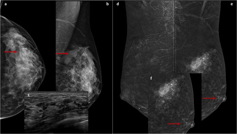
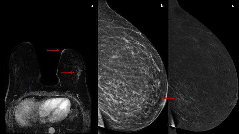
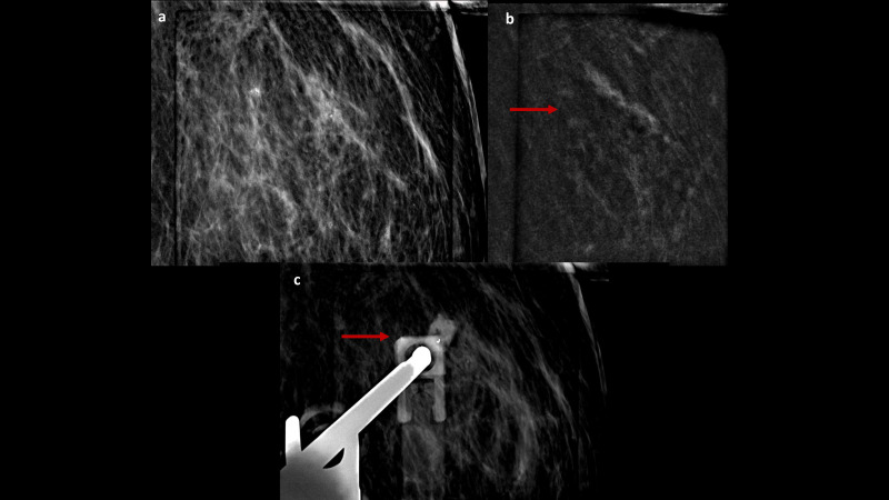
Clinical cases: Two clinical cases of PD diagnosed at our Breast Imaging Division between January and May 2024 were analyzed using CEM. These cases involved imaging techniques, including Digital Mammography (DM), Breast Ultrasound (US), MRI and CEM, alongside histopathological confirmation through nipple-areolar complex (NAC) punch biopsies. CEM identified disease extensions and NAC involvement that was not evident in conventional imaging in both cases. CEM findings influenced surgical decisions, leading to total mastectomies with reconstruction instead of conservative approaches. The cases highlighted CEM's sensitivity and ability to delineate the disease extent comparable to MRI.
Discussion and conclusions: PD often presents diagnostic challenges due to frequent associations with underlying malignancies that are undetectable by standard imaging. While MRI is the gold standard, its limitations, such as costs, contraindications, and false positives, warrant alternative methods. CEM demonstrated utility in diagnosing and staging PD, offering benefits in patient acceptability, cost, and sensitivity comparable to MRI. CEM is a promising diagnostic and planning tool for PD management, especially in MRI-infeasible cases. More extensive multicentric studies will be needed to validate CEM's role in this context. CEM could enhance PD diagnostic workflows and treatment strategies, significantly impacting clinical outcomes.
Keywords: CEM; Paget carcinoma; breast cancer; breast imaging; contrast enhanced mammography.
Copyright © 2025 Mariano, Nicosia, Bozzini, Pesapane, Magnoni, Mazzarol, Meneghetti, Sorce and Cassano.
Conflict of interest statement
The authors declare that the research was conducted in the absence of any commercial or financial relationships that could be construed as a potential conflict of interest.
Figures
Similar articles
-
The Correlation between Morpho-Dynamic Contrast-Enhanced Mammography (CEM) Features and Prognostic Factors in Breast Cancer: A Single-Center Retrospective Analysis.Cancers (Basel). 2024 Feb 22;16(5):870. doi: 10.3390/cancers16050870. Cancers (Basel). 2024. PMID: 38473232 Free PMC article.
-
The performance of contrast-enhanced mammography and breast MRI in local preoperative staging of invasive lobular breast cancer.Eur J Radiol. 2023 Jul;164:110881. doi: 10.1016/j.ejrad.2023.110881. Epub 2023 May 13. Eur J Radiol. 2023. PMID: 37201248
-
Dynamic contrast-enhanced mammography and breast MRI in the diagnosis of breast cancer and detection of tumor size.Turk J Med Sci. 2023 Dec 11;54(1):249-261. doi: 10.55730/1300-0144.5786. eCollection 2024. Turk J Med Sci. 2023. PMID: 38812642 Free PMC article.
-
Contrast-enhanced mammography in high-dense breasts: a narrative review.Transl Breast Cancer Res. 2025 Mar 10;6:15. doi: 10.21037/tbcr-24-64. eCollection 2025. Transl Breast Cancer Res. 2025. PMID: 40421154 Free PMC article. Review.
-
Contrast enhanced mammography (CEM) versus magnetic resonance imaging (MRI) for staging of breast cancer: The pro CEM perspective.Eur J Radiol. 2021 Sep;142:109883. doi: 10.1016/j.ejrad.2021.109883. Epub 2021 Jul 30. Eur J Radiol. 2021. PMID: 34358810 Review.
References
Publication types
LinkOut - more resources
Full Text Sources