Case Reports
. 2025 Jan 8:14:1420597.
doi: 10.3389/fonc.2024.1420597. eCollection 2024.

Angiomatoid fibrous histiocytoma with EWSR1-CREB1 gene fusion occurs in lungs and ribs with systemic multiple metastases: a case report and review of the literature

Affiliations
Case Reports

Angiomatoid fibrous histiocytoma with EWSR1-CREB1 gene fusion occurs in lungs and ribs with systemic multiple metastases: a case report and review of the literature

Dongmei Feng et al. Front Oncol. .

Abstract

Angiomatoid fibrous histiocytoma (AFH) is a rare soft tissue tumor with intermediate malignant potential, and it rarely metastasizes. We encountered a unique AFH case where, the tumor was discovered initially in unusual locations-the left lung and the left 4th rib. Combined histological features with FISH and NGS analysis, the diagnosis of AFH was supported, however, it is difficult to determine which of these two is the primary lesion. Eight months after the initial surgery, multiple systemic metastases were detected, eventually leading to the patient's death 18 months later due to widespread metastasis. Our case signifies the first reported occurrence of systemic metastasis in either bone-originating or pulmonary-originating AFH, and it is the initial instance of mortality resulting from multifocal metastasis originating from an atypical site.

Keywords: EWSR1-CREB1; angiomatoid fibrous histiocytoma; bone; lung; metastasis.


Conflict of interest statement

The authors declare that the research was conducted in the absence of any commercial or financial relationships that could be construed as a potential conflict of interest.

Figures

Figure 1
Figure 1
The imaging data of this patient’s disease progression. (A) In August 2022, chest CT showed a 2.8cm×2.1cm mass in the upper lobe of the left lung. (B) In August 2022, on chest CT, the red arrow refers to the left fourth rib lesion. (C) In May 2023, PET/CT showed multiple metastases throughout the body. (D) In May 2023, Axial T1 MRI contrast of the pelvis shows metastases in and around the left iliac bone, measuring 9.6 cm×6.4 cm, with a visible fluid-fluid level within it (arrow). (E) In May 2023, coronal T1 MRI enhancement showed a subcutaneous nodule on the right buttock, approximately 1.1 cm×1.0cm in size (arrow). (F) In October 2023, brain CT showed large patches of hyperdense opacities in the right basal ganglia and frontoparial lobe, with the maximum level ranging from about 6.2cm×5.0cm (arrow). (G) In October 2023, chest CT showed a soft tissue nodular opacity at the base of the left pleura, about 1.6 cm×2.6cm in size (arrow).
Figure 2
Figure 2
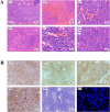
Lung AFH stained with hematoxylin and eosin (A). (A1) There is a distinct fibrous pseudocapsule and pericapsular sleeve of lymphoplasma cells around the tumor nodule (×40). Inset: Lymphoplasma cells densely distributed around the fibrosham capsule (×400), the scale represents 20μm. (B1) A pseudohematous hemorrhagic sac cavity may be seen within the tumor (×100). Slit hemorrhagic areas with hemosiderin deposition and lymphocyte plasmacytic infiltration may be seen within the tumor (×200). (D1) Tumors are mainly composed of fat spindle cells and ovoid cells (×200). (E1) There is more plasma cell infiltration within the tumor (×400). (F1) A mitotic image is visible within the tumor, indicated by arrows (×400). (B) Immunohistochemical staining and fluorescence in situ hybridization of AFH. Tumor cells were positive for Vimentin, CD99, CD68, Desmin (A2-D2), and negative for S-100 (E2). (F2) Fluorescence in situ hybridization analysis revealed fusion of EWSR1 and CREB1. The red fluorescence signal is the CREB1 (2q33) probe, and the green fluorescence signal is the EWSR1 (22q12) probe. The arrow indicates the yellow fluorescence signal, which indicates fusion of EWSR1 and CREB1. (magnification, ×200 for all, tumor cells with brownish−yellow staining are immunohistochemically positive).

Similar articles

Cited by

  • Dermatofibroma: Reappraisal and Updated Review.
    Wan L, Park A, Almatroud L, Khachemoune A. Wan L, et al. Clin Cosmet Investig Dermatol. 2025 Aug 4;18:1873-1887. doi: 10.2147/CCID.S526191. eCollection 2025. Clin Cosmet Investig Dermatol. 2025. PMID: 40785832 Free PMC article. Review.

References

    1. Michcik A, Bień M, Wojciechowska B, Polcyn A, Garbacewicz Ł, Kowalski J, et al. . Difficulties in diagnosing angiomatoid fibrous histiocytoma of the head and neck region: A case report. World J Clin cases. (2023) 11:6252–61. doi: 10.12998/wjcc.v11.i26.6252 - DOI - PMC - PubMed
    1. Xiang Y, Carreon CK, Guerrero J, Putra J. Tle-1 immunoreactivity in angiomatoid fibrous histiocytoma: A potential diagnostic pitfall. Pathology. (2020) 52:722–5. doi: 10.1016/j.pathol.2020.06.017 - DOI - PubMed
    1. Ding J, Zhou G, Dong Y, Li X, Wang L, Guo B, et al. . Angiomatoid fibrous histiocytoma in the spinal canal of T3-T4: A case report and literature review. Br J Neurosurg. (2023) 37:1069–73. doi: 10.1080/02688697.2020.1854686 - DOI - PubMed
    1. Çetin M, Katipoglu K, Türk İ, Özkara Ş, Kosemehmetoglu K, Bıçakçıoğlu P. Endobronchial primary pulmonary angiomatoid fibrous histiocytoma in a patient with testicular germ cell tumor: an evidence against somatic transformation. Int J Surg Pathol. (2022) 30:662–7. doi: 10.1177/10668969221076548 - DOI - PubMed
    1. Fanburg-Smith JC, Miettinen M. Angiomatoid “Malignant” Fibrous histiocytoma: A clinicopathologic study of 158 cases and further exploration of the myoid phenotype. Hum Pathol. (1999) 30:1336–43. doi: 10.1016/s0046-8177(99)90065-5 - DOI - PubMed

Publication types

LinkOut - more resources