. 2025 Jan 21;15(1):e70038.
doi: 10.1002/pul2.70038. eCollection 2025 Jan.

Multiscale Three-Dimensional Evaluation and Analysis of Murine Lung Vasculature From Macro- to Micro-Structural Level

Affiliations

Multiscale Three-Dimensional Evaluation and Analysis of Murine Lung Vasculature From Macro- to Micro-Structural Level

Birger Tielemans et al. Pulm Circ. .

Abstract

The pulmonary vasculature plays a pivotal role in the development and progress of chronic lung diseases. Due to limitations of conventional two-dimensional histological methods, the complexity and the detailed anatomy of the lung blood circulation might be overlooked. In this study, we demonstrate the practical use of optical serial block face imaging (SBFI), ex vivo microcomputed tomography (micro-CT), and nondestructive optical tomography for visualization and quantification of the pulmonary circulation's 3D architecture from macro- to micro-structural levels in murine lung samples. We demonstrate that SBFI can provide rapid, cost-effective, and label-free visualization of mouse lung macrostructures and large pulmonary vessels. Micro-CT offers high-resolution imaging and captures microvascular and (pre)capillary structures, with microstructural quantification. Optical microscopy techniques such as optical projection tomography (OPT) and light sheet fluorescence microscopy, allows noninvasive, mesoscopic imaging of optically cleared mouse lungs, still enabling 3D microscopic reconstruction down to the precapillary level. By integrating SBFI, micro-CT, and nondestructive optical microscopy, we provide a framework for detailed and 3D understanding of the pulmonary circulation, with emphasis on vascular pruning and rarefaction. Our study showcases the applicability and complementarity of these techniques for organ-level vascular imaging, offering researchers flexibility in selecting the optimal approach based on their specific requirements. In conclusion, we propose 3D-directed approaches for a detailed whole-organ view on the pulmonary vasculature in health and disease, to advance our current knowledge of diseases affecting the pulmonary vasculature.

Keywords: 3D visualization and evaluation; microcomputed tomography; optical imaging; pulmonary vasculature; serial block face imaging.


Conflict of interest statement

Image acquisition and data collection at the Kratoscope were performed at Kaer Labs company, Nantes, France. P.D.V. and P.D. are employees of the company Kaer Labs.

Figures
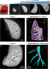
Figure 1
Figure 1
Overview of the Kratoscope workflow and 3D segmentation of arteries and airways. (A) Samples of interest embedded in Oil red O‐colored paraffin. (B) Representative images of the different zoom levels, scale bars 1 mm. See Video S1 also. (C) One block face image taken at zoom 1 indicating airways (cyan arrowheads), arteries (red arrowheads) and veins (yellow arrows). (D) semi‐automated 3D segmented view of Zoom 1 using Imaris software presenting airways, lung arteries and veins. (E) One block face image taken at zoom 2 indicating airways (cyan arrowheads), arteries (red arrowheads) and veins (yellow arrows). (F) Manual 3D segmentation of zoom 2 presenting airways aligned by the lung arteries (See Video S2 also).
Figure 2
Figure 2
Overview of micro‐CT setup and visualization of the arterial bed. (A) heart‐lung complex of Vascupaint‐lime perfused lungs demonstrating a uniform filling of the lungs with contrast agent. (B) Isolated lungs presenting visible contrast‐filled blood vessels reaching distal lung regions. (C) sample mounting before scanning. (D) Tomographic image showing perfused blood vessels in white pixels. (E) 3D volume rendering of a lung complex showing uniform vascular perfusion with a reconstructed voxel size of 4.7 µm and colors presenting density variations (See Video S3).
Figure 3
Figure 3
Arterial analysis of contrast‐perfused mouse lung for micro‐CT. (A) 3D view of the tissue mask of the perfused mouse lung, scale: 5000 µm. (B) 3D visualization of the representative local diameter and (C) local separation of the lung vasculature, scale: 5000 µm. (D) Close‐up of the distal regions of the lung vasculature, scale: 700 µm (E) Visualization of the extracted skeleton of region shown in D), scale: 700 µm. (F) Analysis of micro‐CT perfused lungs with quantification of branch length distribution, vessel thickness distribution, and vessel separation distribution.
Figure 4
Figure 4
Visualization of the vasculature with OPT and LSFM. (A) OPT projection of a mouse lung (top) cleared with BABB and reconstructed tomographic slices (middle and bottom) showing 3D airway structures (470/40 nm excitation filter, 535/50 nm emission filter) in green and blood vessels (EB fluorescent dye, 620/60 nm excitation filter, 700/75 nm emission filter) in red. Airways (cyan arrowhead), arteries (red arrowhead) and veins (yellow arrows). See Video S4. (B) LCS‐SPIM slices from stitched stacks covering the full left lung lobe at 2.2× magnification. See Video S5. (C) representative images at 8.8× magnification. (D) MuViSPIM images at 20× magnification, highlighting EB stained microvessels (red arrowhead) at precapillary level aligning terminal bronchioles and alveolar sacs (cyan arrowhead) (See Video S6 also).

References

    1. Tielemans B., Dekoster K., Verleden S. E., et al., “From Mouse to Man and Back: Closing the Correlation Gap Between Imaging and Histopathology for Lung Diseases,” Diagnostics 10, no. 9 (2020): 636. - PMC - PubMed
    1. Huertas A., Guignabert C., Barberà J. A., et al., “Pulmonary Vascular Endothelium: The Orchestra Conductor in Respiratory Diseases: Highlights From Basic Research to Therapy,” European Respiratory Journal 51, no. 4 (2018): 1700745. - PubMed
    1. May J., Mitchell J. A., and Jenkins R. G., “Beyond Epithelial Damage: Vascular and Endothelial Contributions to Idiopathic Pulmonary Fibrosis,” Journal of Clinical Investigation 133, no. 18 (2023): e172058. - PMC - PubMed
    1. Blanco I., Piccari L., and Barberà J. A., “Pulmonary Vasculature in COPD: The Silent Component,” Respirology 21, no. 6 (2016): 984–994. - PubMed
    1. Humbert M., Guignabert C., Bonnet S., et al., “Pathology and Pathobiology of Pulmonary Hypertension: State of the Art and Research Perspectives,” European Respiratory Journal 53, no. 1 (2019): 1801887. - PMC - PubMed

LinkOut - more resources