. 2025 Jan 21;6(2):e70064.
doi: 10.1002/mco2.70064. eCollection 2025 Feb.

Butyrolactone I blocks the transition of acute kidney injury to chronic kidney disease in mice by targeting JAK1

Affiliations

Butyrolactone I blocks the transition of acute kidney injury to chronic kidney disease in mice by targeting JAK1

Zijun Zhang et al. MedComm (2020). .

Abstract

Chronic kidney disease (CKD) is a disease that affects more than 850 million people. Acute kidney injury (AKI) is a common cause of CKD, and blocking the AKI-CKD transition shows promising therapeutic potential. Herein, we found that butyrolactone I (BLI), a natural product, exerts significant nephroprotective effects, including maintenance of kidney function, inhibition of inflammatory response, and prevention of fibrosis, in both folic acid- and ureteral obstruction-induced AKI-CKD transition mouse models. Notably, BLI showed greater blood urea nitrogen reduction and anti-inflammatory effects than telmisartan. Bioinformatics analysis and target confirmation assays suggested that BLI directly binds to JAK1, and kinase inhibition assay confirmed it is a potent JAK1inhibitor with an IC50 of 0.376 µM. Experiments in JAK1-knockdown mice also proved that BLI targets JAK1 to work. Furthermore, BLI demonstrated nephroprotective effects and safety comparable to ivarmacitinib, the well-known JAK1 inhibitor. Mechanistically, BLI targets JAK1 and inhibits its phosphorylation and JAK-STAT activation, subsequently regulating the downstream signaling pathways to inhibit reactive oxygen species production, inflammation, and ferroptosis, thereby preventing the occurrence of kidney fibrosis and blocking the AKI-CKD transition process. This study demonstrates for the first time that BLI is a JAK1 inhibitor and a promising candidate for delaying CKD progression, which warrants further investigation.

Keywords: AKI–CKD transition; JAK1; butyrolactone I; ferroptosis.


Conflict of interest statement

The authors declare no conflicts of interest.

Figures
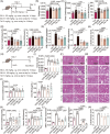
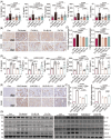
FIGURE 1
FIGURE 1
BLI exerts nephroprotective effects on mouse AKI–CKD transition models. The results from the FA‐induced AKI–CKD transition model are shown in (A–F): (A) Schematic of the experiment. (B) Kidney indices of the mice. Kidney index = kidney weight/body weight (n ≥10). (C) The levels of urinary Cr and MAU (n ≥10). (D) The levels of serum BUN and Cr (n ≥10). (E) mRNA levels of several kidney injury biomarkers (n = 6). (F) and (G) Representative H&E staining and pathological scores of the kidneys (n = 5). The results from the UUO‐induced AKI–CKD transition model are shown in (H–N): (H) Scheme of the experiment. (I) Kidney indices of the mice. (J) The levels of urinary Cr and MAU. (K) The levels of serum Cr and BUN (n ≥ 8). (L) mRNA levels of several kidney injury biomarkers (n = 6). (M) and (N) Representative H&E staining and pathological scores of the kidneys (n = 5). Significant p values are indicated on figure panels. Scale bars were as shown in the figure.
FIGURE 2
FIGURE 2
BLI inhibits the inflammatory response in mouse AKI–CKD transition models. The results from the FA‐induced AKI–CKD transition model are shown in (A–C): (A) Serum levels of multiple chemokines (n ≥ 10). (B) and (C) Results of immunohistochemical staining for CD68 and F4/80 in the kidney (n = 5). The results from the UUO‐induced AKI–CKD transition model are shown in (D‐F): (D) Serum levels of multiple chemokines (n ≥ 8). (E) and (F) Immunohistochemical staining for CD68 and F4/80 in the kidney (n = 5). (G) Immunoblotting results of chemokine proteins in mouse kidneys (n = 3). Significant p values are indicated on figure panels. Scale bars were as shown in the figure.
FIGURE 3
FIGURE 3
BLI blocks kidney fibrosis in mouse AKI–CKD transition models. (A) and (C) Representative images of Masson or immunohistochemical staining and quantification of fibrosis, Col‐I, Fn, and α‐SMA in kidney sections (n = 5). (B) and D) Immunoblotting results of pro‐fibrotic proteins in mouse kidneys (n = 3). Significant p values are indicated on figure panels. Scale bars were as shown in the figure.
FIGURE 4
FIGURE 4
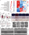
BLI inhibits kidney ferroptosis. RNA sequencing analysis were conducted using the kidney tissues from the FA‐induced mouse model as indicated in Figure 1A. (A) Results of KEGG analysis of the differentially expressed genes from RNA‐seq (n = 3). (B) Results of RT‐qPCR (n = 3). (C) MDA and GSH levels in kidneys (n ≥ 8). (D) Representative images of 4‐HNE, Diaminobenzidine (DAB)‐enhanced Prussian blue and FTH1 staining of the kidneys (n = 5). (E) Representative images obtained from transmission electron microscopy of kidney tissues in the FA‐induced mouse model. (F) Immunoblotting results of the proteins in mouse kidneys (n = 3). Significant p values are indicated on figure panels. Scale bars were as shown in the figure.
FIGURE 5
FIGURE 5
BLI inhibits ferroptosis in HK2 and NRK‐52E cells. (A) Cell viability. The cells were treated for 24 h (Era: 20 µM; BLI: 100 µM; Fer‐1: 2 µM; n = 3). (B) Intracellular iron and ROS levels (n = 3). (C) MDA and GSH levels in HK2 cells (n = 3). (D) Immunoblotting results of the proteins. (E) Results of RT‐qPCR (n = 3). Significant p values are indicated on figure panels. Scale bars were as shown in the figure.
FIGURE 6
FIGURE 6
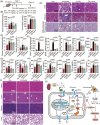
BLI exerts a nephroprotective effect by targeting JAK1. (A) Transcription factor enrichment analysis of the RNA‐seq data (n = 3). (B) Results of immunoblotting for p‐STAT1 and p‐STAT3 (n = 3). (C) The binding affinities of STAT1, STAT3, JAK1, JAK2, and JAK3, for BLI were assessed by molecular dynamics simulation. (D) 3D and 2D images revealing the binding mode of BLI with JAK1. (E) Immunoblotting results for p‐JAK1 (n = 3). (F) Results of CETSA showing the thermal stability of JAK1 after treatment with BLI. (G) Results of DARTS showing the protease stability of JAK1 after treatment with BLI. (H) A cell viability assay was used to analyze the biological activity of bio‐BLI (n = 3). (I) Pull‐down assay showing the binding of Bio‐BLI to JAK1 (n = 2). (J) The IC50 of BLI inhibiting JAK1 at 3.92 µM ATP concentration (n = 3). Significant p values are indicated on figure panels.
FIGURE 7
FIGURE 7
JAK1 knockdown attenuated the nephroprotective effect of BLI. (A) Scheme of the experiment. (B) Representative images of JAK1 staining of the kidneys (n = 4). (C) Urine levels of Cr and MAU (n = 4). (D) Serum levels of Cr and BUN (n = 4). (E) RT‐qPCR results of several kidney injury biomarkers (n = 4). (F) and (G) Representative images of H&E, Masson and DAB‐enhanced Prussian blue staining of the kidneys (n = 4). (H) Levels of MDA andGSH in kidneys (n = 4). (I) Serum levels of multiple chemokines (n = 4). Significant p values are indicated on figure panels. Scale bars were as shown in the figure.
FIGURE 8
FIGURE 8
BLI is as effective and safe as ivarmacitinib in the AKI–CKD transition mouse model. (A) Scheme of the experiment. (B) Urine levels of Cr and MAU (n ≥ 5). (C) Serum levels of Cr and BUN (n ≥ 5). (D) RT‐qPCR results of several kidney injury biomarkers (n = 5). (E) and (F) Representative images of H&E, Masson, and DAB‐enhanced Prussian blue staining of the kidney (n = 5). (G) MDA and GSH levels in kidney tissues (n ≥ 5). (H) Serum levels of multiple chemokines (n ≥ 5). (I) H&E staining of mouse heart, liver, spleen, and lung tissues (n = 3). (J) Detailed molecular mechanism by which BLI blocks the AKI–CKD transition by targeting JAK1. Significant p values are indicated on figure panels. Scale bars were as shown in the figure.

Similar articles

Cited by

References

    1. Kalantar‐Zadeh K, Jafar TH, Nitsch D, Neuen BL, Perkovic V. Chronic kidney disease. Lancet. 2021;398(10302):786‐802. - PubMed
    1. Kellum JA, Romagnani P, Ashuntantang G, Ronco C, Zarbock A, Anders H‐J. Acute kidney injury. Nat Rev Dis Primers. 2021;7(1):52. - PubMed
    1. Levin A, Ahmed SB, Carrero JJ, et al. Executive summary of the KDIGO 2024 clinical practice guideline for the evaluation and management of chronic kidney disease: known knowns and known unknowns. Kidney Int. 2024;105(4):684‐701. - PubMed
    1. Sundström J, Bodegard J, Bollmann A, et al. Prevalence, outcomes, and cost of chronic kidney disease in a contemporary population of 2·4 million patients from 11 countries: the CaReMe CKD study. Lancet Regional Health—Europe. 2022;20:100438. - PMC - PubMed
    1. Lv J‐C, Zhang L‐X. Prevalence and disease burden of chronic kidney disease. In: Liu B‐C, Lan H‐Y, Lv L‐L, eds. Renal Fibrosis: Mechanisms and Therapies. Springer; 2019:3‐15. - PubMed

LinkOut - more resources