. 2025 Jan;7(1):e11785.
doi: 10.1002/acr2.11785.

Citrullinated Autoantigen-Specific T and B Lymphocytes in Rheumatoid Arthritis: Focus on Follicular T Helper Cells and Expansion by Coculture

Affiliations

Citrullinated Autoantigen-Specific T and B Lymphocytes in Rheumatoid Arthritis: Focus on Follicular T Helper Cells and Expansion by Coculture

Tim Ammon et al. ACR Open Rheumatol. 2025 Jan.

Abstract

Objective: Rheumatoid arthritis (RA) is characterized by circulating anti-cyclic citrullinated peptide (CCP) autoantibodies (ACPAs), resulting in inflammation of the joints and other organs. We have established novel assays to assess immune cell subpopulations, including citrullinated antigen-specific (CAS) autoreactive B and T lymphocytes, in patients with RA.

Methods and results: We found that activated CD25+ T cells were markedly increased in patients with RA compared to healthy controls. Novel combinations of major histocompatibility complex class II citrulline epitope tetramers were developed, which enabled robust detection of CAS T cells and showed increases of CAS-naive T helper cells, Th1.17 cells, CAS total circulating T follicular helper (cTfh) cells, and cTfh1 cells in ACPA+ patients with RA. In addition, an innovative assay using dual labeling with CCP-biotin probes allowed for reproducible identification of primary CAS B cells after enrichment with advantages over existing detection methods. Furthermore, patient-derived immune cells were successfully expanded. Primary RA B cells were successfully cultured on novel feeder cell lines, whereas T cells were expanded ex vivo in the presence of interleukin-2 and citrullinated peptides, and subsequent alterations in cell frequencies were assessed.

Conclusion: Novel assays were established to reliably detect CAS T and B cells in patients with RA, and specific CAS-naive T helper cells, Th1.17 cells, cTfh cells, and cTfh1 cells were observed more frequently in RA. Based on these results, new coculture systems of disease-relevant cells are developed to simulate human secondary lymphoid tissues ex vivo. This technology will serve as a platform to identify therapies that modulate disease-specific immune cells.


Figures

Figure 1
Figure 1
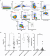
Frequencies of CD4+ T cell subsets in PBMCs derived from patients with RA and ACPA+ patients with RA compared to HCs. (B) Activated CD25+ cells were gated as depicted in panel. Data are expressed as the percentage of activated CD25+ cells among viable CD4+ T cells; HC: n = 22, RA: n = 32, RA ACPA+: n = 15; the Kruskal–Wallis test with Dunn's multiple comparison test: HC versus RA: P = 0,0002, HC versus RA ACPA+: P < 0.0001. (A) Representative gating strategy for the assessment of CD4+ T cell subsets and pMHCII tetramer analysis of T lymphocytes in PBMC samples after thawing. (C) Frequencies of CD4+ T cell subsets among HCs and patients with RA assessed by flow cytometry after thawing. HC: n = 22, ACPA+ RA: n = 15. ACPA, anti–cyclic citrullinated peptide autoantibody; CP, citrullinated peptide; cTfh, circulating T follicular helper; FSC‐A, forward scatter‐area; FSC‐H, forward scatter‐height; HC, healthy control; LD, live/dead; PBMC, peripheral blood mononuclear cell; pMHCII, peptide major histocompatibilty complex‐II; RA, rheumatoid arthritis; SSC‐A, side scatter‐area; Tcm, T central memory cells; Tem, T effector memory cells.
Figure 2
Figure 2
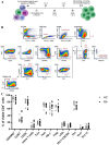
In vitro stimulation of PBMCs derived from patients with RA and HCs and CD4+ T cell subtyping. (A) Schematic overview showing the experimental procedure for in vitro stimulation of PBMCs with a CP mix. (B) Representative gating strategy for the assessment of CD4+ T cell subsets and pMHCII tetramer analysis of T lymphocytes in PBMC samples after 12 days of in vitro stimulation with IL‐2 and CP mix. (C) Frequencies of CD4+ T cell subsets among HCs and patients with RA assessed by flow cytometry on day 12 of in vitro stimulation. HC: n = 7, RA: n = 12. CP, citrullinated peptide; cTfh, circulating T follicular helper; DC, dendritic cells; FSC‐A, forward scatter‐area; FSC‐H, forward scatter‐height; HC, healthy control; IL‐12, interleukin‐12; LD, live/dead; NK, natural killer cells; PBMC, peripheral blood mononuclear cell; pMHCII, peptide major histocompatibilty complex‐II; Poly‐IC, poly‐inosin‐cytosin; RA, rheumatoid arthritis; SSC‐A, side scatter‐area; Tcm, T central memory cells; Tem, T effector memory cells.
Figure 3
Figure 3
(A) Frequencies and (B) numbers of cTfh subset+ cells from patients with RA and HCs on day 0 and after 12 days of in vitro stimulation with a CP mix. PBMCs from patients with RA and HCs were cultured in vitro for 12 days in medium containing IL‐2 and a CP mix as shown in Figure 1B. Frequencies and numbers of cTfh cell subsets were assessed on day 0 and on day 12 of in vitro stimulation by flow cytometry using the gating strategies shown in Figures 2A and 3B, respectively. Data are expressed (A) as percentages of cTfh1/cTfh2/cTfh17/cTfh1.17 cells among viable CD4+ T cells or (B) as numbers of cTfh1/cTfh2/cTfh17/cTfh1.17 cells per million viable CD4+ T cells. CP, citrullinated peptide; cTfh, circulating T follicular helper; HC, healthy control; IL‐12, interleukin‐12; IVS, in vitro stimulation; PBMC, peripheral blood mononuclear cell; RA, rheumatoid arthritis.
Figure 4
Figure 4
Assessment of citrulline‐reactive T helper cells in PBMCs derived from ACPA+ patients with RA compared to HCs. Frequencies of citrullinated antigen–reactive CD4+ T cells were examined in PBMCs derived from ACPA+ patients with RA and HCs using MHCII‐RA tetramers. MHCII‐RA tetramer+ cells were gated as shown in Figure 1B (viable CD3+CD4+tetramer+). (A) Data are expressed as the percentage of MHCII‐RA tetramer+ cells among total viable CD4+ T cells. (B) Frequencies of MHCII‐RA tetramer+ cells are increased among naive CD4+ T cells. (C) Frequencies of MHCII‐RA tetramer+ cells are increased among Th1.17 cells. (D) Frequencies of MHCII‐RA tetramer+ cells are increased among cTfh cell subsets in PBMCs derived from ACPA+ patients with RA compared to HCs. HC: n = 22, ACPA+ RA: n = 15; Mann–Whitney test: * P < 0.0. ACPA, anti–cyclic citrullinated peptide autoantibody; cTfh, circulating T follicular helper; HC, healthy control; MHCII, major histocompatibility complex class II; PBMC, peripheral blood mononuclear cell; RA, rheumatoid arthritis.
Figure 5
Figure 5
(A) Representative example and gating strategy of primary B cells of a patient with RA. Representative images show gating of CD19+ B cell subsets and CCP dual staining of enriched B lymphocytes from RA and HC PBMC samples. CCP‐PE and CCP‐PEVio770 double‐positives (CCP‐dPos) within the CD19+CD24lowCD38low non‐PC population are considered CAS B cells. (B) Average major subpopulations of primary B cells in patients with RA. B cells from PBMCs were purified and stained for characteristic subset markers to discriminate Bmem cells, naive B cells, and TransB cells. Data represent comparison of major subsets in PBMCs derived from patients with RA compared to HCs. HC: n = 15, RA: n = 37; Mann–Whitney test: * P < 0.05. ASC, antibody‐secreting cells; Bmem, memory B; CAS, citrullinated antigen–specific; CCP, cyclic citrullinated peptide; CCP‐PEVio770, cyclic citrullinated peptide‐Vio770; CCP‐PE, cyclic citrullinated peptide‐phycoerythrin; FSC‐A, forward scatter‐area; FSC‐H, forward scatter‐height; HC, healthy control; NIR, amine reactive fluorescent dye; PBMC, peripheral blood mononuclear cell; PC, plasma cell; RA, rheumatoid arthritis; SSC‐A, side scatter‐area; SSC‐W, side scatter‐width; TransB, transitional B.
Figure 6
Figure 6
CCP–biotin double positively stained cells in ACPA+ patients with RA versus HCs. B cells from PBMCs were purified, and CCP dual staining was performed followed by B cell subset marker staining (representative image and gating strategy, upper left panel). (A) CCP‐double‐positive cells were identified in pan B cells. (B) CCP‐double‐positive cells in more specified B cell subsets of ACPA+ patients: Bmem cells, naive B cells, and TransB cells. Data represent comparison of major subsets in PBMCs derived from patients with RA compared to HCs. HC: n = 12, RA: n = 37, ACPA+ RA: n = 16; Mann–Whitney test: * P < 0.05. (C) B feeder cell systems for RA patient cells. Description of the novel feeder cell systems (Bsupp) for B cell culture propagation and differentiation of RA patient cells. (D) Screening for optimal coculture conditions and BSupp feeder lines using cell proliferation assays (CellTrace) to monitor cell divisions in healthy primary B cells. The figure shows cell proliferation profiles of B cells from healthy donors under various culture conditions, including without feeders (top, black), B cell coculture with parental cell line A (light blue), successful coculture of B cells and BSuppA (dark blue) as well as B cell cocultures with parental cell line B (light green), and B cell cocultures with BSuppB (dark green). With parental cell lines as well as without feeder cells, no proliferation was detected. BSupp feeder lines BSuppA and BSuppB provided stimuli to promote B cell proliferation and increased cell viability. (E) Cell proliferation and division index for RA patient B cells in BSupp cocultures for seven days. B cells derived from patients with RA and HC donors were examined under various BSupp coculture conditions. The figure shows CellTrace‐determined cell division index of B cells, which is markedly increased for both BSupp and OP‐9 cells compared to no support (n = 3, technical biologic replicates and SEM). ACPA, anti–cyclic citrullinated peptide autoantibody; Bmem, memory B; CCP, cyclic citrullinated peptide; CCP‐PEVio770, cyclic citrullinated peptide‐Vio770; CCP‐PE, cyclic citrullinated peptide‐phycoerythrin; FSC‐A, forward scatter‐area; FSC‐H, forward scatter‐height; HC, healthy control; IL‐21, interleukin‐21; NIR, amine reactive fluorescent dye; PBMC, peripheral blood mononuclear cell; RA, rheumatoid arthritis; SEM, standard error of the means; SSC‐A, side scatter‐area; SSC‐W, side scatter‐width; TransB, transitional B.

References

    1. McInnes IB, Schett G. The pathogenesis of rheumatoid arthritis. N Engl J Med 2011;365(23):2205–2219. - PubMed
    1. Alamanos Y, Drosos AA. Epidemiology of adult rheumatoid arthritis. Autoimmun Rev 2005;4(3):130–136. - PubMed
    1. Mysler E, Caubet M, Lizarraga A. Current and emerging DMARDs for the treatment of rheumatoid arthritis. Open Access Rheumatol 2021;13:139–152. - PMC - PubMed
    1. Smolen JS, Aletaha D, McInnes IB. Rheumatoid arthritis. Lancet 2016;388(10055):2023–2038. - PubMed
    1. Albrecht K, Müller‐Ladner U. Side effects and management of side effects of methotrexate in rheumatoid arthritis. Clin Exp Rheumatol 2010;28(5 suppl 61):S95–S101. - PubMed

LinkOut - more resources