Review
. 2025 Jan 8;15(1):9.
doi: 10.3390/jox15010009.

Cardiotoxicity of Chemotherapy: A Multi-OMIC Perspective

Affiliations
Review

Cardiotoxicity of Chemotherapy: A Multi-OMIC Perspective

Yan Ma et al. J Xenobiot. .

Abstract

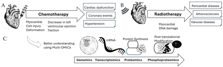
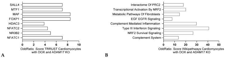
Chemotherapy-induced cardiotoxicity is a critical issue in cardio-oncology, as cancer treatments often lead to severe cardiovascular complications. Approximately 10% of cancer patients succumb to cardiovascular problems, with lung cancer patients frequently experiencing arrhythmias, cardiac failure, tamponade, and cardiac metastasis. The cardiotoxic effects of anti-cancer treatments manifest at both cellular and tissue levels, causing deformation of cardiomyocytes, leading to contractility issues and fibrosis. Repeated irradiation and chemotherapy increase the risk of valvular, pericardial, or myocardial diseases. Multi-OMICs analyses reveal that targeting specific pathways as well as specific protein modifications, such as ubiquitination and phosphorylation, could offer potential therapeutic alternatives to current treatments, including Angiotensin converting enzymes (ACE) inhibitors and beta-blockers that mitigate symptoms but do not prevent cardiomyocyte death, highlighting the need for more effective therapies to manage cardiovascular defects in cancer survivors. This review explores the xenobiotic nature of chemotherapy agents and their impact on cardiovascular health, aiming to identify novel biomarkers and therapeutic targets to mitigate cardiotoxicity.

Keywords: cancer; cardiotoxicity; cardiovascular diseases; chemotherapy.


Conflict of interest statement

The authors declare no conflict of interest.

Figures

Figure 1
Figure 1
Cardiovascular events in cancer patients after chemotherapy or radiotherapy (A,B) (adapted from [17]) as well as relevance of multi-OMICs analyses to improve their understanding (C).
Figure 2
Figure 2
TrancriptOMICs analysis of cardiac disease initiation in cardiomyocytes (A) Pathway analysis of RNA sequencing from isolated cardiomyocytes after exposition to doxorubicin (GSE226116) comparing differentially expressed genes (q value < 0.005) using ENRICHR (Hallmark analysis). N = 3. Odds ratios are graphed, and the bars sorted from the most significant adjusted p value. (B) CRISPR screen comparing inhibited sgRNA (CRISPRi) when comparing the Doxocyclin-treated vs. Vehicle IPSC-derived cardiomyocytes conditions (Dataset: GSE276161). Positively and negatively enriched sgRNAs in red and blue respectively, grey indicate sgRNAs that do not show effects. (C,D) Reactome analysis performed on the positively and negatively enriched sgRNAs. The color of the dot corresponds to the adjusted p-value for each pathway (green for the most significant adjusted p value to dark blue for the less significant ones) and the size of the dot corresponds to the number of inhibited sgRNA for each group.
Figure 3
Figure 3
Transcriptomic analysis of cardiac disease initiation in endothelial cells. (A,B) Pathway analysis of RNA sequencing from isolated cardiac ECs after exposition to doxorubicin (GSE226116) comparing differentially expressed genes (q value < 0.005) using ENRICHR N = 3. Odds ratios are graphed, and the bars are sorted from the most significant corrected p value. TRRUST analysis and Wikipathways analysis respectively.
Figure 4
Figure 4
Transcriptomic analysis of cardiac disease initiation upon Sorafenib treatment in cardiomyocytes adapted from Series GSE222642 comparing differentially expressed genes (q value < 0.005), where male rats were gavaged with 50 mg/kg sorafenib (heart tissues collected at 14 days after treatment). ENCODE analysis (A) and Wikipathways analysis (B) respectively (ENRICHR, software developed by Ma’ayan lab, Computational Systems Biology, New York, NY, USA).
Figure 5
Figure 5
Transcriptomic analysis of cardiac disease initiation upon Indisulam treatment in cardiomyocytes from Query DataSets for GSE213311 comparing differentially expressed genes (q value < 0.005), RNA-seq analysis on cardiomyocytes treated with vehicle or indisulam. ENCODE analysis (A) and Wikipathways analysis (B) respectively (ENRICHR, Ma’ayan lab).
Figure 6
Figure 6
Transcriptomic analysis of cardiac disease initiation upon Trastuzumab treatment in cardiomyocytes adapted from GSE264120 comparing differentially expressed genes (q value < 0.005), ENCODE analysis (A) and MCF7 GEO UP signatures analysis (B) respectively (ENRICHR, Ma’ayan lab).
Figure 7
Figure 7
Transcriptomic analysis of cardiac disease initiation in rats upon 5-FU treatment in cardiomyocytes adapted from GSE166957 comparing differentially expressed genes (q value < 0.005), TRRUST analysis (A) and Wikipathways analysis (B) respectively (ENRICHR, Ma’ayan lab).
Figure 8
Figure 8
Transcriptomic analysis of cardiomyocyte response to doxorubicin in mice with OTUB1 heterozygous knockout according to Data obtained from GSE240959 and comparing differentially expressed genes (q value < 0.005), using ENRICHR. Odds ratios are graphed, and corrected p values are indicated. TRRUST analysis (A) and Wikipathways analysis (B) respectively.
Figure 9
Figure 9
Transcriptomic analysis of cardiomyocyte response to doxorubicin in mice with ADAM17 knockout. Data obtained from GSE276325 comparing differentially expressed genes (q value < 0.005), Odds ratios are graphed, and bars are sorted from the most significant corrected p-value. TRRUST analysis (A) and Wikipathways analysis (B) respectively.

Similar articles

References

    1. Zaborowska-Szmit M., Krzakowski M., Kowalski D.M., Szmit S. Cardiovascular Complications of Systemic Therapy in Non-Small-Cell Lung Cancer. J. Clin. Med. 2020;9:1268. doi: 10.3390/jcm9051268. - DOI - PMC - PubMed
    1. Dos Santos V.M., Dos Santos L.A.M. Cardiac and lymphatic metastases from lung cancer. Arch. Iran. Med. 2018;21:82–83. - PubMed
    1. Sosinska-Mielcarek K., Senkus-Konefka E., Jassem J., Kulczycka J., Jendrzejewski J., Jaskiewicz K. Cardiac involvement at presentation of non-small-cell lung cancer. J. Clin. Oncol. 2008;26:1010–1011. doi: 10.1200/JCO.2007.14.9328. - DOI - PubMed
    1. Ciurus T., Maciejewski M., Lelonek M. Cardiac involvement of lung cancer mimicking myocardial infarction. Kardiol. Pol. 2013;71:994. doi: 10.5603/KP.2013.0245. - DOI - PubMed
    1. Mizutani H., Kume H., Kimura T., Ogawa M., Yoshida N., Yoshida M., Ito Y., Suzuki R., Yamaki K. Primary lung cancer (adenocarcinoma) associated with cardiac sarcoidosis. Nihon Kokyuki Gakkai Zasshi = J. Jpn. Respir. Soc. 1999;37:489–494. - PubMed

LinkOut - more resources