. 2024 Dec 30;14(1):1.
doi: 10.3390/biotech14010001.

Peptide Inhibitor Assay for Allocating Functionally Important Accessible Sites Throughout a Protein Chain: Restriction Endonuclease EcoRI as a Model Protein System

Affiliations

Peptide Inhibitor Assay for Allocating Functionally Important Accessible Sites Throughout a Protein Chain: Restriction Endonuclease EcoRI as a Model Protein System

Joji M Otaki. BioTech (Basel). .

Abstract

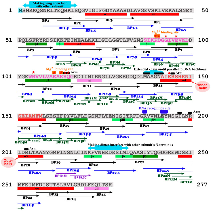
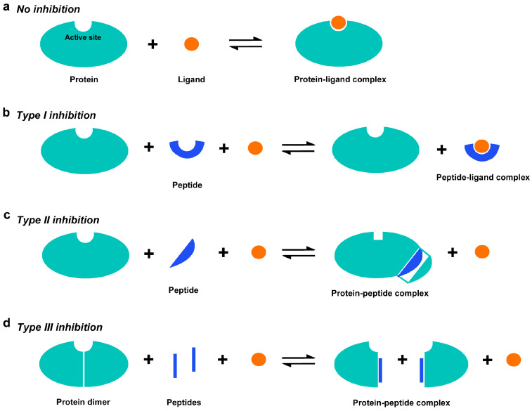
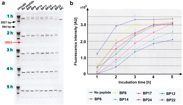
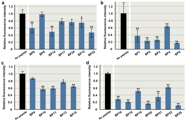
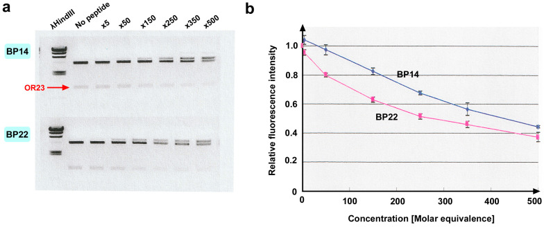
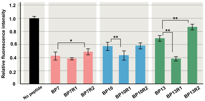
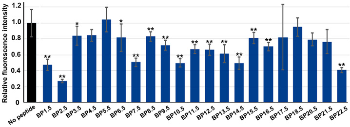
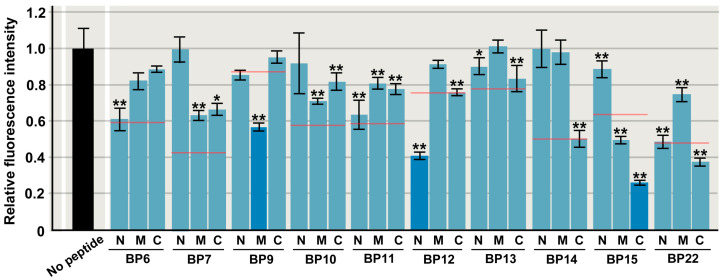
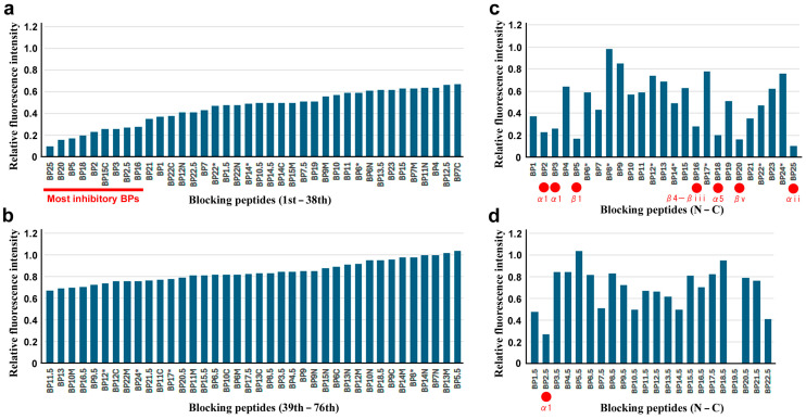
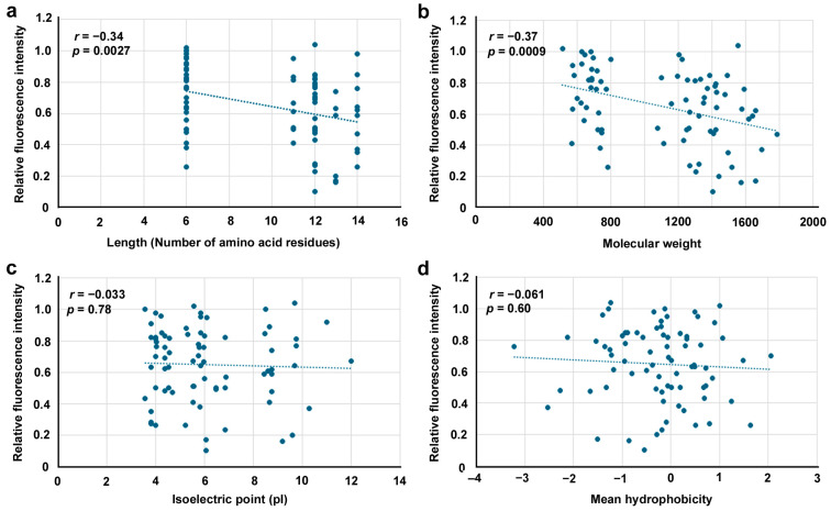
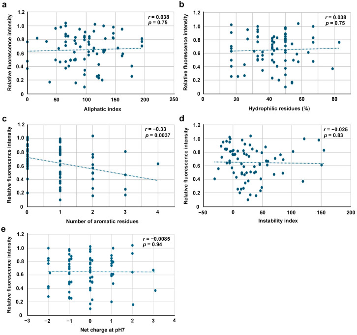
Functionally important amino acid sequences in proteins are often located at multiple sites. Three-dimensional structural analysis and site-directed mutagenesis may be performed to allocate functional sites for understanding structure‒function relationships and for developing novel inhibitory drugs. However, such methods are too demanding to comprehensively cover potential functional sites throughout a protein chain. Here, a peptide inhibitor assay (PIA) was devised to allocate functionally important accessible sites in proteins. This simple method presumes that protein‒ligand interactions, intramolecular interactions, and dimerization interactions can be partially inhibited by high concentrations of competitive "endogenous" peptides of the protein of interest. Focusing on the restriction endonuclease EcoRI as a model protein system, many endogenous peptides (6mer-14mer) were synthesized, covering the entire EcoRI protein chain. Some of them were highly inhibitory, but interestingly, the nine most effective peptides were located outside the active sites, with the exception of one. Relatively long peptides with aromatic residues (F, H, W, and Y) corresponding to secondary structures were generally effective. Because synthetic peptides are flexible enough to change length and amino acid residues, this method may be useful for quickly and comprehensively understanding structure‒function relationships and developing novel drugs or epitopes for neutralizing antibodies.

Keywords: EcoRI; drug design; molecular accessibility; peptide and protein engineering; peptide inhibitor; protein structure and function.


Conflict of interest statement

The author declares no conflicts of interest. The funders had no role in the design of the study; in the collection, analyses, or interpretation of data; in the writing of the manuscript; or in the decision to publish the results.

Figures

Figure 2
Figure 2
Amino acid sequence of EcoRI (M1-K277). The first M is removed after translation in this protein. The secondary structures and functional sites are indicated below and above the EcoRI sequence, respectively, according to Kim et al. (1990) [63], Heitman (1992) [64], and Watrob et al. (2001) [78]. The blocking peptides (BPs) designed and tested in the present study are indicated below the EcoRI sequence. Major active site sequences are indicated in pink letters (SIKPDGGIVEVKD and WRVVLVAEAKHQG). Red asterisks indicate important sites identified via site-directed mutagenesis. A red dot (E144) indicates salt bridge formation. A sequence of EcoRI corresponding to a helical peptide (α4) that has been demonstrated for its inhibitory effect on EcoRI by Brickner and Chmielewski (1998) [81] is indicated in red letters (AIERSHKNISEIANFM). Two parallel helices, α4 and α5, that form the inner and outer helices, respectively, to hold DNA are indicated.
Figure 1
Figure 1
Three possible types of peptide inhibition via synthetic endogenous peptides in the peptide inhibitor assay (PIA). (a) No inhibition. A ligand binds to the active site (functional site) of a protein, forming a protein‒ligand complex. (b) Type I inhibition: competitive binding of a peptide to a ligand (substrate) because a peptide mimics the active site of the protein. (c) Type II inhibition: competitive binding of a peptide to a site of intramolecular interactions. (d) Type III inhibition: competitive binding of a peptide to a dimerization site or a site of protein‒protein interaction (PPI).
Figure 3
Figure 3
Results of initially chosen seven peptides. (a) Agarose gel electrophoresis sampled every hour until 5 h. NC: negative control without EcoRI (shown only at 1 h and 5 h). Excised OR23 bands are indicated. (b) Fluorescence intensity versus incubation time. Mean values (±standard deviation) (n = 3) are shown. Fluorescence intensity is shown in arbitrary unit (AU).
Figure 4
Figure 4
Relative fluorescence intensity of original peptides (except BP7). Mean values (±standard deviation) are shown (n = 3 for each peptide). *: p < 0.05, **: p < 0.01 (Dunnett’s test by comparison to “no peptide”). (a) Initially chosen seven peptides. (b) N-terminus five peptides. (c,d) Remaining 13 peptides.
Figure 5
Figure 5
Concentration dependence of inhibitory effects of BP14 and BP22. (a) Agarose gel electrophoresis. Concentration (×5 to ×500) indicates molar equivalence to EcoRI. Excised OR23 bands are indicated. (b) Relative fluorescence intensity plotted against concentration (molar equivalence). Mean values (±standard deviation) (n = 3) are shown.
Figure 6
Figure 6
Relative fluorescence intensity of randomized peptides of BP7, BP10, and BP13. Two randomized peptides from each original peptide were tested. Mean values (±standard deviation) are shown (n = 3 for each peptide). *: p < 0.05, **: p < 0.01 (Student’s t-test by comparison to each endogenous BP).
Figure 7
Figure 7
Relative fluorescence intensity of staggered peptides. BP19.5 is not included in this figure. Mean values (±standard deviation) are shown (n = 3 for each peptide). *: p < 0.05, **: p < 0.01 (Dunnett’s test by comparison to “no peptide”).
Figure 8
Figure 8
Relative fluorescence intensity of short peptides. N, M, and C indicate N-terminal, middle, and C-terminal sequences of original peptide, respectively. Mean values (±standard deviation) are shown (n = 3 for each peptide). Red lines indicate relative fluorescence intensity values of original peptides. BP9M, BP12N, and BP15C are shown as dark blue bars to indicate a substantial decrease in relative fluorescence intensity values of original peptides. *: p < 0.05, **: p < 0.01 (Dunnett’s test by comparison to “no peptide”).
Figure 9
Figure 9
Overview of inhibitory effects. Asterisks indicate initially chosen seven BPs. (a) Ranks 1st to 36th. The nine most inhibitory BPs are indicated. (b) Rank 37th to 72nd. (c) Original BP series ordered from the N-terminal to the C-terminal regions. The seven most inhibitory BPs in (a) are indicated by red circles. (d) Staggered BP series ordered from N-terminal to C-terminal regions. One of the most inhibitory BPs in (a) is indicated by a red circle. This graph shows the same results as Figure 7 but was aligned with (c) for visual comparison. BP19.5 did not show any fluorescence intensity of the DNA fragment.
Figure 10
Figure 10
Scatter plots of four physicochemical factors of 76 BPs used in this study. Pearson correlation coefficients and their associated p values are shown. (a) Peptide length (number of amino acid residues). (b) Molecular weight. (c) Isoelectric point (pI). (d) Mean hydrophobicity.
Figure 11
Figure 11
Scatter plots of additional five physicochemical factors of 76 BPs used in this study. Pearson correlation coefficients and their associated p values are shown. (a) Aliphatic index. (b) Hydrophilic residues (%). (c) Number of aromatic residues. (d) Instability index. (e) Net charge at pH7.

Similar articles

References

    1. Anfinsen C.B. Principles that govern the folding of protein chains. Science. 1973;181:223–230. doi: 10.1126/science.181.4096.223. - DOI - PubMed
    1. Liddington R.C. Structural basis of protein-protein interactions. Methods Mol. Biol. 2004;261:3–14. doi: 10.1385/1-59259-762-9:003. - DOI - PubMed
    1. Kieber-Emmons T., Murali R., Greene M.I. Therapeutic peptides and peptidomimetics. Curr. Opin. Biotechnol. 1997;8:435–441. doi: 10.1016/S0958-1669(97)80065-1. - DOI - PubMed
    1. Ripka A.S., Rich D.H. Peptidomimetic design. Curr. Opin. Chem. Biol. 1998;2:441–452. doi: 10.1016/S1367-5931(98)80119-1. - DOI - PubMed
    1. Ko E., Liu J., Burgess K. Minimalist and universal peptidomimetics. Chem. Soc. Rev. 2011;40:4411–4421. doi: 10.1039/c0cs00218f. - DOI - PubMed

LinkOut - more resources