Review
. 2025 Jan 14;9(1):6.
doi: 10.3390/vision9010006.

Orienting Gaze Toward a Visual Target: Neurophysiological Synthesis with Epistemological Considerations

Affiliations
Review

Orienting Gaze Toward a Visual Target: Neurophysiological Synthesis with Epistemological Considerations

Laurent Goffart. Vision (Basel). .

Abstract

The appearance of an object triggers an orienting gaze movement toward its location. The movement consists of a rapid rotation of the eyes, the saccade, which is accompanied by a head rotation if the target eccentricity exceeds the oculomotor range and by a slow eye movement if the target moves. Completing a previous report, we explain the numerous points that lead to questioning the validity of a one-to-one correspondence relation between measured physical values of gaze or head orientation and neuronal activity. Comparing the sole kinematic (or dynamic) numerical values with neurophysiological recordings carries the risk of believing that the activity of central neurons directly encodes gaze or head physical orientation rather than mediating changes in extraocular and neck muscle contraction, not to mention possible changes happening elsewhere (in posture, in the autonomous nervous system and more centrally). Rather than reducing mismatches between extrinsic physical parameters (such as position or velocity errors), eye and head movements are behavioral expressions of intrinsic processes that restore a poly-equilibrium, i.e., balances of activities opposing antagonistic visuomotor channels. Past results obtained in cats and monkeys left a treasure of data allowing a synthesis, which illustrates the formidable complexity underlying the small changes in the orientations of the eyes and head. The aim of this synthesis is to serve as a new guide for further investigations or for comparison with other species.

Keywords: cat; dynamics; fixation; kinematics; model; monkey; neuro-ophthalmology; neurophysiology; noise; poly-equilibrium; pursuit; saccade; space.


Conflict of interest statement

The author declares no conflicts of interest.

Figures

Figure 1
Figure 1
Orienting movement of a goldfish toward a small pellet of food dropping at different sites of a square tank. The rightmost panel shows the angle of response as a function of the angular eccentricity of the stimulus for a representative animal. Modified from [7] with the permission of the authors and Elsevier.
Figure 2
Figure 2
Effects of suppressing the activity of a small set of collicular neurons on saccades toward a visual target. A small volume of lidocaine (100 nl) was injected two times inside the population of active neurons while a monkey made saccades toward a target. Immediately after each injection (blue arrow), the peak velocity of saccades was reduced (C) while their duration was lengthened (D). In comparison with these changes in velocity, the direction (A) and amplitude (B) of saccades were barely affected. Courtesy of Dr David L. Sparks, modified with his permission.
Figure 3
Figure 3
Poly-equilibrium hypothesis: A saccade or a slow eye movement is not initiated when the visuo-oculomotor system is at equilibrium, i.e., when opposing commands (issued, for instance, by the left and right superior colliculi or by the left and right nuclei of the optic tract) counterbalance each other. For generating saccadic and pursuit eye movements, symmetry breaking involves different groups of neurons. For saccades, premotor neurons are located in the pontomedullary reticular formation whereas, for slow eye movements, they are located in the vestibular nuclei. Bilateral fastigial activity also contributes to neck muscle tone, which specifies the horizontal orientation (yaw) of the head through fastigio-reticular projections. Different colors are used to distinguish the crossed and uncrossed channels. Read text for explanations.
Figure 4
Figure 4
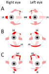
Schematic representation of extraocular muscles. LR: lateral rectus, MR: medial rectus, SR: superior rectus, IR: inferior rectus, SO: superior oblique, IO: inferior oblique, tr: trochlea. The muscles whose contraction rotates the eyes toward the right (A), upward (B) or downward (C) are colored in red. The muscles colored in pink are those that relax during the same rotations, respectively. The muscles outlined by a dashed black line sustain the same contraction level. Note that the SR and IR muscles are not positioned in planes running parallel to the midsagittal plane, as reported in [253,254,255,256].
Figure 5
Figure 5
Neuronal network involved in the generation of leftward saccades. (A) The thickness of arrows attached to the eyeballs schematizes the strength of muscle contraction. LR: lateral rectus, MR: medial rectus, SR: superior rectus, IR: inferior rectus, SO: superior oblique, IO: inferior oblique. (B) Parasagittal section of the brainstem and cerebellum showing the approximate locations of the oculomotor nucleus (OMN), paramedian pontine reticular formation (ppRF), abducens nucleus (ABD), dorsal paragigantocellularis reticular formation (dPGRF), caudal fastigial nucleus (cFN) and superior colliculus (SC). (C) Connecting lines ended by an arrow indicate excitatory connections; those ended by a circle indicate inhibitory synaptic connections. The thickness of connecting lines schematizes the strength with which the neurons fire. SRBNs: saccade-related burst neurons, OPNs: omnipause neurons, EBNs: excitatory burst neurons, IBNs: inhibitory burst neurons, ABD: abducens nucleus, MNs: motoneurons, AINs: abducens internuclear neurons. Read text for explanations.
Figure 6
Figure 6
Network involved in the generation of upward saccades. (A) Same conventions as in Figure 5A. (B) Parasagittal section of the brainstem and cerebellum showing the approximate locations of the oculomotor nucleus (OMN), rostral interstitial nucleus of the medial longitudinal fasciculus (riMLF), interstitial nucleus of Cajal (iNC), trochlear nucleus (TRO) and superior colliculus (SC). (C) Connecting lines ended by an arrow and a circle indicate excitatory and inhibitory synaptic connections, respectively. The thickness of connecting lines schematizes the strength with which the neurons fire. Note the crossing axons of motor neurons innervating the SO and SR muscles. uEBNs: upward excitatory burst neurons, dEBNs: downward excitatory burst neurons, uIBNs: upward inhibitory burst neurons, MNs: motoneurons, SC: superior colliculus. Read text for explanations.
Figure 7
Figure 7
Network involved in the generation of downward saccades. Same conventions as in Figure 6. Read text for explanations.
Figure 8
Figure 8
Neuronal network involved in combined horizontal eye and head movements. Connecting lines ended by an arrow indicate excitatory connections; those ended by a circle indicate inhibitory synaptic connections. Blue color indicates the agonist neuronal oculomotor elements, red color the antagonist ones. Green color indicates the cephalomotor elements. The thickness of connecting lines schematizes the strength with which the neurons fire. EN-RSNs: eye–neck reticulospinal neurons, EBNs: excitatory burst neurons, IBNs: inhibitory burst neurons, E-MNs: abducens motor and internuclear neurons, N-MNs: neck motoneurons, PVNs: primary vestibular neurons, IVNs: inhibitory vestibular neurons, EVNs: excitatory vestibular neurons, VIII: eighth cranial nerve. Read text for explanations.

Similar articles

References

    1. Pavlov I.P. Conditioned Reflexes: An Investigation of the Physiological Activity of the Cerebral Cortex. Dover Publications; New York, NY, USA: 1960. - PMC - PubMed
    1. Sokolov E.N. Higher nervous functions: The orienting reflex. Ann. Rev. Physiol. 1963;25:545–580. doi: 10.1146/annurev.ph.25.030163.002553. - DOI - PubMed
    1. Lynn R. Attention, Arousal and the Orientation Reaction. Pergamon Press; Oxford, UK: 1966.
    1. Bertenthal B., von Hofsten C. Eye, head and trunk control: The foundation for manual development. Neurosci. Biobehav. Rev. 1998;22:515–520. doi: 10.1016/S0149-7634(97)00038-9. - DOI - PubMed
    1. Koceja D.M., Allway D., Earles D.R. Age differences in postural sway during volitional head movement. Arch. Phys. Med. Rehab. 1999;80:1537–1541. doi: 10.1016/S0003-9993(99)90327-1. - DOI - PubMed

LinkOut - more resources