

Novel Integration of Spatial and Single-Cell Omics Data Sets Enables Deeper Insights into IPF Pathogenesis
- PMID: 39846634
- PMCID: PMC11755616
- DOI: 10.3390/proteomes13010003
Novel Integration of Spatial and Single-Cell Omics Data Sets Enables Deeper Insights into IPF Pathogenesis
Abstract
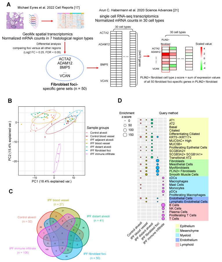
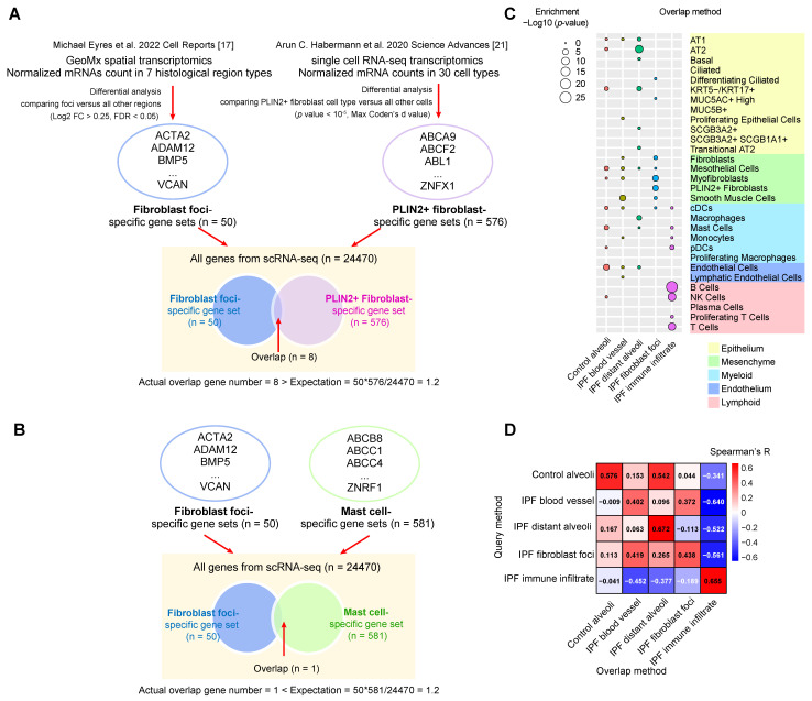
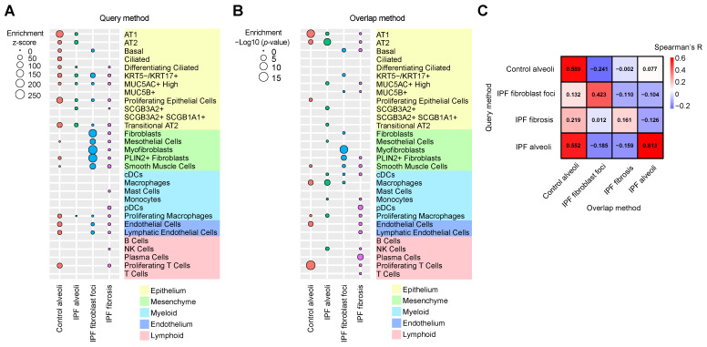
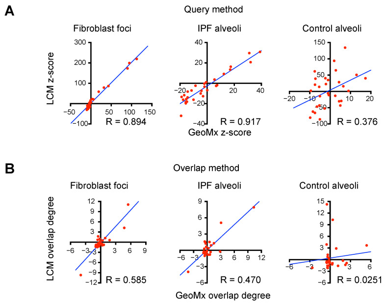
Idiopathic pulmonary fibrosis (IPF) is a progressive lung disease characterized by repetitive alveolar injuries with excessive deposition of extracellular matrix (ECM) proteins. A crucial need in understanding IPF pathogenesis is identifying cell types associated with histopathological regions, particularly local fibrosis centers known as fibroblast foci. To address this, we integrated published spatial transcriptomics and single-cell RNA sequencing (scRNA-seq) transcriptomics and adopted the Query method and the Overlap method to determine cell type enrichments in histopathological regions. Distinct fibroblast cell types are highly associated with fibroblast foci, and transitional alveolar type 2 and aberrant KRT5-/KRT17+ (KRT: keratin) epithelial cells are associated with morphologically normal alveoli in human IPF lungs. Furthermore, we employed laser capture microdissection-directed mass spectrometry to profile proteins. By comparing with another published similar dataset, common differentially expressed proteins and enriched pathways related to ECM structure organization and collagen processing were identified in fibroblast foci. Importantly, cell type enrichment results from innovative spatial proteomics and scRNA-seq data integration accord with those from spatial transcriptomics and scRNA-seq data integration, supporting the capability and versatility of the entire approach. In summary, we integrated spatial multi-omics with scRNA-seq data to identify disease-associated cell types and potential targets for novel therapies in IPF intervention. The approach can be further applied to other disease areas characterized by spatial heterogeneity.
Keywords: Idiopathic pulmonary fibrosis; cell type; gene signature; laser capture microdissection; mass spectrometry; multi-omics integration; protein signature; scRNA-seq; spatial proteomics; spatial transcriptomics.
Conflict of interest statement
All authors are employed by the AbbVie company. AbbVie funded the study and participated in study design, research, data collection, analysis and interpretation of data, writing, reviewing, and approving the publication. There are no additional conflicts of interest to report.
Figures
Similar articles
-
Cross-species integration of single-cell RNA-seq resolved alveolar-epithelial transitional states in idiopathic pulmonary fibrosis.Am J Physiol Lung Cell Mol Physiol. 2021 Sep 1;321(3):L491-L506. doi: 10.1152/ajplung.00594.2020. Epub 2021 Jun 16. Am J Physiol Lung Cell Mol Physiol. 2021. PMID: 34132117
-
Disparate Interferon Signaling and Shared Aberrant Basaloid Cells in Single-Cell Profiling of Idiopathic Pulmonary Fibrosis and Systemic Sclerosis-Associated Interstitial Lung Disease.Front Immunol. 2021 Mar 30;12:595811. doi: 10.3389/fimmu.2021.595811. eCollection 2021. Front Immunol. 2021. PMID: 33859634 Free PMC article.
-
Alveolar Basal Cells Differentiate towards Secretory Epithelial- and Aberrant Basaloid-like Cells In Vitro.Cells. 2022 Jun 2;11(11):1820. doi: 10.3390/cells11111820. Cells. 2022. PMID: 35681516 Free PMC article.
-
Computational solutions for spatial transcriptomics.Comput Struct Biotechnol J. 2022 Sep 1;20:4870-4884. doi: 10.1016/j.csbj.2022.08.043. eCollection 2022. Comput Struct Biotechnol J. 2022. PMID: 36147664 Free PMC article. Review.
-
Insights Into Development and Progression of Idiopathic Pulmonary Fibrosis From Single Cell RNA Studies.Front Med (Lausanne). 2020 Dec 16;7:611728. doi: 10.3389/fmed.2020.611728. eCollection 2020. Front Med (Lausanne). 2020. PMID: 33392232 Free PMC article. Review.
Cited by
-
Integrative Spatial Proteomics and Single-Cell RNA Sequencing Unveil Molecular Complexity in Rheumatoid Arthritis for Novel Therapeutic Targeting.Proteomes. 2025 May 22;13(2):17. doi: 10.3390/proteomes13020017. Proteomes. 2025. PMID: 40559990 Free PMC article.
References
-
- King T.E., Bradford W.Z., Castro-Bernardini S., Fagan E.A., Glaspole I., Glassberg M.K., Gorina E., Hopkins P.M., Kardatzke D., Lancaster L., et al. A Phase 3 Trial of Pirfenidone in Patients with Idiopathic Pulmonary Fibrosis. N. Engl. J. Med. 2014;370:2083–2092. doi: 10.1056/NEJMoa1402582. - DOI - PubMed
-
- Raghu G., Remy-Jardin M., Myers J.L., Richeldi L., Ryerson C.J., Lederer D.J., Behr J., Cottin V., Danoff S.K., Morell F., et al. Diagnosis of Idiopathic Pulmonary Fibrosis. An Official ATS/ERS/JRS/ALAT Clinical Practice Guideline. Am. J. Respir. Crit. Care Med. 2018;198:e44–e68. doi: 10.1164/rccm.201807-1255ST. - DOI - PubMed
LinkOut - more resources
Full Text Sources
Research Materials
Miscellaneous
