. 2025 Jan 20;13(1):6.
doi: 10.3390/proteomes13010006.

DNA Damage-Induced Ferroptosis: A Boolean Model Regulating p53 and Non-Coding RNAs in Drug Resistance

Affiliations

DNA Damage-Induced Ferroptosis: A Boolean Model Regulating p53 and Non-Coding RNAs in Drug Resistance

Shantanu Gupta et al. Proteomes. .

Abstract

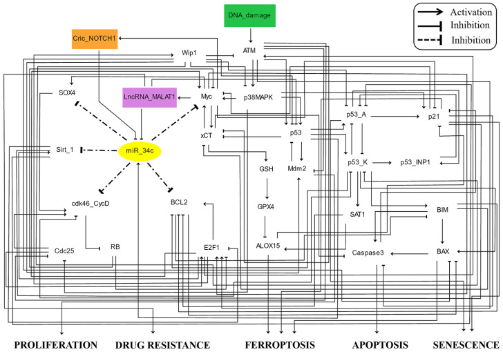
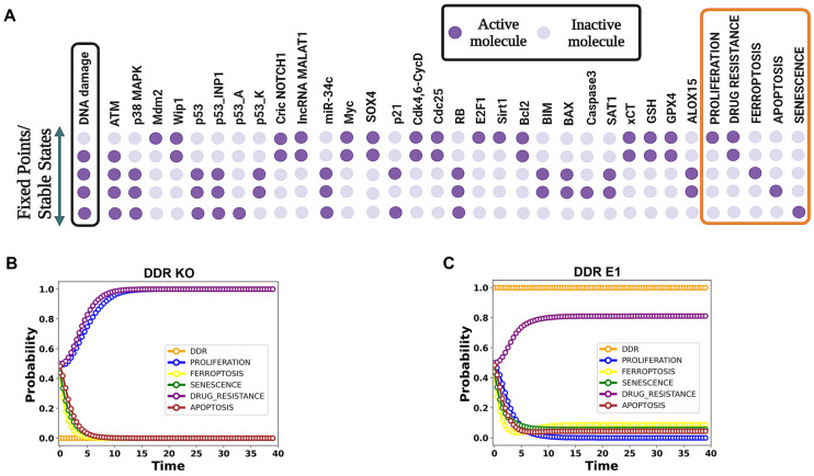
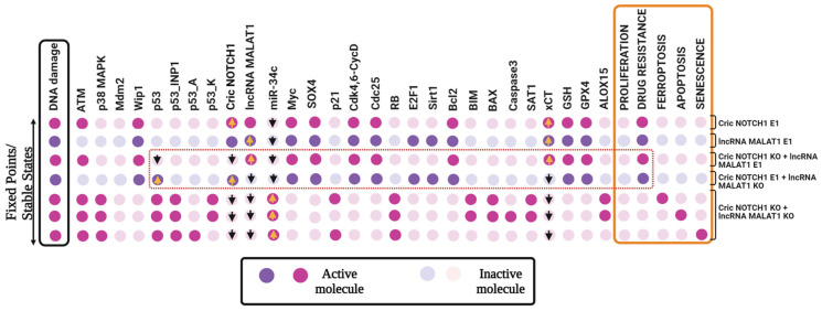
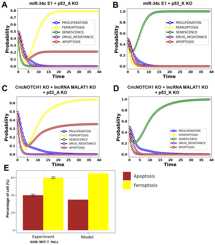
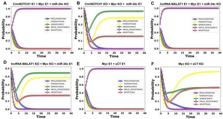
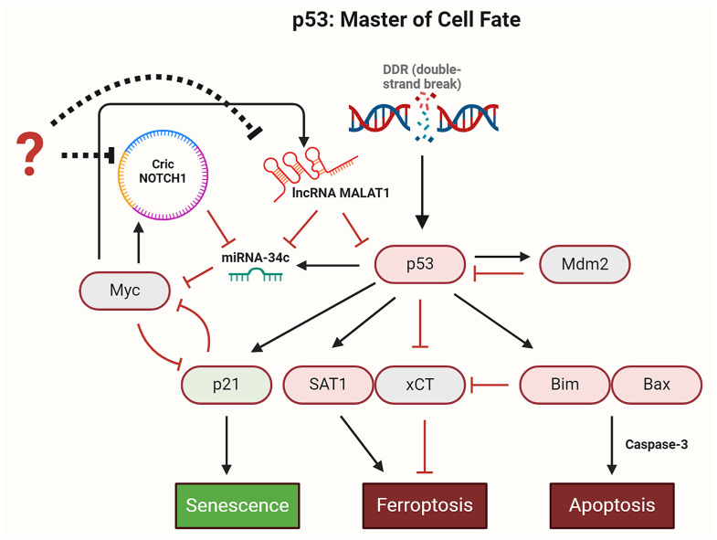
The tumor suppressor p53, in its wild-type form, plays a central role in cellular homeostasis by regulating senescence, apoptosis, and autophagy within the DNA damage response (DDR). Recent findings suggest that wild-type p53 also governs ferroptosis, an iron-dependent cell death process driven by lipid peroxidation. Post-translational modifications of p53 generate proteoforms that significantly enhance its functional diversity in regulating these mechanisms. A key target in this process is the cystine/glutamate transporter (xCT), which is essential for redox balance and ferroptosis resistance. Additionally, p53-induced miR-34c-5p suppresses cancer cell proliferation and drug resistance by modulating Myc, an oncogene further influenced by non-coding RNAs like circular RNA NOTCH1 (CricNOTCH1) and long non-coding RNA MALAT1. However, the exact role of these molecules in ferroptosis remains unclear. To address this, we introduce the first dynamic Boolean model that delineates the influence of these ncRNAs and p53 on ferroptosis, apoptosis, and senescence within the DDR context. Validated through gain- and loss-of-function perturbations, our model closely aligns with experimental observations in cancers such as oral squamous cell carcinoma, nasopharyngeal carcinoma, and osteosarcoma. The model identifies crucial positive feedback loops (CricNOTCH1/miR-34c/Myc, MALAT1/miR-34c/Myc, and Myc/xCT) and highlights the therapeutic potential of using p53 proteoforms and ncRNAs to combat drug resistance and induce cancer cell death.

Keywords: CricRNA NOTCH1; apoptosis; ferroptosis; lncRNA MALAT1; miR-34c-5p; p53.


Conflict of interest statement

The authors declare no conflicts of interest.

Figures

Figure 1
Figure 1
A dynamic Boolean model of ncRNAs and p53 in ferroptosis. Direct black edges ending in arrowheads represent positive regulatory interactions, while those ending in hammerheads indicate negative ones. Dashed black edges ending with hammerheads specify miR-34c targets. Node colors represent function: signaling proteins are white, CricNOTCH1 is an orange rectangle, and lncRNA MALAT1 is a purple rectangle, with miR-34c as a yellow oval. The green rectangle indicates DNA damage. Model outputs are labeled as proliferation, drug resistance, ferroptosis, and apoptosis.
Figure 2
Figure 2
The dynamics of the wild-type case network, illustrating five stable states. (A) The leftmost column depicts the DNA damage levels highlighted in the black box, while the rightmost column presents the model outputs—proliferation, drug resistance, ferroptosis, apoptosis, and senescence—in the orange box. Each line represents an endpoint corresponding to the input. Light violet cells denote an inactivation, whereas dark violet cells denote activation. (B,C) Using MaBoSS simulations, we identified steady states under wild-type conditions with and without DNA damage. (B) For this analysis, DDR input was set to 0 (OFF), representing the absence of DNA damage. Under these conditions, the model predicted 100% proliferation and drug resistance, reflecting the baseline state of the system. (C) When DDR input was fully activated (probability = 1), representing maximal DNA damage, the model predicted distinct probabilities for cell fate outcomes: 81% drug resistance, 9% ferroptosis, 4% apoptosis, and 6% senescence. These outcomes sum to 100%, indicating the comprehensive distribution of cellular states in response to DNA damage. Detailed simulation data are available in Data S1.
Figure 3
Figure 3
The gain or loss-of-function perturbations of the CricNOTCH1 and lncRNA MALAT1. The stable states identified for distinct scenarios were as follows: CricNOTCH1 E1, lncRNA MALAT1 E1, CricNOTCH1 KO + lncRNA MALAT1 E1, CricNOTCH1 E1 + lncRNA MALAT1 KO, CricNOTCH1 KO + lncRNA MALAT1 KO. E1 represents GoF and KO represents the LoF of the corresponding network element. The leftmost column shows DNA damage levels highlighted in black, and the rightmost column presents the model outputs, which are highlighted in orange: proliferation, drug resistance, ferroptosis, apoptosis, and senescence. Each line represents a single stable state corresponding to the input.
Figure 4
Figure 4
Dynamics of p53 and ncRNAs in DDR under the maximized DNA damage input. The DNA damage input (DDR) was initialized with a probability of 1, representing fully active conditions. (A) For the p53_K LoF + miR-34c GoF, we observed 100% senescence. (B) For the p53_A LoF + miR-34c GoF case, we observed 80% ferroptosis and 20% apoptosis. (C) Combined LoF CricNOTCH1 + LoF lncRNA MALAT1 + LoF p53_A led to 65% ferroptosis and 35% apoptosis. (D) Combined LoF CricNOTCH1 + LoF lncRNA MALAT1 + LoF p53_K resulted in 100% senescence, with no ferroptosis or apoptosis observed. (E) A comparison of the LoF CricNOTCH1, LoF lncRNA MALAT1, and LoF p53_A perturbations with in vitro observations. To enhance clarity and precision, fewer than 41 time steps are shown in the panel to highlight the differences among curves. The detailed simulation data can be found in Data S1.
Figure 5
Figure 5
The dynamics of p53 and ncRNAs in DDR under maximized DNA damage input. Here, the DNA damage input (DDR) was initialized with a probability of 1, representing fully active conditions. (A) For the p53_K LoF + miR-34c GoF, we observed 100% senescence. (B) For the p53_A LoF + miR-34c GoF case, we observed 80% ferroptosis and 20% apoptosis. (C) The combined LoF CricNOTCH1 + LoF lncRNA MALAT1 + LoF p53_A led to 65% ferroptosis and 35% apoptosis. (D) The combined LoF CricNOTCH1 + LoF lncRNA MALAT1 + LoF p53_K resulted in 100% senescence, with no ferroptosis or apoptosis observed. (E) The combination of GoF Myc and GoF xCT led to 100% drug resistance. (F) The combination of LoF Myc and LoF xCT resulted in 47% ferroptosis, 27% apoptosis, and 26% senescence. To enhance clarity and precision, fewer than 41 time steps are shown in the panel to highlight the differences among curves. Detailed simulation data are available in Data S1.
Figure 6
Figure 6
p53 Dynamics in the DDR. Upon DNA damage, p53 is activated and induces Mdm2 expression, which subsequently inhibits p53 activity. Once activated, p53 regulates several critical signaling pathways: it induces p21 to trigger senescence, activates the BIM/BAX axis to initiate apoptosis, and promotes ferroptosis by either directly inhibiting xCT or inducing SAT1 expression. Additionally, p53 activates miR-34c, which suppresses Myc, a potent inducer of CricNOTCH1 and lncRNA MALAT1. By inhibiting Myc, miR-34c reduces the expression of CricNOTCH1 and lncRNA MALAT1. Interestingly, lncRNA MALAT1 directly inhibits p53. Therefore, miR-34c promotes sustained p53 activation by inhibiting Myc, which in turn diminishes MALAT1’s inhibitory effect on p53. In addition, inhibitors (indicated by question mark) of CricNOTCH1 and lncRNA MALAT1 trigger p53-mediated signaling pathways such as ferroptosis, apoptosis and senescence in DDR. Black arrows represent activation, hammerhead arrows represent inhibition, and dotted hammerhead arrows indicate indirect inhibition.

Similar articles

Cited by

References

    1. Chen J. The Cell-Cycle Arrest and Apoptotic Functions of p53 in Tumor Initiation and Progression. Cold Spring Harb. Perspect. Med. 2016;6:a026104. doi: 10.1101/cshperspect.a026104. - DOI - PMC - PubMed
    1. Gupta S., Silveira D.A., Mombach J.C.M. Towards DNA-damage induced autophagy: A Boolean model of p53-induced cell fate mechanisms. DNA Repair. 2020;96:102971. doi: 10.1016/j.dnarep.2020.102971. - DOI - PubMed
    1. Kumari R., Jat P. Mechanisms of Cellular Senescence: Cell Cycle Arrest and Senescence Associated Secretory Phenotype. Front. Cell Dev. Biol. 2021;9 doi: 10.3389/fcell.2021.645593. - DOI - PMC - PubMed
    1. Zhang L., Hou N., Chen B., Kan C., Han F., Zhang J., Sun X. Post-Translational Modifications of p53 in Ferroptosis: Novel Pharmacological Targets for Cancer Therapy. Front. Pharmacol. 2022;13:908772. doi: 10.3389/fphar.2022.908772. - DOI - PMC - PubMed
    1. Kang R., Kroemer G., Tang D. The tumor suppressor protein p53 and the ferroptosis network. Free Radic. Biol. Med. 2019;133:162–168. doi: 10.1016/j.freeradbiomed.2018.05.074. - DOI - PMC - PubMed

LinkOut - more resources