. 2025 Jan 5;10(1):23.
doi: 10.3390/jfmk10010023.

Evaluation of Optimized Lumbar Oblique X-Ray Angles with Positioning Assistance for Enhanced Imaging Quality: A Pilot Study in an Asian Cohort

Affiliations

Evaluation of Optimized Lumbar Oblique X-Ray Angles with Positioning Assistance for Enhanced Imaging Quality: A Pilot Study in an Asian Cohort

Yu-Li Wang et al. J Funct Morphol Kinesiol. .

Abstract

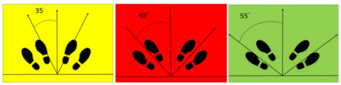
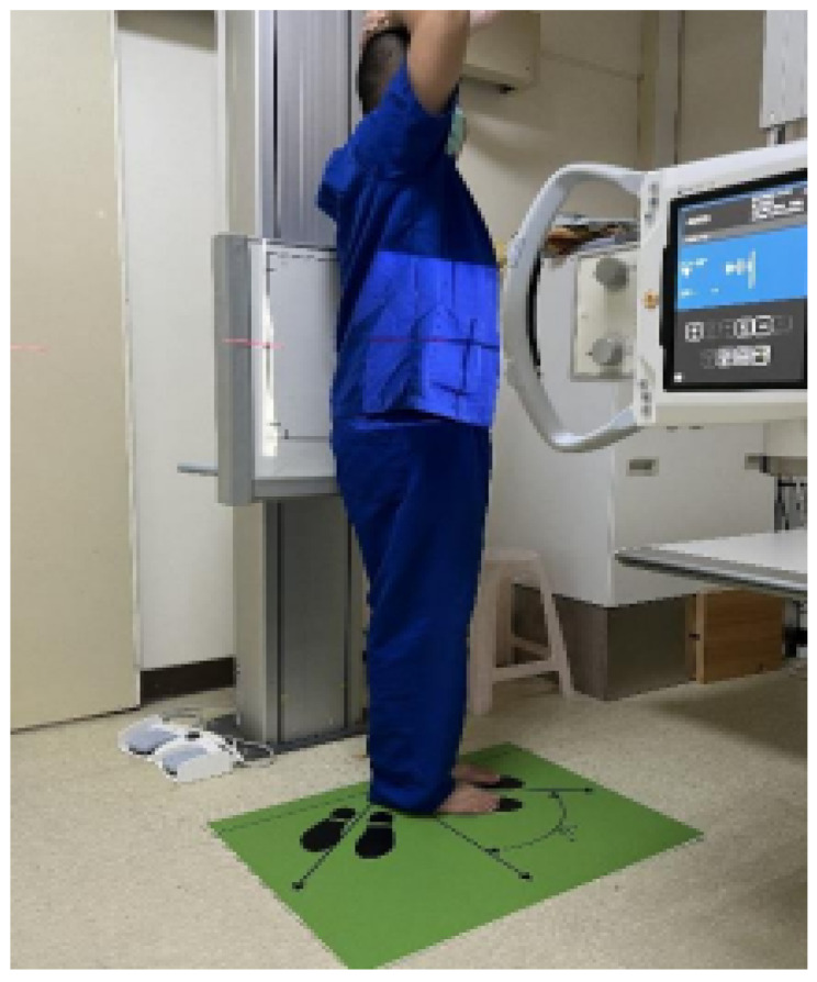
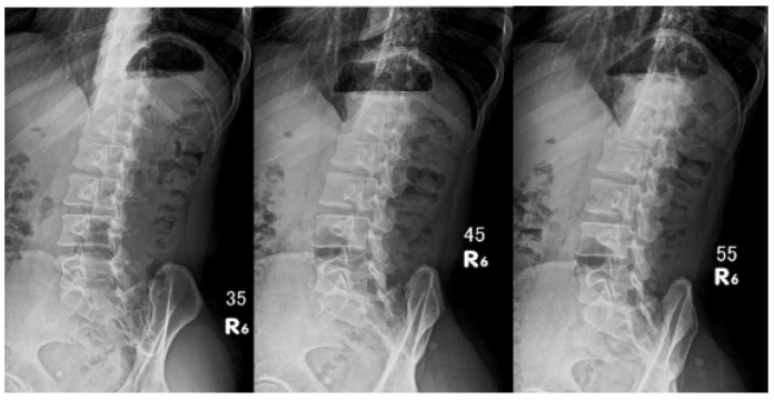
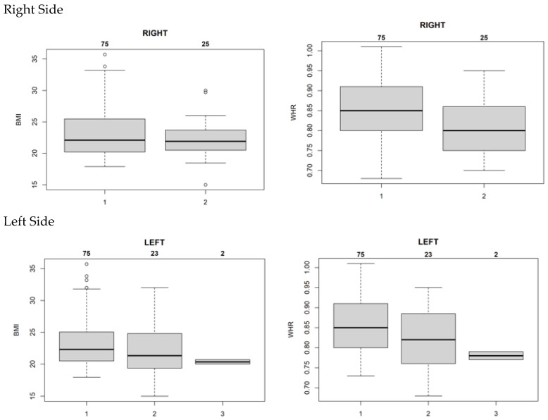
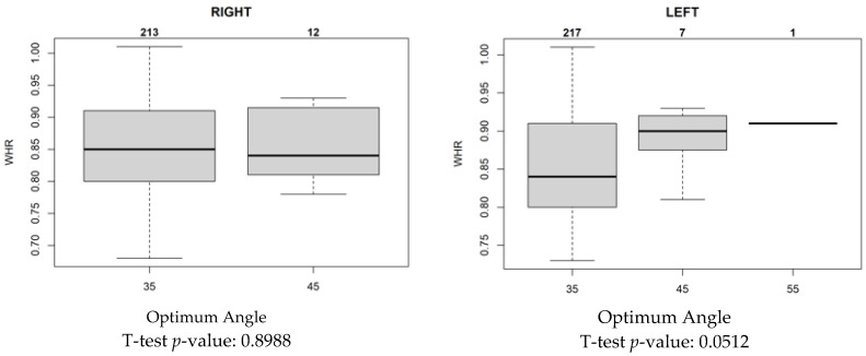
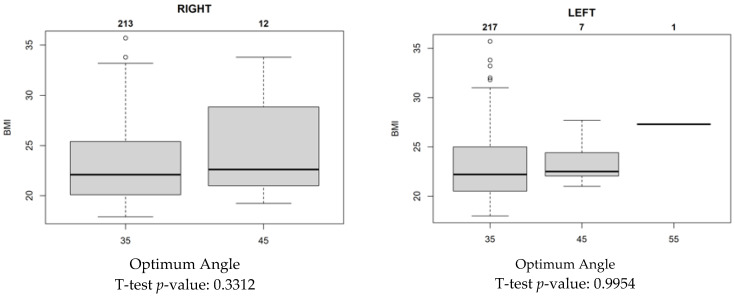
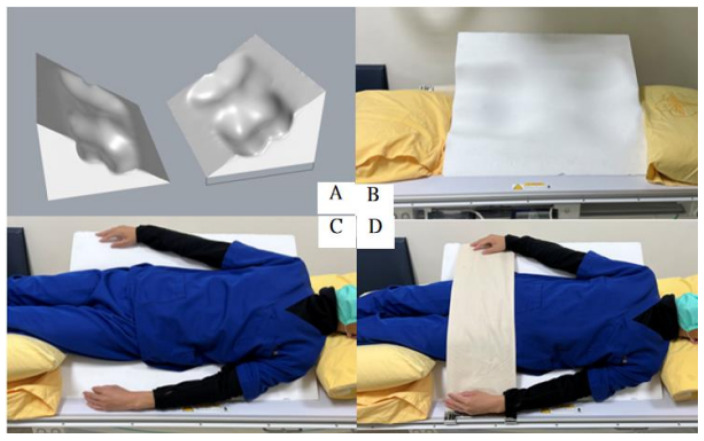
Objective: Pars fractures are a common cause of lower back pain, especially among young individuals. Although computed tomography (CT) and magnetic resonance imaging (MRI) scanning are commonly used in developed regions, traditional radiography remains the main diagnostic method in many developing countries. This study assessed whether the standard radiographic angles suggested in textbooks are optimal for an Asian population since Asian groups have lower lumbar lordosis. This study found a 35° angle to be the most effective angle for lumbar oblique X-ray imaging. Additionally, the potential for a customized positioning auxiliary device was examined to improve image quality and reduce patient discomfort in cost-sensitive healthcare settings like Taiwan's single-payer system. Methods: A total of 100 participants underwent lumbar oblique radiography using a specially designed footboard with angle markings. Radiologists evaluated 600 images based on waist-to-hip ratio (WHR) and body mass index to identify the optimal angulation for various body types. Results: For individuals with a WHR of 0.85, a 35° angle provided superior image quality, while 45° was more effective for slimmer patients. This optimized approach indicates the cost-effectiveness and diagnostic value of traditional X-ray imaging. Conclusions: The 35° angulation standardizes lumbar X-ray imaging for an Asian cohort, reducing repeat scans and improving accuracy. Using a positioning device further enhances image quality and patient comfort, supporting the clinical utility of traditional radiography in resource-limited environments.

Keywords: 3D manufacture; image quality; oblique examination; positioning auxiliary device; spondylolisthesis.


Conflict of interest statement

The authors declare no conflicts of interest.

Figures

Figure 1
Figure 1
Three different angle markings of the footboard are shown. These boards are color-coded to prevent technicians from being confused. Positioned in front of the vertical bucky stand, candidates can easily locate their standing position based on the footprints.
Figure 2
Figure 2
Use of the footboard. This figure illustrates a candidate standing on the footboard and respecting the footprint angle easily. Minor adjustments are conducted following the arrow. This way, the researcher can obtain an oblique examination under the correct shooting angle.
Figure 3
Figure 3
Image from three different angles. The technician marked the actual shooting angle and direction on the film. Thus, the radiologist can clearly distinguish each image. Image files are recorded according to case numbers to protect patient privacy.
Figure 4
Figure 4
Scoring consistency between optimum angle and median WHR. This figure illustrates the sufficiency of the imaging angle compared with BMI and WHR. The number 1 means the ratings are all consistent, 2 means two people are consistent, and 3 means none. When the median WHR is 0.85, the consistency among the three doctors is the best. Meanwhile, BMI did not show an obvious trend to support this consistency.
Figure 5
Figure 5
Relationship between optimum angle and median WHR. This figure points out that for a median WHR of 0.85, 35° was deemed the most suitable angle; however, as the WHR increased to 0.90, the optimal angle increased to 45°. Moreover, there is a large difference in WHR between the two groups at 35° and 45°, but no significant difference in BMI.
Figure 5
Figure 5
Relationship between optimum angle and median WHR. This figure points out that for a median WHR of 0.85, 35° was deemed the most suitable angle; however, as the WHR increased to 0.90, the optimal angle increased to 45°. Moreover, there is a large difference in WHR between the two groups at 35° and 45°, but no significant difference in BMI.
Figure 6
Figure 6
Three-dimensional (3D)-assisted device in action. This figure shows the 3D-assisted device designed and used by the authors. (A) shows a 3D design drawing of our assistive device. (B) illustrates the device placed on the examination table. (C) is a schematic diagram of a medium-sized male imitating an individual placed on an assistive device. Shoulders and hips remain equally aligned. However, pillows needed to be placed on the head and lower limbs to maintain the examination posture. In subsequent modifications, the length of the assistive device can be increased to increase comfort. In (D), the restraint straps attached to the examination equipment are used to assist in positioning and make the inspected patient feel more secure.

Similar articles

References

    1. Leone A., Cianfoni A., Cerase A., Magarelli N., Bonomo L. Lumbar Spondylolysis: A Review. Skeletal Radiol. 2011;40:683–700. doi: 10.1007/s00256-010-0942-0. - DOI - PubMed
    1. Hu S.S., Tribus C.B., Diab M., Ghanayem A.J. Spondylolisthesis and Spondylolysis. J. Bone Joint Surg. Am. 2008;90:656–671. - PubMed
    1. McCleary M.D., Congeni J.A. Current Concepts in the Diagnosis and Treatment of Spondylolysis in Young Athletes. Curr. Sports Med. Rep. 2007;6:62–66. doi: 10.1007/s11932-007-0014-y. - DOI - PubMed
    1. Fadzan M., Bettany-Saltikov J. Etiological Theories of Adolescent Idiopathic Scoliosis: Past and Present. Open Orthop. J. 2017;11:1466–1489. doi: 10.2174/1874325001711011466. - DOI - PMC - PubMed
    1. Cavalier R., Herman M.J., Cheung E.V., Pizzutillo P.D. Spondylolysis and Spondylolisthesis in Children and Adolescents: I. Diagnosis, Natural History, and Nonsurgical Management. J. Am. Acad. Orthop. Surg. 2006;14:417–424. doi: 10.5435/00124635-200607000-00004. - DOI - PubMed

LinkOut - more resources