. 2025 Jan 16;12(1):2.
doi: 10.3390/medicines12010002.

Hypomyelinating Leukodystrophy 14 (HLD14)-Related UFC1 p.Arg23Gln Decreases Cell Morphogenesis: A Phenotype Reversable with Hesperetin

Affiliations

Hypomyelinating Leukodystrophy 14 (HLD14)-Related UFC1 p.Arg23Gln Decreases Cell Morphogenesis: A Phenotype Reversable with Hesperetin

Yuri Ichihara et al. Medicines (Basel). .

Abstract

Introduction: In the central nervous system (CNS), proper interaction between neuronal and glial cells is crucial for the development of mature nervous tissue. Hypomyelinating leukodystrophies (HLDs) are a group of genetic CNS disorders characterized by hypomyelination and/or demyelination. In these conditions, genetic mutations disrupt the biological functions of oligodendroglial cells, which are responsible for wrapping neuronal axons with myelin sheaths. Among these, an amino acid mutation of the ubiquitin-fold modifier conjugating enzyme 1 (UFC1) is associated with HLD14-related disease, characterized by hypomyelination and delayed myelination in the brain. UFC1 is a critical component of the UFMylation system, functioning similarly to E2-conjugating enzymes in the ubiquitin-dependent protein degradation system.

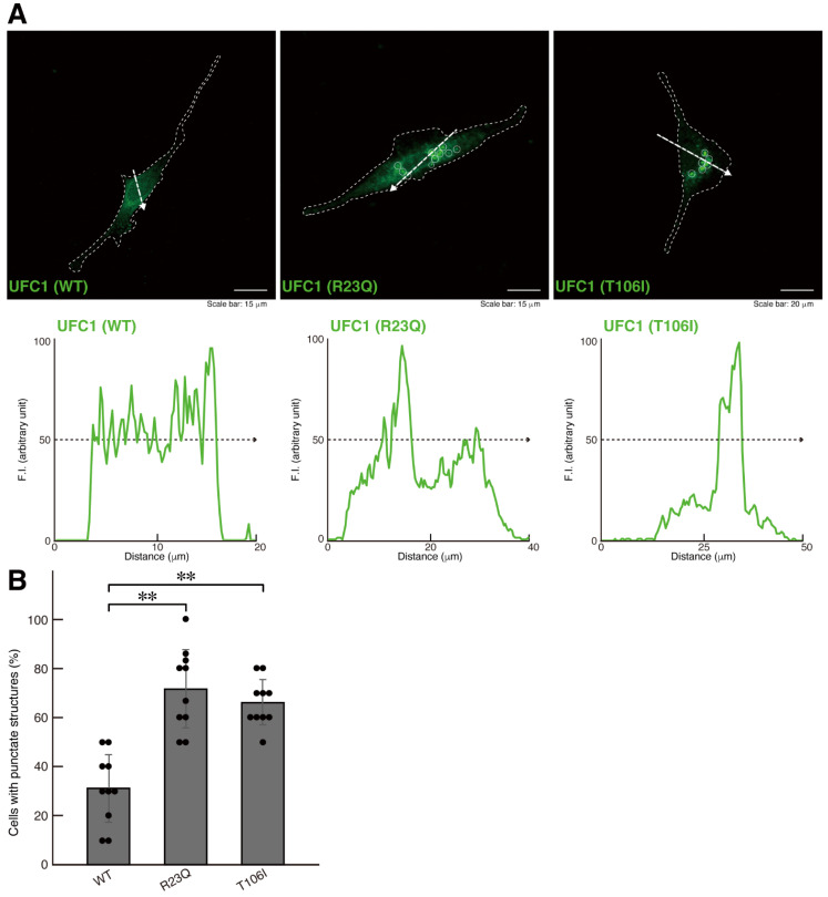
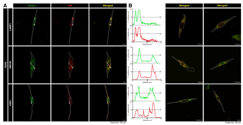
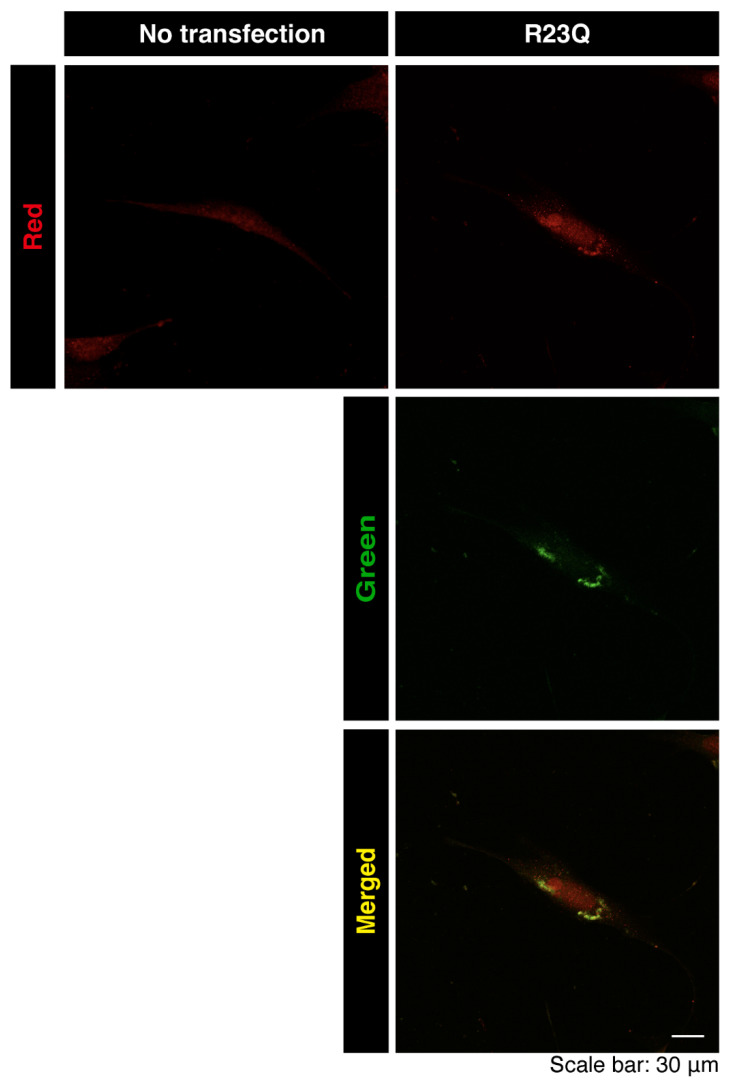
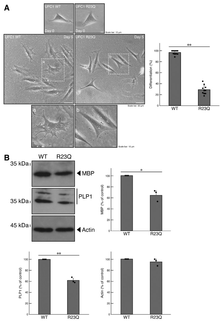
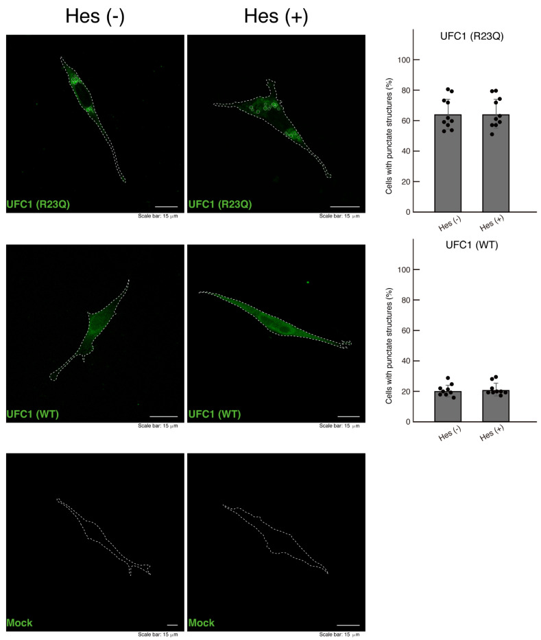
Methodology: We describe how a missense mutation in UFC1 (p.Arg23Gln) leads to the aggregation of UFC1 primarily in lysosomes in FBD-102b cells, which are undergoing oligodendroglial cell differentiation.

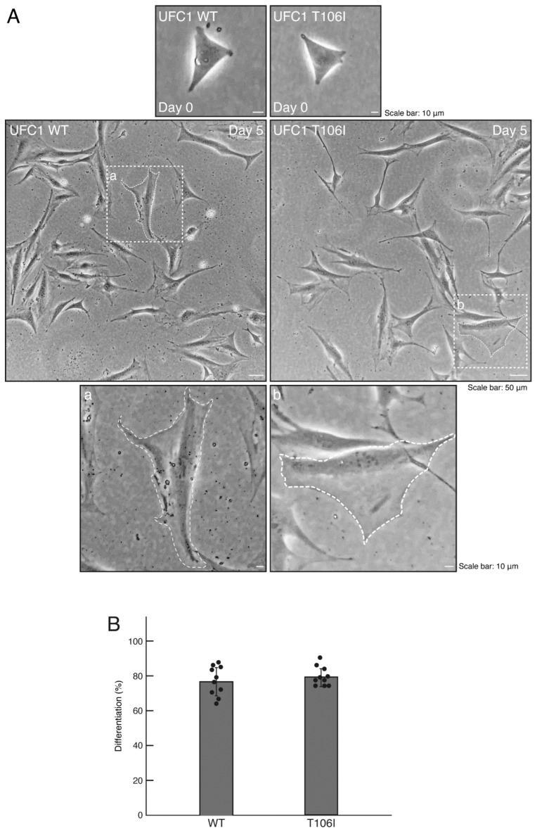
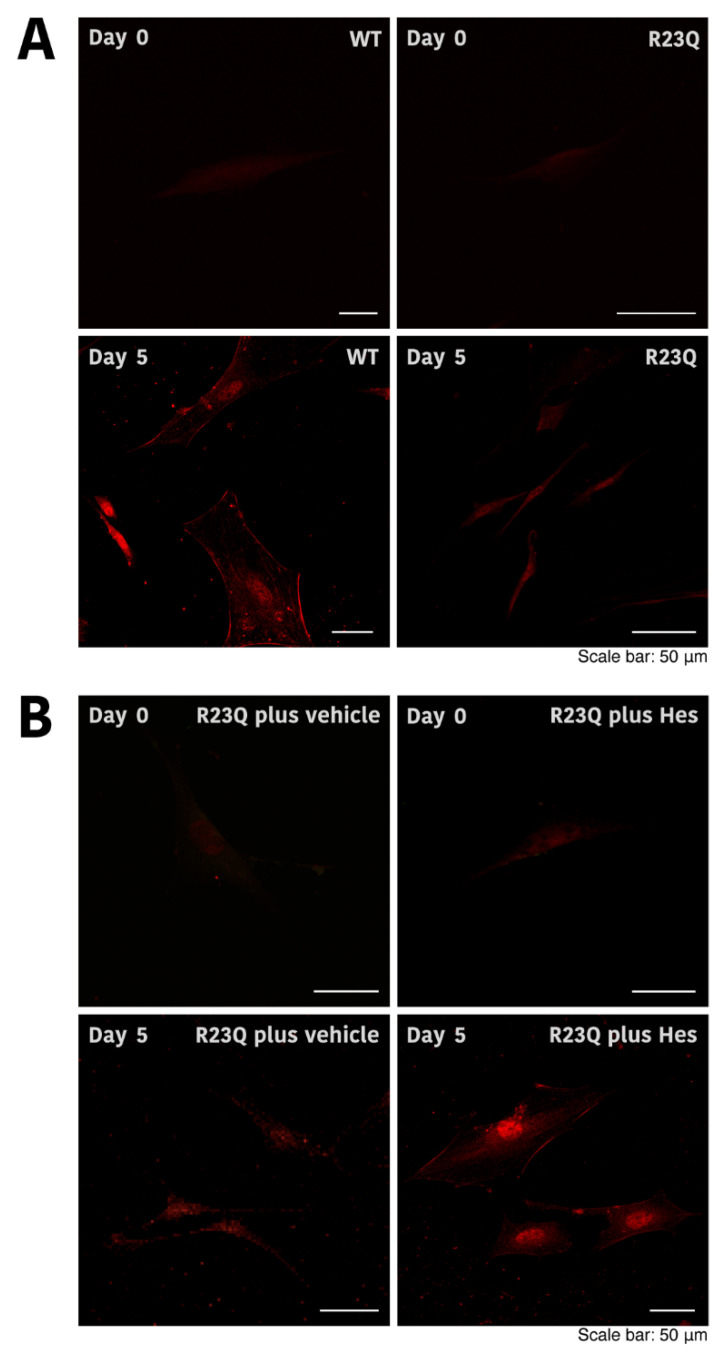
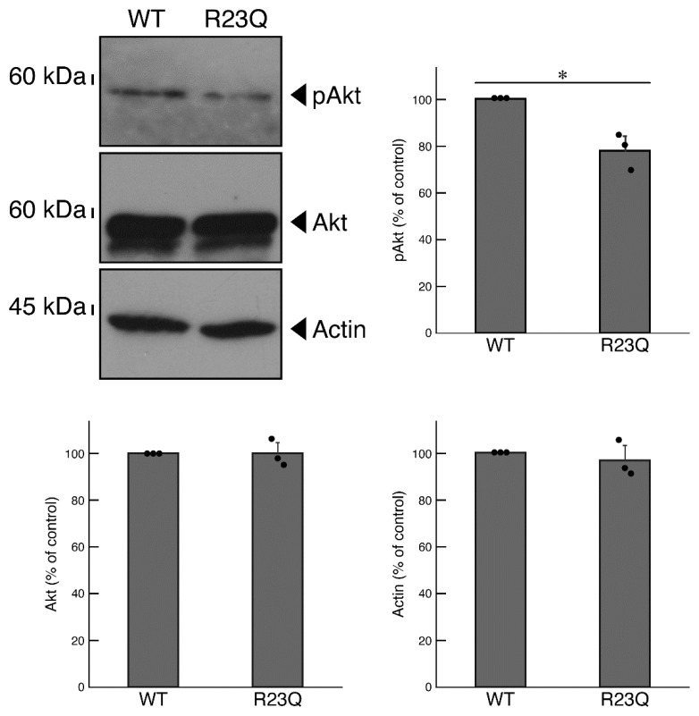
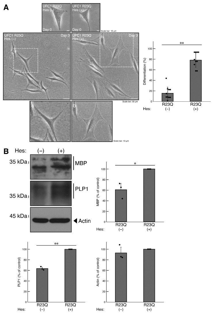
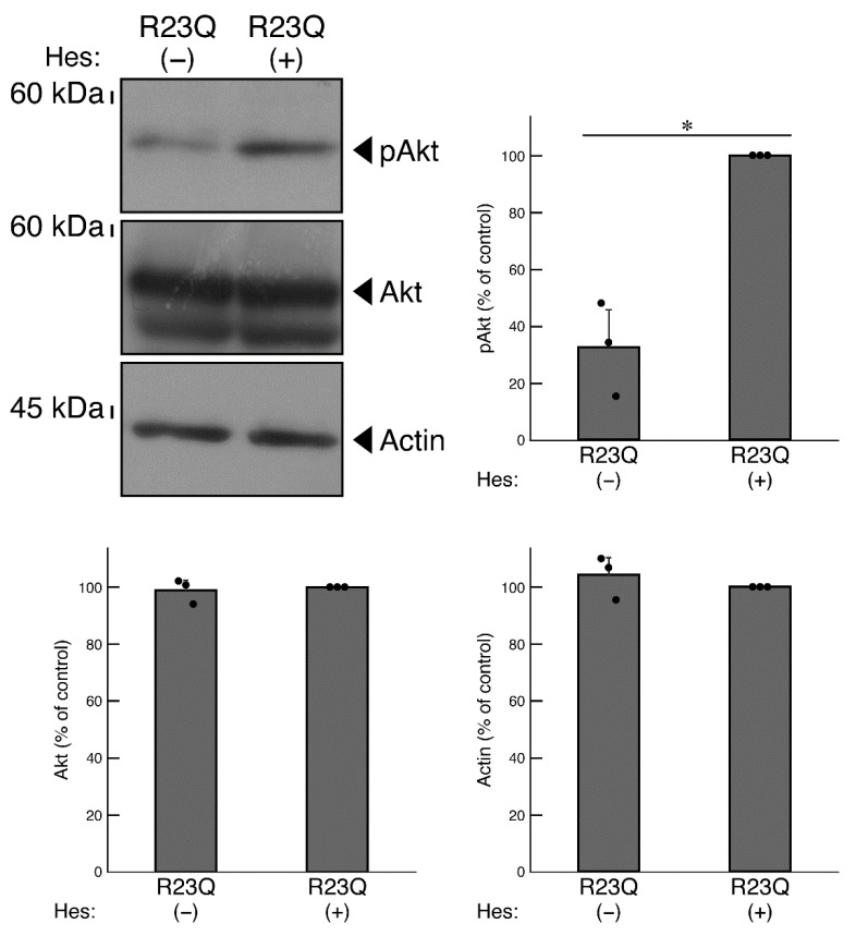
Results: Cells with mutated UFC1 exhibit reduced Akt kinase phosphorylation and reduced expression of differentiation and myelination marker proteins. Consistently, these cells exhibit impaired morphological differentiation with a reduced ability to extend widespread membranes. Interestingly, hesperetin, a citrus flavonoid with known neuroprotective properties, was found to restore differentiation abilities in cells with the UFC1 mutation.

Conclusions: These findings indicate that the HLD14-related mutation in UFC1 causes its lysosomal aggregation, impairing its morphological differentiation. Furthermore, the study highlights potential therapeutic insights into the pathological molecular and cellular mechanisms underlying HLD14 and suggests hesperetin as a promising candidate for treatment.

Keywords: HLD14; UFC1; differentiation; hesperetin; morphogenesis; oligodendrocyte.


Conflict of interest statement

Hiroaki Oizumi, Masahiro Yamamoto, and Katsuya Ohbuchi were employed by Tsumura & Co. The remaining authors declare that the research was conducted in the absence of any commercial or financial relationships that could be construed as a potential conflict of interest.

Figures

Figure 1
Figure 1
The R23Q and T106I mutant proteins of UFC1 are localized in punctate structures. (A,B) FBD-102b cells, delineated by dotted lines, were transfected with the plasmid encoding GFP-tagged wild-type (WT) UFC1 or the R23Q or T106I mutant constructs. Transfected cells were detected using transfected proteins (green). White circles in the images showing mutant proteins indicate representative aggregate-like structures (indicated representatively with white circles). Scan plots were performed along the white dotted lines in the direction of the arrows in the images. Graphs showing the fluorescence intensities (arbitrary units) along the white dotted lines in the direction of the arrows are presented in the bottom panels. Percentages of cells with punctate structures were statistically assessed (** p < 0.01; n = 10 fields [a total of 120 cells per experiment]).
Figure 2
Figure 2
Cells harboring UFC1 with the T106I mutation undergo morphological changes. (A,B) Cells harboring GFP-tagged wild-type (WT) UFC1 or UFC1 (T106I) were allowed to differentiate for 0 or 5 days. The square fields a and b indicated by the dotted lines in the center panels are magnified in the bottom panels a and b. Cells with widespread membranes were statistically assessed (n = 10 fields [a total of 360 cells per experiment]).
Figure 3
Figure 3
The R23Q mutant protein of UFC1 is primarily co-localized with the lysosome antigen. (A,B) FBD-102b cells were transfected with the plasmid encoding GFP-tagged UFC1 (R23Q). Cells were detected with transfected proteins (green) and the organelle antigen (red). Scan plots were performed along the white dotted lines in the direction of the arrows in the representative color images (green and red images). Staining data for the other two cells are depicted as merged images in the two lanes on the right.
Figure 4
Figure 4
Aggregate-like structures of UFC1 with the R23Q mutation are co-localized with endogenous UFC1. Cells were transfected with or without the plasmid encoding GFP-tagged UFC1 (green) with the R23Q mutation and stained with an anti-UFC1 antibody (red). Merged images are shown as representative examples.
Figure 5
Figure 5
Cells harboring UFC1 with the R23Q mutation fail to undergo morphological differentiation. (A) Cells harboring GFP-tagged wild-type (WT) UFC1 or UFC1 (R23Q) were allowed to differentiate for 0 or 5 days. The square fields a and b indicated by the dotted lines in the center panels are magnified in the bottom panels a and b. Cells with widespread membranes were statistically assessed (** p < 0.01; n = 10 fields [a total of 360 cells per experiment]). (B) The lysates of the respective cells were immunoblotted with an antibody against MBP, PLP1, or control actin. Their expression levels were statistically compared to their respective controls (** p < 0.01 or * p < 0.05; n = 3 blots).
Figure 6
Figure 6
Morphological changes in cells harboring UFC1 with the R23Q mutation and the effect of hesperetin. (A,B) Cells harboring GFP-tagged wild-type (WT) UFC1 or UFC1 (R23Q) were allowed to differentiate for 0 or 5 days in the presence or absence (vehicle) of hesperetin (Hes). Cells were stained with an anti-PLP1 antibody (red). Representative images are shown.
Figure 7
Figure 7
Cells harboring UFC1 with the R23Q mutation show decreased Akt phosphorylation levels. The lysates of cells harboring GFP-tagged wild-type (WT) UFC1 or UFC1 (R23Q) were immunoblotted with an antibody specific for (pS473)Akt (pAkt), Akt, or control actin. Their levels were statistically compared to their respective controls (* p < 0.05; n = 3 blots).
Figure 8
Figure 8
Hesperetin recovers phenotypes in cells harboring UFC1 with the R23Q mutation. (A) Cells harboring GFP-tagged UFC1 (R23Q) were allowed to differentiate in the presence (+) or absence (−, vehicle) of 5 micromolar of hesperetin (Hes). The square fields a and b indicated by the dotted lines in the center panels are magnified in the bottom panels a and b. Cells with widespread membranes were statistically assessed (** p < 0.01; n = 10 fields [a total of 340 cells per each experiment]). (B) The lysates of the respective cells were immunoblotted with an antibody against MBP, PLP1, or control actin. Their expression levels were statistically compared to their respective controls (** p < 0.01 or * p < 0.05; n = 3 blots).
Figure 9
Figure 9
Hesperetin recovers the phosphorylation levels of Akt in cells harboring UFC1 with the R23Q mutation. The lysates of cells harboring GFP-tagged UFC1 (R23Q) in the presence (+) or absence (−, vehicle) of hesperetin (Hes) were immunoblotted with an antibody specific for (pS473)Akt (pAkt), Akt, or control actin. Their levels were statistically compared to their respective controls (* p < 0.05; n = 3 blots).
Figure 10
Figure 10
The effect of hesperetin on aggregate-like structures in cells harboring UFC1 with the R23Q mutation. Cells harboring GFP-tagged UFC1 (R23Q) or wild-type (WT, green) or mock-transfected cells were treated with (+) or without (−, vehicle) hesperetin (Hes). Cells with punctate structures (indicated representatively by white circles) were statistically counted (a total of 120 cells per experiment).

Similar articles

References

    1. Nave K.A., Werner H.B. Myelination of the nervous system: Mechanisms and functions. Annu. Rev. Cell Dev. Biol. 2014;30:503–533. doi: 10.1146/annurev-cellbio-100913-013101. - DOI - PubMed
    1. Stadelmann C., Timmler S., Barrantes-Freer A., Simons M. Myelin in the central nervous system: Structure, function, and pathology. Physiol. Rev. 2019;99:1381–1431. doi: 10.1152/physrev.00031.2018. - DOI - PubMed
    1. Peles E., Salzer J.L. Molecular domains of myelinated axons. Curr. Opin. Neurobiol. 2000;10:558–565. doi: 10.1016/S0959-4388(00)00122-7. - DOI - PubMed
    1. Knowles J.K., Batra A., Xu H., Monje M. Adaptive and maladaptive myelination in health and disease. Nat. Rev. Neurol. 2022;18:735–746. doi: 10.1038/s41582-022-00737-3. - DOI - PubMed
    1. Narayanan S.P., Flores A.I., Wang F., Macklin W.B. Akt signals through the mammalian target of rapamycin pathway to regulate CNS myelination. J. Neurosci. 2009;29:6860–6870. doi: 10.1523/JNEUROSCI.0232-09.2009. - DOI - PMC - PubMed

LinkOut - more resources