. 2025 Feb 25;99(2):e0161924.
doi: 10.1128/jvi.01619-24. Epub 2025 Jan 23.

Discovery of three novel neutralizing antibody epitopes on the human astrovirus capsid spike and mechanistic insights into virus neutralization

Affiliations

Discovery of three novel neutralizing antibody epitopes on the human astrovirus capsid spike and mechanistic insights into virus neutralization

Sarah Lanning et al. J Virol. .

Abstract

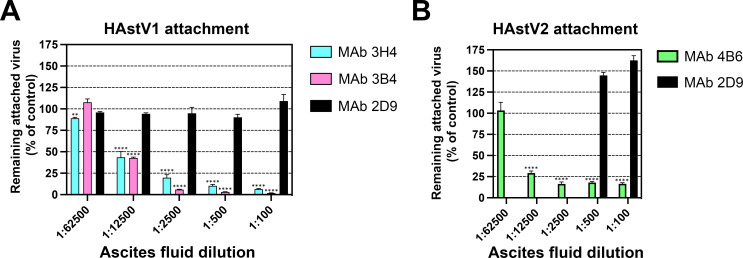
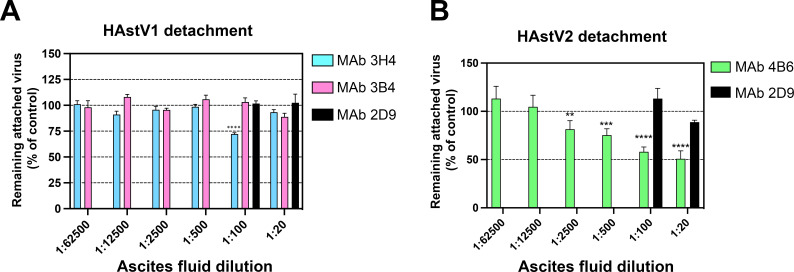
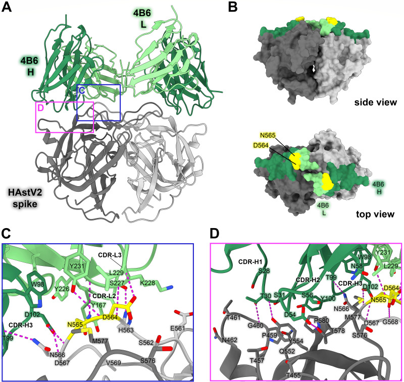
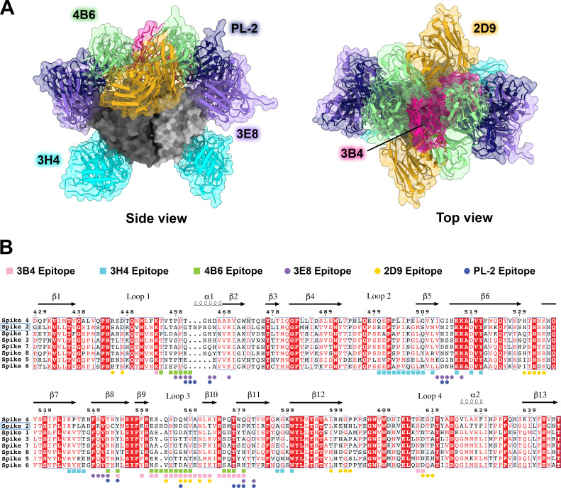
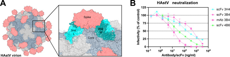
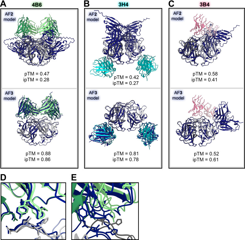
Human astroviruses (HAstVs) are a leading cause of viral childhood diarrhea that infects nearly every individual during their lifetime. Although human astroviruses are highly prevalent, no approved vaccine currently exists. Antibody responses appear to play an important role in protection from HAstV infection; however, knowledge about the neutralizing epitope landscape is lacking, as only three neutralizing antibody epitopes have previously been determined. Here, we structurally define the epitopes of three uncharacterized HAstV-neutralizing monoclonal antibodies: antibody 4B6 with X-ray crystallography to 2.67 Å, and antibodies 3H4 and 3B4 simultaneously with single-particle cryogenic-electron microscopy to 3.33 Å. We assess the epitope locations relative to conserved regions on the capsid spike and find that while antibodies 4B6 and 3B4 target the upper variable loop regions of the HAstV spike protein, antibody 3H4 targets a novel region near the base of the spike that is more conserved. Additionally, we found that all three antibodies bind with high affinity, and they compete with receptor FcRn binding to the capsid spike. These studies inform which regions of the HAstV capsid can be targeted by monoclonal antibody therapies and could aid in rational vaccine design.IMPORTANCEHuman astroviruses (HAstVs) infect nearly every child in the world, causing diarrhea, vomiting, and fever. Despite the prevalence of human astroviruses, little is known about how antibodies block virus infection. Here, we determined high-resolution structures of the astrovirus capsid protein in a complex with three virus-neutralizing antibodies. The antibodies bind distinct sites on the capsid spike domain. The antibodies block virus attachment to human cells and prevent capsid spike interaction with the human neonatal Fc receptor. These findings support the use of the human astrovirus capsid spike as an antigen in a vaccine to prevent astrovirus disease.

Keywords: X-ray crystallography; capsid; cryo-EM; human astrovirus; monoclonal antibodies; neutralization; protein structure-function; surface antigens.


Conflict of interest statement

The authors declare no conflict of interest.

Figures

Fig 1
Fig 1
Monoclonal antibodies to HAstV1 and HAstV2 block attachment of the virus to Caco-2 cells. Different dilutions of ascitic fluids of mAbs 3B4 or 3H4 to HAstV1 (A) or mAb 4B6 to HAstV2 (B) were preincubated with the purified corresponding virus for 1 h at 37°C and then the virus-antibody complexes were added to Caco-2 cell monolayers for 1 h on ice to allow the virus to attach to the cell surface. After removing the unbound virus and washing the cells, the attached virus was determined by RT-qPCR as described in Materials and Methods. MAb 2D9, specific to serotype HAstV8, was used as a negative control. Experiments were performed on ice to prevent virus endocytosis. The assay was performed in biological quintuplicates and carried out in duplicate. The data are expressed as percentages of the virus attached without antibodies and represent the mean ± SEM. Significance was determined using a one-way analysis of variance (ANOVA). *P < 0.05; **P < 0.01; ***P < 0.001; ****P < 0.0001. In both panels, the significance of the various 3H4, 3B4, and 4B6 antibody dilutions is referred to as the 1:100 2D9 ascitic fluid.
Fig 2
Fig 2
Monoclonal antibody detachment of HAstV previously bound to the surface of Caco-2 cells. (A) HAstV1 or (B) HAstV2 was attached to cells on ice to prevent virus endocytosis. Subsequently, ascitic fluid of (A) mAb 3B4 or 3H4 or (B) mAb 4B6 was added to cells and incubated for 1 h on ice. After washing the cells, the remaining attached virus was determined by RT-qPCR as described in Materials and Methods. MAb 2D9, specific to serotype HAstV8, was used as a negative control. In both panels, only the two highest concentrations of Mab 2D9 were used as a control. The assay was performed in biological sextuplicates and carried out in duplicate. The data are expressed as percentages of the virus that remained attached in the absence of antibodies and represent the mean ± SEM. Significance was determined using ANOVA. *P < 0.05; **P < 0.01; ***P < 0.001; ****P < 0.0001. In both panels, the significance of the various 3H4, 3B4, and 4B6 antibody dilutions is referred to as the 1:100 2D9 ascitic fluid.
Fig 3
Fig 3
Neutralizing antibody 4B6 binds to a unique epitope on the top of the HAstV2 spike. (A) Crystal structure of scFv 4B6 bound to the HAstV2 spike homodimer solved to 2.67 Å resolution and displayed as a ribbon model. The spike is colored gray and scFv 4B6 is colored green, with the heavy chain colored dark green and the light chain colored light green. Pink and blue panels show the locations of the focused views shown in panels C and D. (B) Surface model of the HAstV2 spike with residues involved in the 4B6 epitope colored in dark green for heavy chain interactions or light green for light chain interactions. The yellow residues indicate previously identified escape mutation locations to antibody 4B6 (27). (C) Focused view on the light chain interaction, with 4B6 light chain colored light green. Side chains involved in hydrogen bonding are shown, with hydrogen bonds colored magenta. 4B6 light chain predominantly interacts with spike loop 3. (D) Focused view on the heavy chain interaction, with 4B6 heavy chain colored dark green. 4B6 heavy chain predominantly interacts with beta sheets 8 and 11, and the tip of loop 3 on the HAstV spike.
Fig 4
Fig 4
Comparison of all known HAstV-neutralizing antibody epitopes, showing that most target the upper variable region of HAstV spike. (A) Alignment of all existing HAstV neutralizing antibody structures 4B6, 3B4, 3H4, 3E8, 2D9, and PL-2, mapped onto HAstV1 spike. (B) Spike protein sequences of the eight classical HAstV serotypes aligned using EMBL-MUSCLE, with residues colored according to conservation. The following sequences were used for the alignment: HAstV1, GenBank #AAC34717.1; HAstV2, GenBank #KY964327.1; HAstV3, UniProt #Q9WFZ0.1; HAstV4, UniProt #Q3ZN05.1; HAstV5, UniProt #Q4TWH7.1; HAstV6, UniProt #Q67815.1; HAstV7, UniProt #Q96818.2; HAstV8, UniProt #Q9IFX1.2. The residue numbering shown above corresponds with HAstV2. Residues highlighted in red are strictly conserved, residues with red text are semi-conserved, and residues in black text have little to no conservation. Spike residues interacting with the antibodies characterized in this paper, 3H4, 3B4, and 4B6, are indicated with colored squares, and epitope residues for antibodies that were previously characterized, 2D9, 3E8, and PL-2, are indicated as colored circles.
Fig 5
Fig 5
Neutralizing antibody 3H4 binds to a unique epitope at the base of the spike, and neutralizing antibody 3B4 has a unique top epitope in which a single antibody binds the spike dimer interface. (A) Single-particle cryoEM reconstructed map solved to FSC0.143 3.33 Å of neutralizing Fab 3H4 and Fab 3B4 bound simultaneously to the HAstV1 spike, displayed as a ribbon model with 3H4 colored cyan and 3B4 colored pink. The heavy and light chains are colored in dark and light shades, respectively. Pink and blue panels show the locations of the focused views shown in panels C and D. (B) Local resolution estimation of the cryoEM structure of HAstV1 spike bound to 3H4 Fab and 3B4 Fab, with contour level at 0.043 in ChimeraX. (C) Focused view of the 3B4 epitope, with the light chain colored light pink, and the heavy chain colored dark pink, with hydrogen bond interactions colored magenta. Serine 560, which was previously identified as a residue that overcomes the neutralization activity of 3B4 when mutated to proline, is highlighted in yellow. (D) Focused view of the 3H4 epitope, with the light chain colored light cyan, and the heavy chain colored dark teal. Hydrogen bond interactions are colored magenta and salt bridges are colored in orange. Lysine 504, which was previously identified as a residue that overcomes the neutralization activity of 3H4 when mutated to glutamic acid, is highlighted in yellow. (E) Surface view of the HAstV1 spike with antibody interacting residues colored according to antibody chain. Residues interacting with both chains are colored according to the predominant interaction. Residues that confer resistance to the respective antibody when mutated are colored in yellow.
Fig 6
Fig 6
Steric hindrance of antibody 3H4 constant domains may play a role in its ability to neutralize HAstV1. (A) Graphic depicting the full virion capsid, with the core domains colored in gray and the spike domains colored in salmon. The panel shows a focused view of how Fab 3H4 would clash with the HAstV capsid core, using the cryoEM reconstruction of the 3H4 variable domain aligned with an AlphaFold 3 model of the constant domain. (B) Neutralization activity of scFv 3H4, scFv 3B4, and mAb 3B4 against HAstV1, or scFv 4B6 against HAstV2. HAstV was preincubated with the corresponding scFv or mAb at the indicated concentrations. The infectivity of the virus was determined as described in Materials and Methods. The infectivity assay was performed in biological triplicates and carried out in duplicate. The data are expressed as % infectivity of control and represent the mean ± SEM.
Fig 7
Fig 7
AlphaFold 3-predicted models of scFv 4B6 bound to HAstV2 spike and Fab 3H4 bound to HAstV1 are highly accurate. (A) Crystal structure of antibody scFv 4B6 bound to HAstV2 spike (in green and gray) aligned in ChimeraX with the AlphaFold 2 predicted model (in navy blue, above) or AlphaFold 3 predicted model (in navy blue, below). (B) CryoEM structure of the variable domain of Fab 3H4 bound to HAstV1 spike (in cyan and gray) aligned in ChimeraX with the AlphaFold 2 predicted model (in navy blue, above) or AlphaFold 3 predicted model (in navy blue, below). (C) CryoEM structure of the variable domain of Fab 3B4 bound to HAstV1 spike (in pink and gray) aligned in ChimeraX with the AlphaFold 2 predicted model (in navy blue, above) or AlphaFold 3 predicted model (in navy blue, below). (D) Focused view of the crystal structure light chain CDR-L3 loop interacting with the tip of HAstV2 spike loop 3 aligned with the AlphaFold 3 predicted model. (E) Focused view of the opposing spike protomer at the same location as in panel D.

Update of

Similar articles

Cited by

References

    1. Olortegui MP, Rouhani S, Yori PP, Salas MS, Trigoso DR, Mondal D, Bodhidatta L, Platts-Mills J, Samie A, Kabir F, et al. . 2018. Astrovirus infection and diarrhea in 8 countries. Pediatrics 141:e20171326. doi:10.1542/peds.2017-1326 - DOI - PMC - PubMed
    1. Ghosh S, Kumar M, Santiana M, Mishra A, Zhang M, Labayo H, Chibly AM, Nakamura H, Tanaka T, Henderson W, Lewis E, Voss O, Su Y, Belkaid Y, Chiorini JA, Hoffman MP, Altan-Bonnet N. 2022. Enteric viruses replicate in salivary glands and infect through saliva. Nature New Biol 607:345–350. doi:10.1038/s41586-022-04895-8 - DOI - PMC - PubMed
    1. Kurtz JB, Lee TW, Craig JW, Reed SE. 1979. Astrovirus infection in volunteers. J Med Virol 3:221–230. doi:10.1002/jmv.1890030308 - DOI - PubMed
    1. Daniel-Wayman S, Fahle G, Palmore T, Green KY, Prevots DRN. 2018. Norovirus, astrovirus, and sapovirus among immunocompromised patients at a tertiary care research hospital. Diagn Microbiol Infect Dis 92:143–146. doi:10.1016/j.diagmicrobio.2018.05.017 - DOI - PMC - PubMed
    1. Osborne CM, Montano AC, Robinson CC, Schultz-Cherry S, Dominguez SR. 2015. Viral gastroenteritis in children in Colorado 2006-2009. J Med Virol 87:931–939. doi:10.1002/jmv.24022 - DOI - PMC - PubMed

MeSH terms