. 2025 Jan 23;9(1):vlae005.
doi: 10.1093/immhor/vlae005.

Defining a novel DYRK1A-gp130/IL-6R-pSTAT axis that regulates Th17 differentiation

Affiliations

Defining a novel DYRK1A-gp130/IL-6R-pSTAT axis that regulates Th17 differentiation

Matthew Malueg et al. Immunohorizons. .

Abstract

Dysregulated differentiation of naïve CD4+ T cells into T helper 17 (Th17) cells is likely a key factor predisposing to many autoimmune diseases. Therefore, better understanding how Th17 differentiation is regulated is essential to identify novel therapeutic targets and strategies to identify individuals at high risk of developing autoimmunity. Here, we extend our prior work using chemical inhibitors to provide mechanistic insight into a novel regulator of Th17 differentiation, the kinase dual-specificity tyrosine phosphorylation-regulated kinase 1A (DYRK1A). We generated a conditional knockout mouse model to validate DYRK1A as a regulator of Th17 differentiation that acts in a dose-dependent fashion at least in part by modulating interleukin (IL)-6 signaling through multiple mechanisms. We identified a new role for DYRK1A in regulating surface expression of IL-6 receptor subunits in naïve CD4+ T cells, consistent with DYRK1A's impact on Th17 differentiation. Physiologic relevance is supported by findings in people with Down syndrome, in which increased expression of DYRK1A, encoded on chromosome 21, is linked to increased IL-6 responsiveness. Our findings highlight DYRK1A as a druggable target of broad therapeutic and prognostic interest in autoimmunity and immune function.

Keywords: DYRK1A; IL-6 signaling; Th17 differentiation; gp130.


Conflict of interest statement

None declared.

Figures

Figure 1.
Figure 1.
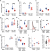
DYRK1A regulates Th17 differentiation. (A) Dose-dependent suppression of Th17 differentiation by the DYRK1A inhibitor (LCTB-21) versus the inactive isomer (iso-LCTB-21). (B) Dose-dependent effect of LCTB-21 on cellular viability in Th17 cultures. (C) Representative flow cytometry analyses showing effect of LCTB-21 on IL-17 and IL-21 expression at the end of Th17 differentiation. (D) Normal thymic subpopulations in CKO versus control mice. (E) Intact development of CD4+ T cell subpopulations in CKO versus control splenocytes. (F) Deletion of Dyrk1a in Dyrk1a CKO mice drives decreased Th17 differentiation in vitro. (G) Dyrk1a expression in naïve CD4+ T cells varies directly with propensity for Th17 differentiation in vitro. (H) Overexpression of Dyrk1a in Dyrk1aBAC (BAC) mice drives increased Th17 differentiation in vitro. (A, B) Error bars show standard deviation. (D, E) Mann-Whitney test with Sidak-Bonferroni correction for multiple comparisons. (F, H) Mann-Whitney test. **P <0.01; ****P < 0.001. Ctrl = control; DN = CD4CD8 double negative; DP = CD4+CD8+ double positive; M = memory; N = naïve; SP = single positive.
Figure 2.
Figure 2.
DYRK1A regulates a subset of the early Th17 program. (A) Mixed CKO and control naïve CD4+ T cells were isolated from Rag1−/− hosts reconstituted with mixed marrow from congenically marked CKO and control mice, then cocultured in pro-Th17 conditions. Four pairs of marrow donors were used, each used to reconstitute 3 hosts. Each line depicts Th17 differentiation in CKO versus control cells from 1 host. (B) Flow cytometric quantitation of RORγt expression in naïve CKO vs control CD4+ T cells stimulated in pro-Th17 conditions for 1 d. (C) Expression of Batf, Mina, and Pou2af1 in naïve CKO vs control CD4+ T cells stimulated in pro-Th17 conditions for 2 h, quantitated by real-time quantitative PCR. (A–C) Mann-Whitney test. *P <0.05; ***P < 0.005. (D) Expression profiling by RNA sequencing was used to identify the early Th17 signature (genes differentially regulated in naïve CD4+ T cells, stimulated in pro-Th17 conditions for 5 h, versus unstimulated). These genes were further queried for dysregulation by co-incubation with harmine (differentially regulated in naïve CD4+ T cells stimulated in pro-Th17 conditions for 5 h, with harmine versus DMSO control). Gene set enrichment analysis was performed on early Th17 signature genes that showed counter-regulation in the presence of harmine. (E) Expression profiling by RNA sequencing of CKO versus control naïve CD4+ T cells, stimulated in pro-Th17 conditions for 5 h. (Left) Standard 1.5-fold change (FC) and false discovery rate (FDR) < 0.05 cutoffs are shown (dotted lines). (Middle) Harmine-counter-regulated Th17 signature genes were queried for concordant directionality of dysregulation in CKO versus control cells; chi-square test was used to calculate the P value compared with the null (equal) distribution. A total of 30 genes were below threshold of detection in the CKO versus control comparison. (Right) Gene set enrichment analysis of the 152 early Th17 signature genes that were concordantly dysregulated by DYRK1A inhibition (harmine) or Dyrk1a deletion (CKO). Ctrl = control; MFI = mean fluorescence intensity; ns = not significant; UV = ultraviolet.
Figure 3.
Figure 3.
DYRK1A regulates Th17 differentiation in part via regulating STAT3S727 phosphorylation. (A–C). Levels of phospho-STAT3Y705 (A), total STAT3 (B), and phospho-STAT3S727 (C) in Dyrk1a CKO versus control naïve CD4+ T cells at baseline (0 min) and after 30-min stimulation in pro-Th17 conditions as measured by phospho-flow cytometry. (D, E) Levels of phospho-STAT3S727 (30 min poststimulation) correlate with levels of phospho-STAT3Y705 (30 min poststimulation) and with Th17 differentiation (4 d poststimulation). Ctrl = control; KO = knockout; MFI = mean fluorescence intensity; ns = not significant.
Figure 4.
Figure 4.
Novel DYRK1A regulation of IL-6 receptor surface levels modulates phospho-STAT response to IL-6. A-D. Levels of phospho-STAT5Y694 (A), phospho-STAT1Y701 (B), total STAT1 (C), and phospho-STAT1S727 (D) in Dyrk1a CKO versus control naïve CD4+ T cells at baseline (0 min) and after 30-min stimulation in pro-Th17 conditions as measured by phospho-flow cytometry. (E) CKO cells show decreased surface levels of gp130 in naïve and memory CD4+ T cells (left). (Right) Representative histograms of gp130 in naïve CD4+ T cells. (F, G) DYRK1A-overexpressing BAC cells show increased surface levels of gp130 in naïve and memory CD4+ T cells (F) and increased phospho-STAT3S727 after stimulation (G) in pro-Th17 conditions versus control cells. (H) Loss of Dyrk1a leads to decreased surface levels of IL-6Ra in naïve CD4+ cells. (I) Real-time quantitative PCR analyses comparing transcription of Il6ra (encodes IL-6R) and Il6st (encodes gp130) in CKO versus control naïve CD4+ T cells. (J) Flow cytometric analysis of phospho-STAT3 in naïve CD4+ T cells from individuals with DS versus control subjects, treated with IL-6 for 30 min. (A–J) Mann-Whitney test (J) with Sidak-Bonferroni correction (A–I) for multiple comparisons. *P < 0.05; **P <0.01; ***P < 0.005; ****P < 0.001. Ctrl = control; GMFI = geometric mean fluorescence intensity; KO = knockout; MFI = mean fluorescence intensity; ns = not significant.

References

    1. Chemin K, Gerstner C, Malmström V.. Effector functions of CD4+ T cells at the site of local autoimmune inflammation—lessons from rheumatoid arthritis. Front Immunol 2019;10:353. - PMC - PubMed
    1. Collier JL, Weiss SA, Pauken KE, Sen DR, Sharpe AH.. Not-so-opposite ends of the spectrum: CD8+ T cell dysfunction across chronic infection, cancer and autoimmunity. Nat Immunol 2021;22:809–819. - PMC - PubMed
    1. Yasuda K, Takeuchi Y, Hirota K.. The pathogenicity of Th17 cells in autoimmune diseases. Semin Immunopathol 2019;41:283–297. - PubMed
    1. Khor B et al. The kinase DYRK1A reciprocally regulates the differentiation of Th17 and regulatory T cells. eLife. 2015;4. - PMC - PubMed
    1. Aranda S, Laguna A, de la Luna S.. DYRK family of protein kinases: evolutionary relationships, biochemical properties, and functional roles. FASEB J. 2011;25:449–462. - PubMed

Publication types

MeSH terms