. 2025 Jan 28;122(4):e2415222122.
doi: 10.1073/pnas.2415222122. Epub 2025 Jan 23.

Molecular glue for phycobilisome attachment to photosystem II in Synechococcus sp. PCC 7002

Affiliations

Molecular glue for phycobilisome attachment to photosystem II in Synechococcus sp. PCC 7002

Zhenggao Zheng et al. Proc Natl Acad Sci U S A. .

Abstract

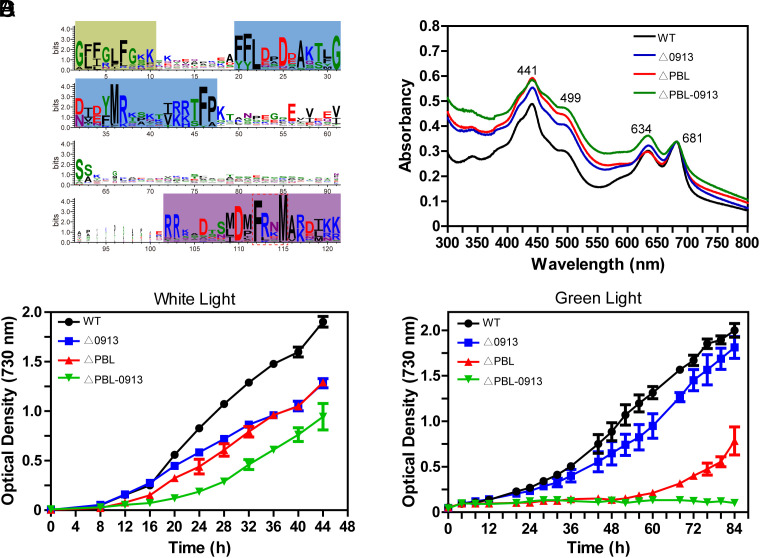
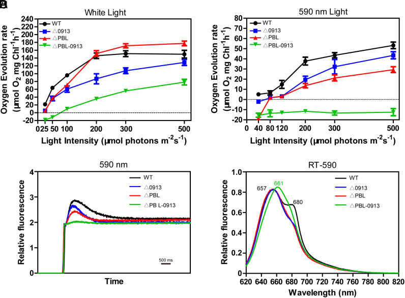
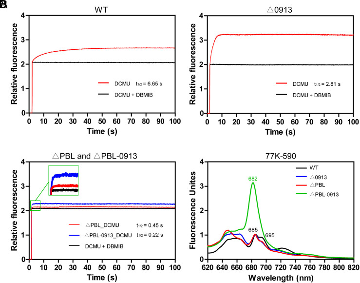
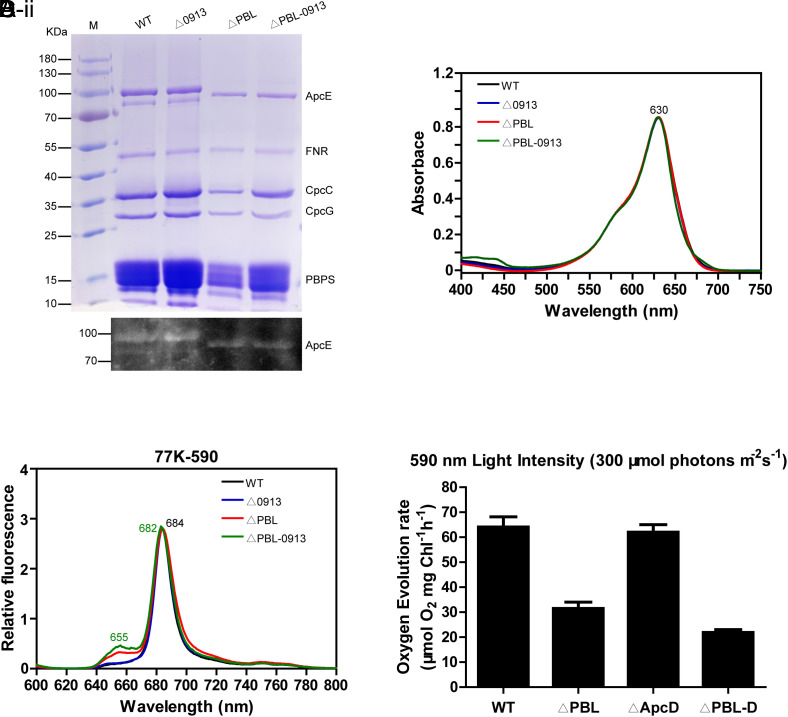
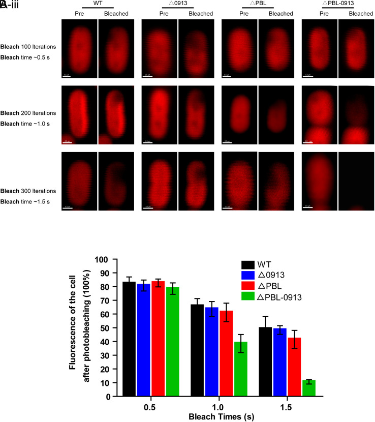
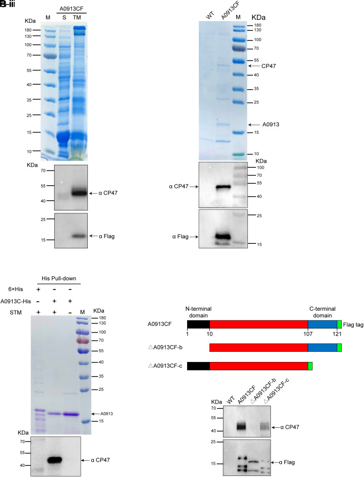
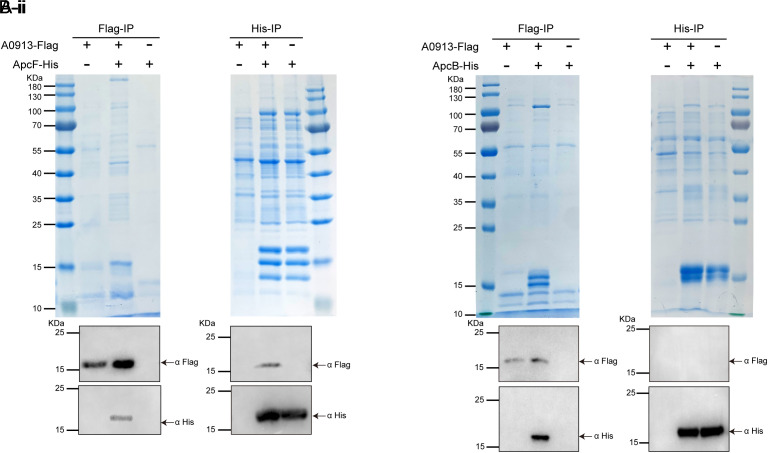
Phycobilisomes (PBS) are the major photosynthetic light-harvesting complexes in cyanobacteria and red algae. While the structures of PBS have been determined in atomic resolutions, how PBS are attached to the reaction centers of photosystems remains less clear. Here, we report that a linker protein (LcpA) is required for the attachment of PBS to photosystem II (PSII) in the cyanobacterium Synechococcus sp. PCC 7002. We also report that the PB-loop of PBS, which is located within the α-APC domain of ApcE, is required for the attachment of PBS to PSII. Deletion of either PB-loop or the gene A0913 led to a decreased rate of photoautotrophic growth under illumination of green light, which is preferentially absorbed by PBS. A double mutant lacking the PB-loop and A0913 (ΔPBL-0913) showed a complete inhibition of O2 evolution under the 590 nm light and could not grow under green light illumination. While assembled PBS could be isolated from ΔPBL-0913, the energy transfer from its PBS to PSII was blocked as measured by fluorescence induction. Photobleaching with intact cells showed that the PBS movement speed in ΔPBL-0913 was 2.5 times as fast as that of the wild type, suggesting that association of its PBS with thylakoids was weakened significantly. The pull-down and coimmunoprecipitation results showed that the LcpA interacts with the CP47 subunit of PSII through its N-terminal region and interacts with ApcB of PBS through its C-terminal α-helix motif. Our results provide insights into the molecular mechanism of PBS-PSII association and shed light on excitation energy transfer from PBS to PSII.

Keywords: cyanobacteria; energy transfer; photosystem II; phycobilisomes.


Conflict of interest statement

Competing interests statement:The authors declare no competing interest.

Figures

Fig. 1.
Fig. 1.
The functions of the gene A0913 of the cyanobacterium Synechococcus 7002. (A) Sequence conservation logo of 83 cyanobacterial A0913 homologs. The numbering refers to residue positions in A0913. The N-terminal conserved region is highlighted in green, the middle β-sheet region in blue, and C-terminal α-helix region in purple. The conserved FxxM motif is highlighted with a dashed box. (B) The absorption spectra of the cell cultures of the WT (black) and the mutant strains Δ0913 (blue), ΔPBL (red), and ΔPBL-0913 (green). Absorption peaks are indicated by their maximum absorption wavelengths (nm). (C and D) Photoautotrophic growth curves of the WT and mutant strains grown under white light (C) and green light (530 nm) (D). In panels C and D, the color representing for the strains is the same as in panel B. Results of BD were the averages of three biological replicates.
Fig. 2.
Fig. 2.
Measurement of the photosynthetic activities of the WT (black) and Δ0913 (blue), ΔPBL (red), and ΔPBL-0913 (green). (A and B) Measurement of oxygen evolution rates of the WT and the three mutant strains under white light (A) and a 590 nm light (B) in different light intensities. (C) Fluorescence induction transients for WT and the three mutant strains. The chlorophyll a fluorescence emission at 685 nm was excited with a 590 nm light at room temperature (RT) and was continuously recorded. Fluorescence kinetics was normalized at F0 Level. (D) RT fluorescence emission spectra (excitation at 590 nm) of the WT and the three mutant strains. Results of AD were the averages of three biological replicates.
Fig. 3.
Fig. 3.
State transitions of the WT and the three mutant strains. Dark incubated cells in the presence of DCMU or DCMU/DBMIB were illuminated with a 590 nm light and fluorescence emission at 695 nm was continuously recorded at RT. (A and B) Fluorescence inductions in the presence of DCMU (red curves) or DCMU/DBMIB (black curves) of the WT (A) and Δ0913 (B). (C) Fluorescence induction curves of ΔPBL (red curve) and ΔPBL-0913 (blue) in the presence of DCMU; and fluorescence induction curve of ΔPBL in the presence of DCMU/DBMIB (black curve). (D) Fluorescence emission spectra (excitation at 590 nm) of the WT (black) and Δ0913 (blue), ΔPBL (red), and ΔPBL-0913 (green) at 77 K. Numbers indicate emission peak wavelengths (nm). Results of AD were the averages of three biological replicates.
Fig. 4.
Fig. 4.
Isolation of PBS and characterization. (A) SDS-PAGE analysis of protein components of the PBS isolated from the WT and the three mutant strains. (A-i) The gel was stained with Coomassie brilliant blue contains. Lanes from Left to Right are molecular mass standard (M), WT, Δ0913, ΔPBL, and ΔPBL-0913. (A-ii) ZnSO4-enhanced fluorescence from the covalently bound bilin chromophore of ApcE protein bands. Protein masses are indicated on the Left and proteins of interest are indicated on the Right. (B) Absorption spectrum of the isolated PBS from WT (black), Δ0913 (blue), ΔPBL (red), and ΔPBL-0913 (green). Numbers indicate absorption peak wavelengths (nm). (C) Fluorescence emission spectra of the PBS excited by a light at 590 nm at 77 K. (D) Measurement of oxygen evolution rates of the WT, ΔPBL, the apcD mutant ΔApcD, and the double mutant of PB-loop and apcD, ΔPBL-D under illumination of a 590 nm light at the intensities indicated. Panel A was a representation of three biological replicates. Results of BD were representative of three biological replicates.
Fig. 5.
Fig. 5.
Photobleaching experiment of the WT and the three mutant strains. (A) Images before (Pre) and after (bleached) photobleaching in defined regions of interest (ROI) of cells are paired in each panel of the strains WT, Δ0913, ΔPBL, and ΔPBL-0913. (A-i, A-ii, and A-iii) Bleaching iteration by 100, 200, and 300 times, corresponding to 0.5, 1.0, and 1.5 sec, respectively. The fluorescence intensity of the cell that remains after photobleaching is quantitated and analyzed in panel B. (B) the percent of the remaining fluorescence intensity of the cells after beached by different number of iterations. The fluorescence intensity before photobleaching in each cell was normalized to 100%. Each bar in panel B was an average of fifteen replicates.
Fig. 6.
Fig. 6.
Interaction of the N-terminal region of A0913 with CP47 of PSII. (A) Cellular localization of A0913. Supernatant proteins (S) and thylakoid membrane proteins (TM) prepared from the strain containing a Flag tagged A0913 protein at its C terminus, A0913CF, were separated by SDS-PAGE and either stained with Coomassie brilliant blue (A-i) or analyzed by immunoblotting with antibodies against CP47 (A-ii) or against the Flag tag (A-iii). (B) Identification of the proteins interacting with A0913 by immunoblotting analysis of the immunoprecipitation eluate. The proteins in gel were either stained with Coomassie brilliant blue (B-i) and the visible bands were identified by mass spectrometry (SI Appendix, Tables S1 and S2), or analyzed by immunoblotting with antibodies against CP47 (B-ii) or against the Flag tag (B-iii). (C) Immunoblotting analysis of His-tag pull-down eluate. Poly-His peptide (6 × His), A0913C-His, and solubilized thylakoid membrane proteins (STM) were added according to the table above the gel. After incubation, His-tag pull-down was performed and the proteins in eluate were separated by SDS-PAGE and immunoblotting with antibodies against CP47. (D) Determination of the site of A0913 protein that interacts with CP47 of PSII. (D-i) Schematic representation of construction of mutant A0913 proteins. The N- and C-terminal regions are shown in black and blue colors, respectively. Flag-tag (green color) is located at the C termini of A0913. (D-ii) Immunoblotting analyses of immunoprecipitation eluate with WT, A0913CF, A0913CF-n, and A0913CF-c. Results of AD were representative of three replicates.
Fig. 7.
Fig. 7.
Interaction of A0913 with allophycocyanin (β-APC) of PBS core. (A-i) Supernatant protein and solubilized thylakoid membrane proteins of A0913CF strain expressing Flag-tagged A0913 (second lane), ApcF-His-A0913CF strain expressing Flag-tagged A0913 and His-tagged ApcF (third lane), and ApcF-His-WT strain expressing His-tagged ApcF (fourth lane) were incubated together with beads for Flag-tag. Flag containing protein and its interacting proteins were purified and analyzed with immunoblotting with antibodies against Flag or against His-tag. (A-ii) The same as in panel A except that beads for His-tag were used for purification before immunoblotting. (B-i and B-ii) The same treatment as in panels (A-i and A-ii) respectively, except that ApcB instead of ApcF was tagged with His-tag. Results of A and B were representative of three replicates.

Similar articles

References

    1. Lewis N. S., Nocera D. G., Powering the planet: Chemical challenges in solar energy utilization. Proc. Natl. Acad. Sci. U.S.A. 103, 15729–15735 (2006). - PMC - PubMed
    1. Martin W. F., Bryant D. A., Beatty J. T., A physiological perspective on the origin and evolution of photosynthesis. FEMS Microbiol. Rev. 42, 205–231 (2018). - PMC - PubMed
    1. Mahir M., Govindjee V., Semenov A., Nadtochenko A., Primary electron transfer processes in photosynthetic reaction centers from oxygenic organisms. Photosynth Res. 125, 51–63 (2015). - PubMed
    1. Bryant D. A., Canniffe D. P., How nature designs light-harvesting antenna systems: Design principles and functional realization in chlorophototrophic prokaryotes. J Phys. B-At Mol. Opt. 51, 033001 (2018).
    1. Lokstein H., Renger G., Götze J. P., Photosynthetic light-harvesting (antenna) complexes-structures and functions. Molecules. 26, 3378 (2021). - PMC - PubMed

MeSH terms

LinkOut - more resources