. 2025 Apr:27:100581.
doi: 10.1016/j.ijpddr.2025.100581. Epub 2025 Jan 18.

Investigation of the threonine metabolism of Echinococcus multilocularis: The threonine dehydrogenase as a potential drug target in alveolar echinococcosis

Affiliations

Investigation of the threonine metabolism of Echinococcus multilocularis: The threonine dehydrogenase as a potential drug target in alveolar echinococcosis

Marc Kaethner et al. Int J Parasitol Drugs Drug Resist. 2025 Apr.

Abstract

Alveolar echinococcosis (AE) is a severe zoonotic disease caused by the metacestode stage of the fox tapeworm Echinococcus multilocularis. We recently showed that E. multilocularis metacestode vesicles scavenge large amounts of L-threonine from the culture medium. This motivated us to study the effect of L-threonine on the parasite and how it is metabolized. We established a novel metacestode vesicle growth assay with an automated readout, which showed that L-threonine treatment led to significantly increased parasite growth. In addition, L-threonine increased the formation of novel metacestode vesicles from primary parasite cell cultures in contrast to the non-proteinogenic threonine analog 3-hydroxynorvaline. Tracing of [U-13C]-L-threonine and metabolites in metacestode vesicles and culture medium resulted in the detection of [U-13C]-labeling in aminoacetone and glycine, indicating that L-threonine was metabolized by threonine dehydrogenase (TDH). EmTDH-mediated threonine metabolism in the E. multilocularis metacestode stage was further confirmed by quantitative real-time PCR, which demonstrated high expression of emtdh in in vitro cultured metacestode vesicles and also in metacestode samples obtained from infected animals. EmTDH was enzymatically active in metacestode vesicle extracts. The compounds disulfiram, myricetin, quercetin, sanguinarine, and seven quinazoline carboxamides were evaluated for their ability to inhibit recombinantly expressed EmTDH. The most potent inhibitors, albeit not very strong or highly specific, were disulfiram, myricetin and sanguinarine. These compounds were subsequently tested for activity against E. multilocularis metacestode vesicles and primary parasite cells and only sanguinarine demonstrated significant in vitro activity. However, TDH is not its only cellular target, and it is also known to be highly toxic. Our findings suggest that additional targets of sanguinarine should be explored, and that it may serve as a foundation for developing more specific compounds against the parasite. Moreover, the EmTDH assay could be a valuable high-throughput, target-based platform for discovering novel anti-echinococcal compounds.

Keywords: Cestode; Disulfiram; Echinococcus multilocularis; Sanguinarine; Target-based screening; Threonine metabolism.


Figures

Image 1
Graphical abstract
Fig. 1
Fig. 1
Pathways of threonine catabolism in the non-parasitic helminth C. elegans. L-Thr can be used as substrate by the three different enzymes threonine deaminase (TD), threonine dehydrogenase (TDH) and threonine aldolase (TA) (Yilmaz and Walhout, 2016). Upon metabolization of L-Thr by TD, α-ketobutyrate and ammonia are generated. TDH metabolizes L-Thr to 2-amino-3-ketobutyrate, which is further metabolized by the 2-amino-3-ketobutyrate coenzyme A ligase (KBL) to glycine and acetyl-coenzyme A. TA generates glycine and acetaldehyde upon degradation of L-Thr. Enzymes are depicted in light grey and metabolites in dark grey. Reactions are represented by arrows (enzymatic) or dashed arrows (non-enzymatic).
Fig. 2
Fig. 2
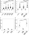
L-Thr stimulates E. multilocularis metacestode vesicle growth and development in vitro. A, E. multilocularis metacestode vesicles were cultured with various concentrations of L-Thr (1 mM, n = 24; 2 mM, n = 23; 4 mM, n = 23), D-Thr (4 mM, n = 23), or water as a control (n = 24). B, E. multilocularis metacestode vesicles were cultured in the presence of 3-HNV (4 mM, n = 21), a combination of 3-HNV and L-Thr (each at 4 mM, n = 23), or water (n = 24). Metacestode vesicle diameters in A and B were measured via an automated script and are given in mm at weeks 0 (grey) and 6 (black). Bonferroni-corrected p-values are displayed as compared to the water control or 4 mM 3-HNV. C, E. multilocularis GL cell cultures were supplemented with L-Thr (4 mM, n = 4), D-Thr (4 mM, n = 4), or water as a control (n = 4). D, E. multilocularis GL cells were cultured with 3-HNV (4 mM, n = 4), a combination of 3-HNV and L-Thr (both at 4 mM, n = 4), or water as a control (n = 4). Newly formed metacestode vesicles in C and D were counted manually in a blinded manner and the mean values, SD and Bonferroni-corrected p-values are shown. Abbreviations: Thr = threonine, 3-HNV = 3-hydroxynorvaline. Two independent experiments were performed for each setup, and the results of one representative experiment is shown, while the results of the second experiment are provided in S3 Fig.
Fig. 3
Fig. 3
Results of the [U-13C]-L-Thr flux assay.E. multilocularis metacestode vesicles were cultured with 5 mM [U-13C]-L-Thr or unlabeled L-Thr for 24 h at 37 °C under a humid, microaerobic atmosphere (n = 4). Shown are graphs with for the metabolites L-Thr, glycine and aminoacetone in vesicle medium (VM) via LC-MS. Raw area under the curve (AUC) values and fractional enrichment for each isotopologue are shown in S3 Table.
Fig. 4
Fig. 4
Functional TDH assays with E. multilocularis metacestode vesicle tissue crude extracts and recombinant EmTDH. Enzymatic assays were performed by measuring the increase in NADH formation via the increase in absorbance at 340 nm at 37 °C in technical triplicates. A, E. multilocularis metacestode vesicle tissue crude extract (80 μg protein per well) with 10 mM NAD+ and varying concentrations of L-Thr or D-Thr (0, 1, 2, 4, 8 and 16 mM). B, recEmTDH (0.05 μg protein per well) was tested with 10 mM NAD+ in combination with various amino acids (3-HNV, D-threonine (D-thr), glycine (gly), L-alanine (L-Ala), L-cysteine (L-Cys), L-serine (L-Ser), L-threonine (L-Thr)) and L-Thr with NADP+. C, recEmTDH (0.05 μg protein per well) was tested with varying concentrations of NAD+ (0, 0.125, 0.25, 0.5, 1, 2, 4 and 8 mM) and 16 mM L-Thr. Grey line shows the fitted Michaelis-Menten curve. D, recEmTDH (0.05 μg protein per well) was tested with varying concentrations of L-Thr (0, 0.25, 0.5, 1, 2, 4, 8 and 16 mM) and 2 mM NAD+. Grey line shows the fitted Michaelis-Menten equation.
Fig. 5
Fig. 5
Effect of various potential TDH inhibitors on recEmTDH and recMmTDH and structures of potential TDH inhibitors. TDH activity was assessed as in Fig. 4. Compounds were tested at 20 μM and shown are mean values and SDs of three technical replicates. Shown are relative activities of recEmTDH (A) or recMmTDH (B) treated with different compounds at 20 μM compared to their respective DMSO control with Bonferroni-corrected p-values. C, structures of potential TDH inhibitors tested against recEmTDH and recMmTDH in this study. The synthesis for QC1 to QC7 is described in S1 File.
Fig. 6
Fig. 6
Effect of recEmTDH inhibitors on E. multilocularis metacestode vesicles and GL cell cultures. A, E. multilocularis metacestode vesicles were incubated with the negative control 0.1% DMSO, the positive control 0.1% Triton X-100, or the three recEmTDH inhibitors disulfiram, myricetin and sanguinarine at 20 μM (n = 3 for each condition). Metacestode vesicles were incubated under a humid, microaerobic atmosphere and after five days, pictures were taken and damage marker release was assessed relative to Triton X-100 treatment. Shown are photos of metacestode vesicles upon treatment (scale bars = 2 mm), as well as PGI assay results with individual values (empty circles), mean values (filled circles), SDs and Bonferroni-corrected p-values of one representative experiment. The second independent experiment is shown in S6 Fig. B, E. multilocularis GL cell cultures were incubated with recEmTDH inhibitors disulfiram, myricetin and sanguinarine at 20 μM, the negative control 0.2% DMSO and the positive control 0.1% Triton X-100, (n = 4 for each condition). GL cell cultures were incubated under a humid, microaerobic atmosphere. After five days, pictures were taken (scale bars = 200 μm), and cell viability was measured and calculated relative to DMSO. Shown are individual values (empty circles) mean values (filled circles), SDs and Bonferroni-corrected p-values of one representative experiment. The other experiment is shown in S6 Fig.
figs1
figs1
figs2
figs2
figs3
figs3
figs4
figs4
figs5
figs5
figs6
figs6

Similar articles

Cited by

References

    1. Adjogatse E., Erskine P., Wells S.A., Kelly J.M., Wilden J.D., Chan A.W.E., Selwood D., Coker A., Wood S., Cooper J.B. Structure and function of L-threonine-3-dehydrogenase from the parasitic protozoan Trypanosoma brucei revealed by X-ray crystallography and geometric simulations. Acta Crystallogr. Sect. Struct. Biol. 2018;74:861–876. doi: 10.1107/S2059798318009208. - DOI - PubMed
    1. Adjogatse E.K. Structure-based drug design for the discovery of new treatments for trypanosomiasis. Dr. Thesis UCL Univ. Coll. Lond. UCL (University College London) 2015 https://discovery.ucl.ac.uk/id/eprint/1467152/
    1. Alexander P.B., Wang J., McKnight S.L. Targeted killing of a mammalian cell based upon its specialized metabolic state. Proc. Natl. Acad. Sci. U.S.A. 2011;108:15828–15833. doi: 10.1073/pnas.1111312108. - DOI - PMC - PubMed
    1. Babich H., Zuckerbraun H.L., Barber I.B., Babich S.B., Borenfreund E. Cytotoxicity of sanguinarine chloride to cultured human cells from oral tissue. Pharmacol. Toxicol. 1996;78:397–403. doi: 10.1111/j.1600-0773.1996.tb00225.x. - DOI - PubMed
    1. Balestrini L., Di Donfrancesco A., Rossi L., Marracci S., Isolani M.E., Bianucci A.M., Batistoni R. The natural compound sanguinarine perturbs the regenerative capabilities of planarians. Int. J. Dev. Biol. 2017;61:43–52. doi: 10.1387/ijdb.160169rb. - DOI - PubMed

Publication types

MeSH terms

Supplementary concepts