. 2025 Feb:112:105566.
doi: 10.1016/j.ebiom.2025.105566. Epub 2025 Jan 22.

Enhancing the efficacy of near-infrared photoimmunotherapy through intratumoural delivery of CD44-targeting antibody-photoabsorber conjugates

Affiliations

Enhancing the efficacy of near-infrared photoimmunotherapy through intratumoural delivery of CD44-targeting antibody-photoabsorber conjugates

Yuichi Adachi et al. EBioMedicine. 2025 Feb.

Abstract

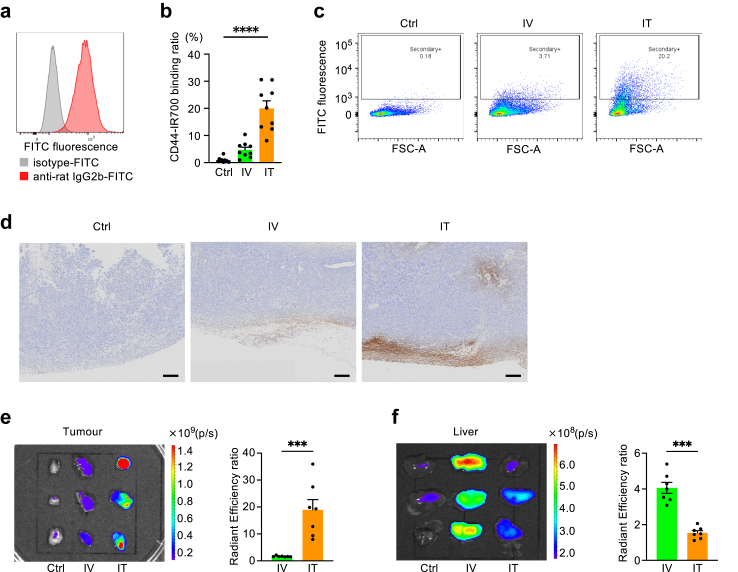
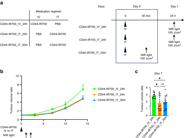
Background: Photoimmunotherapy (PIT) is a potent modality for cancer treatment. The conventional PIT regimen involves the systemic delivery of an antibody-photoabsorber conjugate, followed by a 24-h waiting period to ensure adequate localisation on the target cells. Subsequent exposure to near-infrared (NIR) light selectively damages the target cells. We aimed to improve the efficacy of PIT in vivo by evaluating the effects of the different routes of conjugate administration on treatment outcomes.

Methods: Subcutaneous Lewis lung carcinoma tumours were established in mice, targeting cluster of differentiation (CD)44 with an anti-CD44 antibody conjugated to IRDye700DX (IR700). The conjugate was administered via the intravenous or intratumoural route followed by the assessment of antibody binding and therapeutic effects of PIT.

Findings: Compared to intravenous administration, intratumoural delivery of the CD44-IR700 conjugate significantly increased the number of cells binding to the conjugate by >five-fold. This method, combined with NIR light irradiation, halved tumour growth when compared to intravenous delivery. Reducing the interval between intratumoural injection and NIR light exposure to 30 min did not diminish efficacy, thereby demonstrating the feasibility of a 1-h procedure.

Interpretation: Intratumoural administration of the antibody-photoabsorber conjugate enhanced the efficacy of PIT in vivo. A simplified, 1-h procedure involving conjugate tumour injection followed by irradiation emerged as a potent cancer treatment strategy.

Funding: This study was supported by the Japan Society for the Promotion of Science, the Japan Agency for Medical Research and Development, Japan Science and Technology Agency, and the Osaka Medical Research Foundation for Intractable Diseases.

Keywords: Antibody–photoabsorber conjugate; Intratumoural administration; Lung cancer; Photoimmunotherapy.


Conflict of interest statement

Declaration of interests The authors declare that they have no known competing financial interests or personal relationships that could have appeared to influence the work reported in this paper.

Figures

Fig. 1
Fig. 1
CD44-IR700 conjugate generation and in vitro effects of NIR light photoimmunotherapy using CD44-IR700. (a) Generation of the CD44-IR700 conjugate as confirmed by sodium dodecyl sulphate polyacrylamide gel electrophoresis. The left panel indicates Coomassie blue staining for protein detection and the right panel displays fluorescence at 700 nm. (b) Flow cytometry analysis demonstrating CD44-IR700 binding to Lewis lung carcinoma (LLC) cells. The panel shows LLC cells after incubation with CD44-IR700. In the blocking experiment, cells were pre-incubated with an excess amount of unconjugated anti-CD44 antibody. (c) Assessment of cell viability using a colourimetric assay following photoimmunotherapy with CD44-IR700 in LLC cells. Cells were cultured in a medium with or without CD44-IR700 and subsequently exposed to NIR light. Viability was measured and normalised against cells grown in a medium without CD44-IR700 and not exposed to NIR light (n = 4 for each bar). (d) Evaluation of luciferase activity post-photoimmunotherapy in LLC-luc cells treated with CD44-IR700. LLC-luc cells were cultured with or without CD44-IR700 and subjected to NIR light exposure, after which luciferase activity was quantified (n = 4 for each bar). CD44-IR700, IR700-conjugated anti-CD44 monoclonal antibody; CD, cluster of differentiation; and NIR, near-infrared.
Fig. 2
Fig. 2
Intratumoural administration enhances the delivery of CD44-IR700 to tumours. (a) Flow cytometry analysis demonstrating CD44-IR700 binding to Lewis lung carcinoma cells in vitro. The cells were visualised using a secondary antibody against CD44-IR700 (anti-rat IgG2b-FITC). (b) Quantification of CD44-IR700 binding to allograft tumour cells for each treatment group: Ctrl, IV (100 μg), and IT (100 μg) CD44-IR700 administration (n = 9 per group). CD44-IR700 binding to isolated tumour cells was detected using the secondary antibody. (c) Representative flow cytometry images from figure (b) illustrating the binding of CD44-IR700 to cells in the allograft tumour. Treatments were administered as described in (b). (d) Representative immunohistochemical staining of CD44-IR700 at 40 × magnification. Subcutaneous tumours were induced in mice, followed by treatment in each group: Ctrl, IV (50 μg), and IT (50 μg) CD44-IR700 administration. CD44-IR700 was visualised using a secondary antibody. Scale bar, 200 μm. (e and f) Biodistribution of CD44-IR700 in tumour (e) and liver (f), visualised using IVIS imaging system the day after administration of CD44-IR700. Treatments were administered as described in (d). The bar graphs on the right shows the fluorescence values of the intravenous and intratumoural administration groups compared to the Ctrl group (n = 7 per group). Data are presented as mean + standard error of the mean. Statistical difference was evaluated using Mann–Whitney U tests for comparisons between two groups and the Kruskal–Wallis test for comparisons between three groups, with asterisks denoting the levels of significance (∗∗∗p < 0.001 and ∗∗∗∗p < 0.0001). CD44-IR700, IR700-conjugated anti-CD44 monoclonal antibody; FITC, fluorescein isothiocyanate; IgG, immunoglobulin G; FSC-A, forward scatter area; Ctrl, control; IV, intravenous; and IT, intratumoural.
Fig. 3
Fig. 3
Intratumoural delivery of CD44-IR700 enhanced efficacy of photoimmunotherapy in vivo. (a) Schematic of experimental schedule. Mice bearing subcutaneous Lewis lung carcinoma tumours were treated with either no CD44-IR700 (Ctrl), CD44-IR700 via IV injection (CD44-IR700_IV), or CD44-IR700 via IT injection (CD44-IR700_IT), followed by NIR light irradiation on subsequent days. (b) The ratio of tumour-infiltrating CD8-positive T cells in live cells, as determined by flow cytometry on day 7 post-treatment, from the CD44-IR700_IV and CD44-IR700_IT groups (n = 6 per group). (c) Immunohistochemical staining of tumours 7 days after photoimmunotherapy. CD8-positive T cells were stained. The scale bar represents 50 μm. The numbers of CD8-positive T cells per view field were counted (n = 5 mice for each group). (d) Cytokine expression in tumours, as determined by quantitative PCR analysis on day 3 post-treatment with photoimmunotherapy, from the CD44-IR700_IV and CD44-IR700_IT groups (n = 5 per group). (e) Tumour volume monitoring. Tumour size was measured starting from the day of CD44-IR700 administration (day 0), and the change in size was expressed as a ratio relative to the initial volume on day 0 (n = 12 per group). (f) Tumour volume ratio on day 7. The graph shows the fold increase in tumour volume on day 7 compared with that on day 0 (n = 12 per group). (g) Representative images displaying tumours from each treatment group on day 7 post-administration. (h) Representative images of tumours excised on day 7 post-treatment from each therapeutic group. Data are presented as mean + standard error of the mean. Statistical difference was assessed using Mann–Whitney U tests, with significance denoted by asterisks (∗p < 0.05 and ∗∗∗∗p < 0.0001). CD44-IR700, IR700-conjugated anti-CD44 monoclonal antibody; CD, cluster of differentiation; NIR, near-infrared; Ctrl, control; IV, intravenous; IT, intratumoural; Ifnγ, interferon-γ; and Tnf, tumour necrosis factor-alpha.
Fig. 4
Fig. 4
The interval between intratumoural administration of CD44-IR700 and NIR irradiation, whether 30 min or 24 h, yields the same effect. (a) Schematic of experimental schedule. Lewis Lung carcinoma cell-induced subcutaneous tumours were established in mice. The mice were divided into three groups for the following treatments: CD44-IR700_IV_24h: IV administration of CD44-IR700 and IT of PBS, with NIR light irradiation 24 h after administration; CD44-IR700_IT_24h: IT administration of CD44-IR700 and IV injection of PBS, followed by NIR light irradiation 24 h later; and CD44-IR700_IT_30m: IT administration of CD44-IR700 and IV injection of PBS, with NIR light irradiation conducted 30 min post-administration. (b) Tumour volume measurement. The tumour volume was monitored from the day of CD44-IR700 administration (designated as day 0), and the change in tumour size was presented as a ratio to the volume on the day of administration (n = 14–18 per group). (c) Tumour volume ratio on day 7 relative to day 0 (n = 14–18 per group). Data are expressed as mean + standard error of the mean. Statistical difference was assessed using Kruskal–Wallis test, with asterisks denoting the levels of significance (∗p < 0.05). ‘ns’ indicates a lack of statistical significance. CD44-IR700, IR700-conjugated anti-CD44 monoclonal antibody; PBS, phosphate-buffered saline; CD, cluster of differentiation; NIR, near-infrared; Ctrl, control; IV, intravenous; and IT, intratumoural.

References

    1. Mitsunaga M., Ogawa M., Kosaka N., Rosenblum L.T., Choyke P.L., Kobayashi H. Cancer cell-selective in vivo near infrared photoimmunotherapy targeting specific membrane molecules. Nat Med. 2011;17:1685–1691. - PMC - PubMed
    1. Watanabe R., Hanaoka H., Sato K., et al. Photoimmunotherapy targeting prostate-specific membrane antigen: are antibody fragments as effective as antibodies? J Nucl Med. 2015;56:140–144. - PMC - PubMed
    1. Cognetti D.M., Johnson J.M., Curry J.M., et al. Phase 1/2a, open-label, multicenter study of RM-1929 photoimmunotherapy in patients with locoregional, recurrent head and neck squamous cell carcinoma. Head Neck. 2021;43:3875–3887. - PMC - PubMed
    1. Sato K., Choyke P.L., Hisataka K. Selective cell elimination from mixed 3D culture using a near infrared photoimmunotherapy technique. J Vis Exp. 2016 doi: 10.3791/53633. - DOI - PMC - PubMed
    1. Isobe Y., Sato K., Nishinaga Y., et al. Near infrared photoimmunotherapy targeting DLL3 for small cell lung cancer. eBioMedicine. 2020;52 - PMC - PubMed

LinkOut - more resources