Review
. 2025 Jan 23;13(1):15.
doi: 10.1038/s41413-024-00397-7.

Role of macrophage in intervertebral disc degeneration

Affiliations
Review

Role of macrophage in intervertebral disc degeneration

Yiming Dou et al. Bone Res. .

Abstract

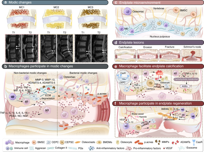
Intervertebral disc degeneration is a degenerative disease where inflammation and immune responses play significant roles. Macrophages, as key immune cells, critically regulate inflammation through polarization into different phenotypes. In recent years, the role of macrophages in inflammation-related degenerative diseases, such as intervertebral disc degeneration, has been increasingly recognized. Macrophages construct the inflammatory microenvironment of the intervertebral disc and are involved in regulating intervertebral disc cell activities, extracellular matrix metabolism, intervertebral disc vascularization, and innervation, profoundly influencing the progression of disc degeneration. To gain a deeper understanding of the inflammatory microenvironment of intervertebral disc degeneration, this review will summarize the role of macrophages in the pathological process of intervertebral disc degeneration, analyze the regulatory mechanisms involving macrophages, and review therapeutic strategies targeting macrophage modulation for the treatment of intervertebral disc degeneration. These insights will be valuable for the treatment and research directions of intervertebral disc degeneration.


Conflict of interest statement

Competing interests: The authors declare no competing interests.

Figures

Fig. 1
Fig. 1
Schematic diagram of healthy IVD, degenerated IVD, and inflammatory pathological microenvironment. a Overview of the IVD. The IVD consists of three parts: the NP, the AF, and the CEP. b Cells and ECM in healthy IVD. In healthy IVD, the NP mainly consists of notochordal cells and nucleus pulposus cells. The AF primarily consists of annulus fibrosus cells. The CEP mainly consists of chondrocytes. c Mechanical function of the IVD. The primary function of the IVD is to provide mechanical support for the vertebrae and allow movement of the spine (flexion, extension, and rotation). d IVDD and IVD herniation. Severe IVDD leads to NP degeneration, AF rupture, and endplate calcification, resulting in IVD herniation that compresses the spinal cord and nerve roots. e Change of cells and ECM in degenerated IVD. In degenerated IVD, notochordal cells disappear, and due to cell senescence and PCD, the number of IVD cells significantly decreases, and their function markedly declines. f Macrophage infiltration and polarization. Under the induction of chemokines, macrophages infiltrate the IVD via neovascularization. Macrophages can polarize into M1 and M2 subtypes under different stimuli
Fig. 2
Fig. 2
Role of macrophages in IVDD. a Macrophages amplify inflammation in the IVD. Macrophages upregulate the expression of pro-inflammatory cytokines, creating an inflammatory microenvironment and causing a series of effects on cellular activities and ECM metabolism. b Macrophage-related cytokines regulate PCDs of IVD cells, causing a significant loss of IVD cells. c Macrophages accelerate the senescence of IVD cells. d Macrophages regulate the proliferation of IVD cells. e Macrophages regulate ECM metabolism. Macrophages phagocytize disc fragments and regulate ECM metabolism by upregulating the expression of MMPs and ADAMTs. This activity can lead to the resorption of herinated NP or exacerbate the progression of IVDD. f Macrophages regulate discogenic LBP. Macrophages can promote nerve growth by upregulating NGF, increasing IVD sensitization, and causing discogenic LBP through inflammatory factors. Macrophages can also promote the formation of new blood vessels in the IVD by upregulating VEGF expression, bringing more immune cells and exacerbating the immune response
Fig. 3
Fig. 3
Role of macrophages in endplate degeneration. a Classification and MRI characteristics of Modic changes. MC1 (low T1 and high T2 signals) is associated with edema and inflammation. MC2 (high T1 and T2 signals) indicates a progression to fatty involution. MC3 (low T1 and T2 signals) signify vertebral endplate bone sclerosis. b Macrophages participate in MCs. In non-bacterial MCs, macrophages accelerate endplate degeneration by promoting the expressions of pro-inflammatory cytokines, MMPs, and ADAMTs. In bacterial MCs, a wider variety of tissue-derived macrophages, including peripheral blood macrophages and osteal macrophages, contribute to endplate inflammation, leading to more severe endplate degeneration. c Schematic of a healthy endplate microenvironment. CEPCs synthesize an extracellular matrix primarily composed of collagen II and ACAN. CEPSCs are stem cells for intrinsic repair of the endplate. d Schematic of endplate lesions. Endplate lesions includes calcification, erosion, fractures, and Schmorl’s nodes. e Macrophages promote endplate calcification. Macrophages enhance CASR expression in CEPCs and regulate carbohydrate metabolism to increase AGEs expression by releasing pro-inflammatory cytokines, thereby promoting calcification. Additionally, macrophages promote calcification by increasing ROS production, which induces osteogenic differentiation of CEPSCs. f Macrophages in endplate regeneration. Macrophages promote neovascularization of the endplate by releasing VEGF. Macrophages participate in bone remodeling by regulating osteoclast formation and bone resorption activity

References

    1. Disease, G. B. D., Injury, I. & Prevalence, C. Global, regional, and national incidence, prevalence, and years lived with disability for 354 diseases and injuries for 195 countries and territories, 1990-2017: a systematic analysis for the Global Burden of Disease Study 2017. Lancet392, 1789–1858 (2018). - PMC - PubMed
    1. Yang, S., Zhang, F., Ma, J. & Ding, W. Intervertebral disc ageing and degeneration: The antiapoptotic effect of oestrogen. Ageing Res. Rev.57, 100978 (2020). - PubMed
    1. Lawson, L. Y. & Harfe, B. D. Developmental mechanisms of intervertebral disc and vertebral column formation. Wiley Interdiscip. Rev. Dev. Biol.6, e283 (2017). - PubMed
    1. Dou, Y., Sun, X., Ma, X., Zhao, X. & Yang, Q. Intervertebral disk degeneration: the microenvironment and tissue engineering strategies. Front. Bioeng. Biotechnol.9, 592118 (2021). - PMC - PubMed
    1. Hunter, C. J., Matyas, J. R. & Duncan, N. A. The notochordal cell in the nucleus pulposus: a review in the context of tissue engineering. Tissue Eng.9, 667–677 (2003). - PubMed

MeSH terms

LinkOut - more resources