. 2025 Jan 23;23(1):41.
doi: 10.1186/s12951-025-03096-5.

Folic acid-modified ginger-derived extracellular vesicles for targeted treatment of rheumatoid arthritis by remodeling immune microenvironment via the PI3K-AKT pathway

Affiliations

Folic acid-modified ginger-derived extracellular vesicles for targeted treatment of rheumatoid arthritis by remodeling immune microenvironment via the PI3K-AKT pathway

Ruina Han et al. J Nanobiotechnology. .

Abstract

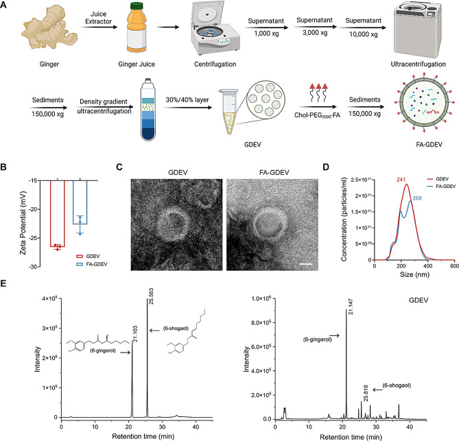
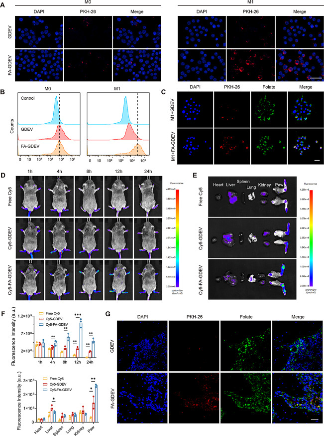
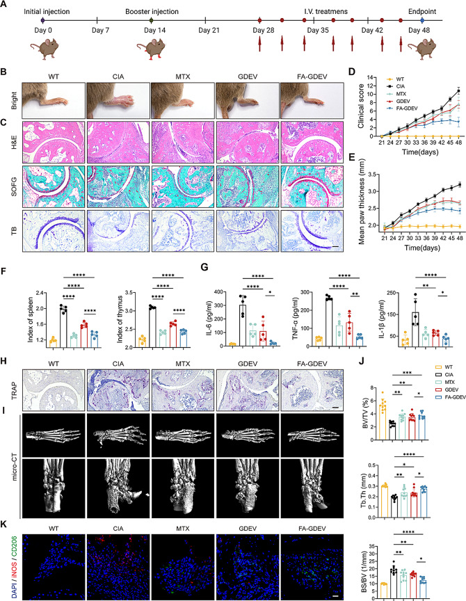
Rheumatoid arthritis (RA), a form of autoimmune inflammation, is marked by enduring synovial inflammation and the subsequent impairment of joint function. Despite the availability of conventional treatments, they are often marred by significant side effects and the associated high costs. Plant-derived extracellular vesicles (PEVs) offer a compelling alternative, owing to their abundant availability, affordability, low immunogenicity, high biocompatibility, and feasibility for large-scale production. These vesicles enhance intercellular communication by transferring intrinsic bioactive molecules. In our research, we delve into the capacity of PEVs to treat RA, highlighting the role of ginger-derived extracellular vesicles (GDEVs). By conjugating GDEVs with folic acid (FA), we have developed FA-GDEVs that maintain their inherent immunomodulatory properties. FA-GDEVs are designed to selectively target M1 macrophages in inflamed joints via the folate receptors (FRs). Our in vitro findings indicate that FA-GDEVs promote the polarization towards a reparative M2 macrophage phenotype by modulating the PI3K-AKT pathway. Further corroboration comes from in vivo studies, which demonstrate that FA-GDEVs not only concentrate efficiently in the affected joints but also markedly reduce the manifestations of RA. Synthesizing these findings, it is evident that FA-GDEVs emerge as a hopeful candidate for RA treatment, offering benefits such as safety, affordability, and therapeutic efficacy.

Keywords: Folate receptor targeting; Immune microenvironment; PI3K-AKT pathway; Plant-derived extracellular vesicles; Rheumatoid arthritis.


Conflict of interest statement

Declarations. Ethics approval and consent to participate: All animal procedures were performed in accordance with the Guidelines for Care and Use of Laboratory Animals of Shanghai University and approved by the Animal Ethics Committee of Shanghai University. Competing interests: The authors declare no competing interests.

Figures

Fig. 1
Fig. 1
Isolation and characterization of GDEVs and FA-GDEVs. (A) Schematic diagram of GDEVs extracted from ginger and preparation of FA-GDEVs by coupling FA-PEG2000-Chol with GDEV. (B) Zeta potential of GDEVs and FA-GDEVs. (C) Representative TEM images of GDEVs and FA-GDEVs. Scale bar = 100 nm. (D) Size distribution and number of particles per unit volume of GDEVs and FA-GDEVs measured by NTA. (E) Confirmation of 6-Gingerol and 6-Shogaol in GDEVs by HPLC. Error bars represent the SD (n = 3)
Fig. 2
Fig. 2
In vitro and in vivo targeted delivery of FA-GDEVs. (A) Cellular uptake behavior of GDEVs and FA-GDEVs in BMDMs. (B) Flow cytometry was used to measure the fluorescence intensity of PKH26-stained GDEVs and FA-GDEVs within BMDMs (C) Fluorescence microscopy showing co-localization of nuclei (blue), PKH26-labeled GDEVs and FA-GDEVs (red) with FRs (green). (D) Full-body fluorescence imaging of Free-Cy5, Cy5-stained GDEVs and FA-GDEVs in CIA mouse models. (E) Images of ex vivo organ distribution of Free-Cy5, Cy5-stained GDEVs and FA-GDEVs. (F) Quantitative analysis of the fluorescence intensity and its temporal changes in the whole body of CIA mice injected with Cy5, GDEVs and FA-GDEVs, as well as the fluorescence intensity in the paws and major organs. (G) Co-localization of GDEVs and FA-GDEVs with FRs in the synovium of inflamed joints in CIA mice. Scale bar = 20 μm. Error bars represent the SD (n = 3). **P < 0.01, and ***P < 0.005
Fig. 3
Fig. 3
In vitro macrophage polarization by FA-GDEVs. (A) Immunofluorescence staining of different groups of treated BMDMs and LPS + IFN-γ activated BMDMs after 48 h. Fluorescence microscopy showed nuclei (blue), iNOS (red) and CD206 (green). (B and C) The ratio (B) and quantification (C) of M1 and M2 markers was analyzed by flow cytometry. (D) Protein levels of iNOS and CD206. (E) qRT-PCR analysis of the expression levels of M1 and M2 macrophage marker genes. (F) Immunofluorescence staining of BMDMs (IL-4 + IL-13) after 48 h of treatment with different groups in the presence of LPS + IFN-γ. Fluorescence microscopy showed nuclei (blue), iNOS (red) and CD206 (green). Scale bar = 20 μm. Error bars represent the SD (n = 3). *P < 0.05, ***P < 0.005, and ****P < 0.001
Fig. 4
Fig. 4
MicroRNA profiling of FA-GDEVs. (A and B) Analysis of GO (A) and KEGG (B) The terms that were significantly enriched in the predicted target genes of both GDEVs and FA-GDEVs are presented. (C and D) WB results of p-PI3K, PI3K, p-AKT, AKT. Additionally, (C) shows the levels of iNOS and CD206, while (D) presents the levels of other proteins, in LPS and IFN-γ stimulated BMDMs that were treated with different experimental groups. (E) Immunofluorescence staining of LPS + IFN-γ activated BMDMs after 48 h of treatment in different groups. Fluorescence microscopy showed nuclei (blue), iNOS (red) and CD206 (green). Scale bar = 20 μm
Fig. 5
Fig. 5
In vivo therapeutic efficacy of FA-GDEVs in CIA mice. (A) Schematic of RA treatment protocol. (B) Representative images of the hind limbs at the end of the experiment in mice of different treatment groups. (C) Images of H&E staining, SOFG staining and TB staining of the ankle joints. (D and E) Arthritis clinical visual scores (D) and paw thickness measurements (E) were recorded on mice every three days. (F) Indexes of spleen and thymus in different treatment groups of mice. (G) Levels of pro-inflammatory cytokines in serum were measured by ELISA. (H) Levels of TRAP-stained osteoclasts in the ankle joints of mice. (I) Representative micro-CT images of the posterior lateral aspect of the ankle joints of mice. (J) Quantitative micro-CT analysis of BV/TV, BS/BV and Tb.Th of the ankle joints. (K) Immunofluorescence staining of the ankle joints. Fluorescence microscopy showed nuclei (blue), iNOS (red) and CD206 (green). Scale bar = 20 μm. Error bars represent the SD (n = 5). *P < 0.05, **P < 0.01, ***P < 0.005, and ****P < 0.001
Fig. 6
Fig. 6
In vivo toxicity assay of FA-GDEVs. (A) H&E staining images of major organs from each treatment group. (B) Hemolysis rates and representative images of each treatment group. (C) Changes in body weight in each treatment group. Scale bar = 50 μm. Error bars represent the SD (n = 5)

References

    1. Tobón GJ, Youinou P, Saraux A. The environment, geo-epidemiology, and autoimmune disease: rheumatoid arthritis. Autoimmun Rev. 2010;9:A288–292. - PubMed
    1. Smolen JS, Aletaha D, Barton A, Burmester GR, Emery P, Firestein GS, Kavanaugh A, McInnes IB, Solomon DH, Strand V, Yamamoto K. Rheumatoid arthritis. Nat Rev Dis Primers. 2018;4:18001. - PubMed
    1. Smolen JS, Aletaha D, McInnes IB. Rheumatoid arthritis. Lancet. 2016;388:2023–38. - PubMed
    1. Sparks JA. Rheumatoid arthritis. Ann Intern Med. 2019;170:Itc1–16. - PubMed
    1. Wynn TA, Chawla A, Pollard JW. Macrophage biology in development, homeostasis and disease. Nature. 2013;496:445–55. - PMC - PubMed

MeSH terms

Substances

LinkOut - more resources