. 2025 Jan 24:31:e946710.
doi: 10.12659/MSM.946710.

Characteristics and Associated Risk Factors of Broad Ligament Hernia: A Systematic Review

Affiliations

Characteristics and Associated Risk Factors of Broad Ligament Hernia: A Systematic Review

Kelsey Dowers et al. Med Sci Monit. .

Abstract

The broad ligament, a double-layered peritoneum attaching the lateral uterus to the pelvic sidewall, plays a vital role in pelvic anatomy. Small bowel herniation through a defect in the broad ligament, known as broad ligament herniation, involving protrusion of viscera through defects in this ligament, is rare but can lead to severe complications. This systematic review aims to evaluate the presentation, diagnosis, management, and factors associated with broad ligament herniation. Following PRISMA guidelines, a systematic search was conducted in PubMed and Cumulative Index to Nursing and Allied Health Literature databases using the terms "broad ligament AND hernia" and "broad ligament AND herniation". Case reports and series with detailed anatomical descriptions were included. Articles not in English or without full-text access were excluded. Extracted data included patient demographics, history of abdominal surgeries, herniated organs, and classification. Results were synthesized to identify patterns and risk factors. A total of 71 articles met the inclusion criteria, with patients predominantly aged 30 to 49 years. A history of abdominal surgery and multiparity were noted to be key risk factors. The small bowel was the most herniated organ (90% of cases). The fenestra type defect accounted for 88.9% of cases, and CT imaging emerged as the preferred diagnostic modality. Detailed surgical and medical histories are crucial in diagnosing broad ligament herniation. Future research should focus on pathogenesis and standardized classification systems to improve management strategies.


Conflict of interest statement

Conflict of interest: None declared

Figures

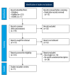
Figure 1
Figure 1
PRISMA flowchart showing breakdown of identification and screening in the literature search. CINAHL – Cumulative Index to Nursing and Allied Health Literature.
Figure 2
Figure 2
Illustration of Cilley’s classification of broad ligament herniation. The classification is divided into 3 distinct types based on the location and extent of the herniation: Type 1: A defect occurs in the broad ligament’s mesosalpinx, through which bowel loops may herniate. Type 2: A defect is found in the entire broad ligament but spares the uterine attachment, allowing organs such as the bowel to pass through. Type 3: A complete trans-ligamentous defect where the herniation occurs through the broad ligament’s entire thickness, often involving large segments of bowel. In the figure, arrows point to the sites of herniation, with numbers identifying each type of defect: Type 1 defect, type 2 defect, and type 3 defect. Each type is visually represented by shaded areas to distinguish between different defect locations within the broad ligament. The anatomical landmarks, including the uterus and adjacent structures, are indicated to clarify the relation between the defect and surrounding tissues. BL – broad ligament; UT – uterus. The image was prepared using professional image-editing software. This image was created by Janine Murta, BA, MSc, a faculty medical illustrator at St. George’s University School of Medicine. No AI or generative features were used in the process of creation. This illustration was developed from scratch using Adobe Illustrator and Photoshop version 25.12 (industry standard software for art and asset creation). The image creation process involved sketching various angles of the anatomy and selecting the best view to encompass all the types of broad ligament herniation. Once the best angle was chosen, further refinement, color, and details were rendered. The final image demonstrates the culmination of this refinement and rendering. [ © 2024, Janine Murta, BA, MSc].
Figure 3
Figure 3
Illustration of Hunt’s classification of broad ligament herniation. The classification is divided into 3 distinct types based on the location and extent of the herniation: Fenestra type: A defect occurs in the broad ligament’s mesosalpinx, through which bowel loops can herniate. Pouch type: A defect is found in the entire broad ligament but spares the uterine attachment, allowing organs such as the bowel to pass through. Hernia sac type: A complete trans-ligamentous defect where the herniation occurs through the broad ligament’s entire thickness, often involving large segments of bowel. The image was prepared using professional image-editing software. This image was created by Janine Murta, BA, MSc, a faculty medical illustrator at St. George’s University School of Medicine. No AI or generative features were used in the process of creation. This illustration was developed from scratch using Adobe Illustrator and Photoshop version 25.12 (industry standard software for art and asset creation). [ © 2024, Janine Murta, BA, MSc].

References

    1. Craig ME, Sudanagunta S, Billow M. StatPearls [Internet] Treasure Island (FL): StatPearls Publishing; 2024. Jan, Anatomy, abdomen and pelvis: Broad ligaments. [Updated 2023 Jul 24] Available from: https://www.ncbi.nlm.nih.gov/books/NBK499943/ - PubMed
    1. Choi PW. Strangulated small bowel obstruction caused by broad ligament hernia: Report of a case and review of literature. Am J Med Case Rep. 2017;5(2):38–40.
    1. Chaudhry SR, Imonugo O, Jozsa F, et al. StatPearls [Internet] Treasure Island (FL): StatPearls Publishing; 2024. Anatomy, abdomen and pelvis: Ligaments. [Updated 2023 Jan 13[ Available from: https://www.ncbi.nlm.nih.gov/books/NBK493215/ - PubMed
    1. Park JH, Kim S, Cho Y. Internal hernia through a defect in the broad ligament of uterus: Laparoscopic management using a self-anchoring barbed suture. J Minim Invasive Surg. 2018;21(3):130–32.
    1. Lanzetta MM, Masserelli A, Addeo G, et al. Internal hernias: A difficult diagnostic challenge. Review of CT signs and clinical findings. Acta Biomed. 2019;90(5):20–37. - PMC - PubMed

Publication types