. 2024 Dec 19:35:e2409004.
doi: 10.4014/jmb.2409.09004.

Comprehensive Analysis of the Fourteen Complete Genome Sequences of Buchnera aphidicola Isolated from Aphis Species

Affiliations

Comprehensive Analysis of the Fourteen Complete Genome Sequences of Buchnera aphidicola Isolated from Aphis Species

Jin-Ho Yun et al. J Microbiol Biotechnol. .

Abstract

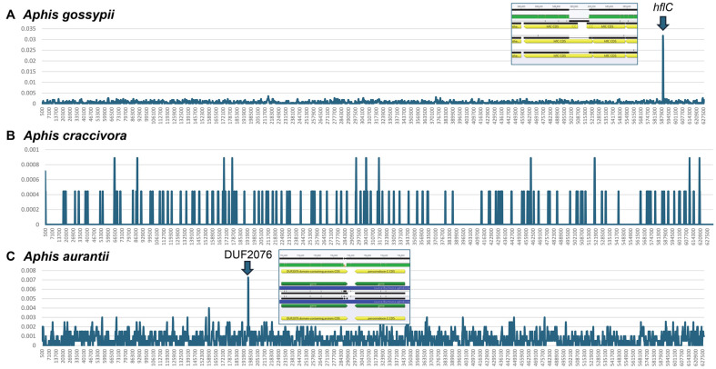
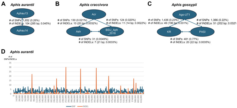
Endosymbionts are important for insect species as they provide essential substances to the host. Due to the technical advance of NGS technology and de novo assemblers, many endosymbionts bacterial genomes are available now. Here, we analysed fourteen endosymbiont bacterial genomes of Aphis genius, one of notorious pest species. Fourteen genomes displayed the length between 628,098 bp to 634,931 bp; GC ratio was from 24.2 % to 25.6 % with no structural variation found. The nucleotide diversity distribution across the 14 endosymbiont genomes revealed three distinct regions, each separated by varying levels of nucleotide diversity. Intraspecific variations identified from endosymbiont bacterial genomes of the same host species revealed numbers of SNPs ranging from 31 (0.0049%) to 1,652 (0.26%) and those of INDELs ranging from 7 (21 bp; 0.0033%) to 104 (285 bp; 0.0045%). 250 unique SSRs, 28 different common SSR groups, and one different SSR group in two genomes were identified and used as a potential molecular marker to distinguish intraspecific population. Phylogenetic analysis further showed congruence between the endosymbiont bacterial genomes and the host species phylogeny, except Aphis nasturtii, Aphis helianth, and Aphis auranti, which require additional endosymbiont genomes for clarification. This comparative analysis result could serve as a cornerstone for understanding the relationship between host and endosymbiont species from a genomic perspective.

Keywords: Aphis; Buchnera aphidicola; endosymbiont bacterial genome; intraspecific variations; nucleotide diversity; phylogenetic analysis; simple sequence repeats.


Conflict of interest statement

Conflict of Interest

The authors have no financial conflicts of interest to declare.

Figures

Fig. 1
Fig. 1. Multiple sequence alignment of fourteen endosymbiont bacterial genomes of Aphis genus.
Black bar on the top represents endosymbiont bacterial genome with genomic coordination. Below the black bar, dark-yellow graph presented the nucleotide identity in each coordination. Accession of endosymbiont bacterial genomes was displayed in the left part. Red arrows indicate ribosomal RNAs.
Fig. 2
Fig. 2. Genome size, GC ratio, number of CDs and tRNAs of the fourteen endosymbiont bacterial genomes.
(A) X-axis means fourteen endosymbiont bacterial genomes and Y-axis indicates genome size and GC ratios presented by blue bars and yellow line, respectively. (B) X-axis means fourteen endosymbiont bacterial genomes and Y-axis indicates the number of CDs exhibited as green bars and number of tRNAs displayed as purple line.
Fig. 3
Fig. 3. Nucleotide diversity of the fourteen endosymbiont bacterial genomes.
(A) X-axis indicates genomic coordination of multiple sequence alignment of fourteen endosymbiont bacterial genomes and Y-axis presents nucleotide diversity. Orange allows indicate the border of different nucleotide diversity levels and blue arrows exhibit peaks of nucleotide diversity with genes around the position. (B-E) presented the multiple sequence alignments in specific genes containing nucleotide diversity peak.
Fig. 4
Fig. 4. Nucleotide diversity distribution at border of different regions.
X-axis indicates genomic coordinates and Yaxis indicates nucleotide diversity. Transparent light-blue boxes present the region where nucleotide diversity dramatically changed. Below graph, multiple sequence alignments were presented with yellow arrow as CDs.
Fig. 5
Fig. 5. Nucleotide diversity of three multiple sequence alignments of endosymbiont bacterial genomes of three Aphis species.
X-axis presented genomic coordination of multiple sequence alignment and y-axis presented nucleotide diversity. Blow arrows mean peaks of nucleotide diversity and multiple sequence alignment results around peak were presented in the blue-bordered boxes.
Fig. 6
Fig. 6. Intraspecific variations identified from three multiple sequence alignments of endosymbiont bacterial genomes of three Aphis species.
(A-C) Presents intraspecific variations of pair-wise comparison of endosymbiont bacterial genomes. Blue round boxes indicate endosymbiont bacterial genomes and arrows mean pair-wise comparison with number of SNPs and INDELs. (D) Exhibits the distribution of intraspecific variations along with bacterial genome. X-axis genomic coordination of sequence alignment and Y-axis indicate number of SNPs (blue line) and INDELs (yellow line).
Fig. 7
Fig. 7. Simple sequence repeats identified from fourteen endosymbiont bacterial g genomes of Aphis genus.
(A) Pie graph displays number of normal SSRs (blue), potential SSRs (orange), and extended SSRs (green) from 14 endosymbiont bacterial genomes. (B) X-axis indicates types of SSRs and Y-axis displays number of SSRs (blue bar) and ratio between standard deviation and average number of SSRs (orange line). (C) X-axis indicates 14 endosymbiont bacterial genomes and Y-axis indicates number of SSRs. Blue lines upper the graph mean the host species of endosymbiont bacterial genomes is same.
Fig. 8
Fig. 8. SSR groups identified from endosymbiont bacterial genomes of three Aphis species.
X-axis indicates three Aphis host species and Y-axis means number of SSRs starting with 2,000.
Fig. 9
Fig. 9. Phylogenetic tree of endosymbiont bacterial genomes with host phylogeny.
(A) display phylogenetic tree of fifteen endosymbiont bacterial genomes including outgroup species. Phylogenetic tree was drawn based on BI tree. The numbers above the branches are posterior probabilities from the BI tree and the bootstrap support values of the NJ. (B) phylogenetic tree was drawn based on ML tree. The number above the branches is the bootstrap support values of the ML. Grey dotted lines indicate the same phylogenetic position between endosymbiont bacterial genome and host species and black dotted lines means incongruent cases.

Similar articles

References

    1. Wernegreen JJ. Genome evolution in bacterial endosymbionts of insects. Nat. Rev. Genet. 2002;3:850–861. doi: 10.1038/nrg931. - DOI - PubMed
    1. Vega FE, Dowd PF. The role of yeasts as insect endosymbionts. Insect-Fungal Associations: Ecology and Evolution. Oxford University Press, New York; 2005. pp. 211–243.
    1. Najar-Rodríguez AJ, McGraw EA, Mensah RK, Pittman GW, Walter GH. The microbial flora of Aphis gossypii: patterns across host plants and geographical space. J. Invertebr. Pathol. 2009;100:123–126. doi: 10.1016/j.jip.2008.10.005. - DOI - PubMed
    1. Zhao Y, Zhang S, Luo JY, Wang CY, Lv LM, Cui JJ. Bacterial communities of the cotton aphid Aphis gossypii associated with Bt cotton in northern China. Sci. Rep. 2016;6:22958. doi: 10.1038/srep22958. - DOI - PMC - PubMed
    1. Pérez-Brocal V, Gil R, Ramos S, Lamelas A, Postigo M, Michelena JM, et al. A small microbial genome: the end of a long symbiotic relationship? Science. 2006;314:312–313. doi: 10.1126/science.1130441. - DOI - PubMed