. 2024 Dec 17:35:e2410001.
doi: 10.4014/jmb.2410.10001.

Exploring the Proteomic Landscape and Immunomodulatory Functions of Edwardsiella piscicida Derived Extracellular Vesicles

Affiliations

Exploring the Proteomic Landscape and Immunomodulatory Functions of Edwardsiella piscicida Derived Extracellular Vesicles

Mawalle Kankanamge Hasitha Madhawa Dias et al. J Microbiol Biotechnol. .

Abstract

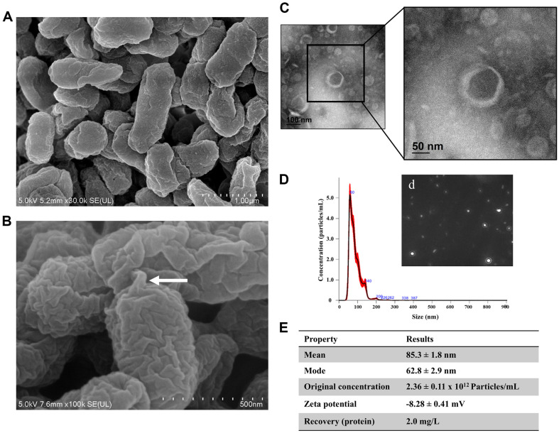
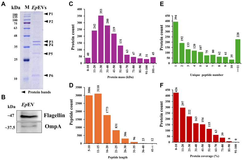
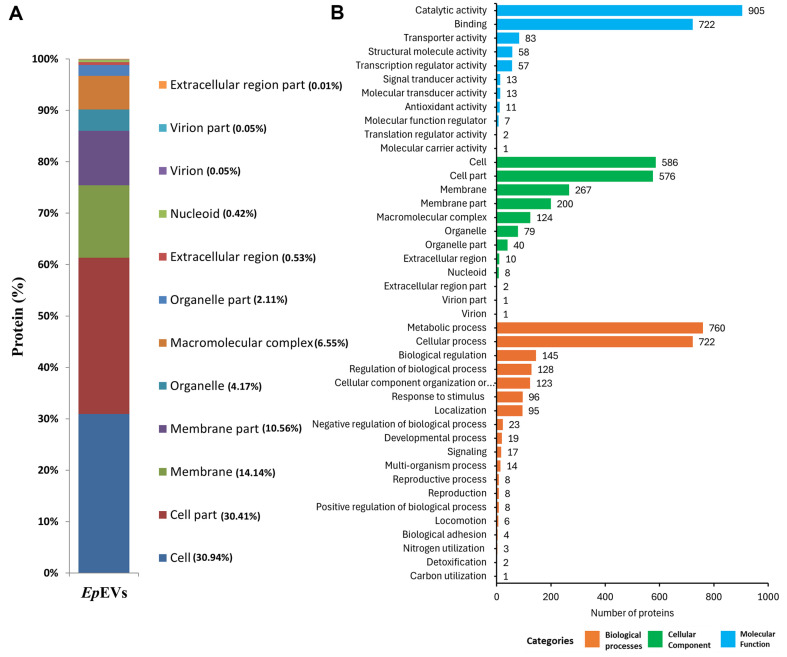
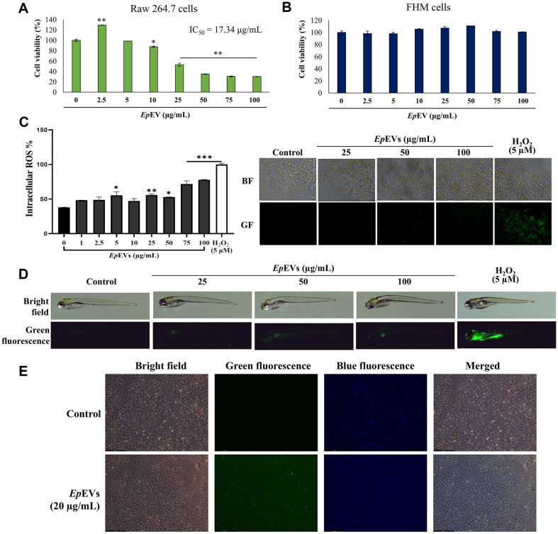
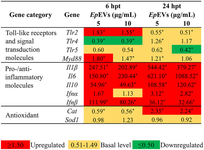
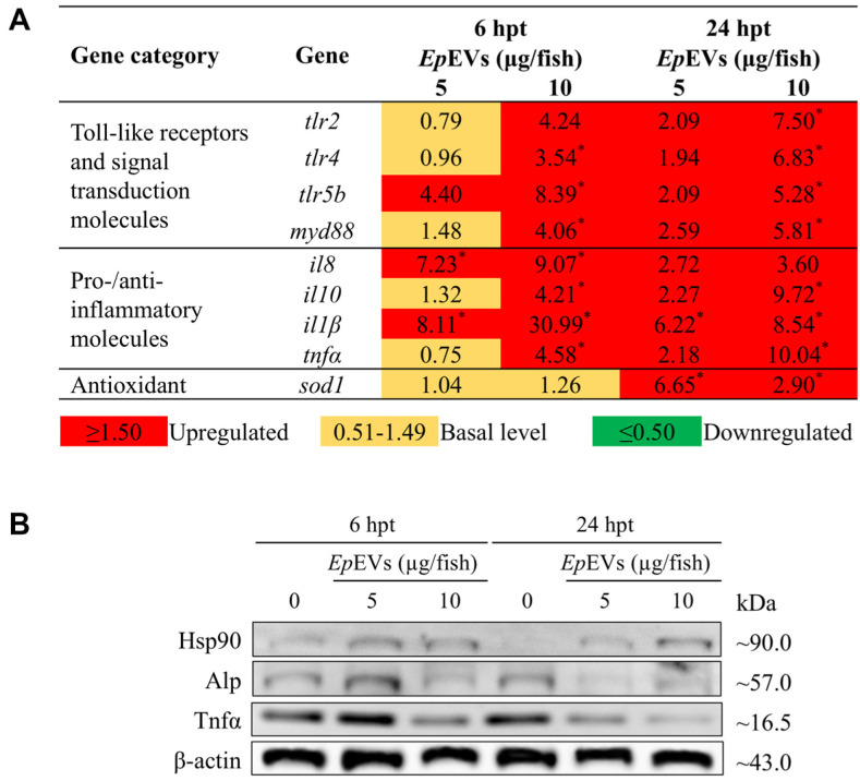
Extracellular vesicles (EVs) have garnered attention in research for their potential as biochemical transporters and immune modulators, crucial for regulating the host immune system. The present study was conducted to isolate and characterize EVs from Gram negative bacteria Edwardsiella piscicida (EpEVs) and investigate their proteomic profile and immune responses. Isolation of EpEVs was carried out using ultracentrifugation method. Transmission electron microscopy results confirmed the spherical shape of EpEVs. The average size and zeta potential were 85.3 ± 1.8 nm and -8.28 ± 0.41 mV, respectively. EpEVs consisted of 1,487 distinct proteins. Subcellular localization analysis revealed that "cell" and "cell part" were the most predominant areas for protein localization. Proteins associated with virulence, along with several chaperones that facilitate protein folding and stability, were also present. No toxicity was detected when EpEVs were treated to fathead minnow (FHM) cells up to 100 μg/ml. Fluorescent-labeled EpEVs showed cellular internalization in FHM cells at 24 h post treatment (hpt). In-vitro gene expression in Raw 264.7 cells showed upregulation of interleukin (Il)6, Il1β, and interferon (Ifn)β with simultaneous upregulation of anti-inflammatory Il10. In vivo, gene expression revealed that except for heat shock protein (hsp)70, all other genes were upregulated suggesting that EpEVs induced the expression of immune-related genes. Western blot analysis showed increased protein levels of tumor necrosis factor (Tnf)α in EpEVs-treated spleen tissue of zebrafish. Our results confirm that EpEVs can be successfully isolated using the ultracentrifugation method. Furthermore, exploring immunomodulatory mechanism of EpEVs is essential for their potential use as novel therapeutics in fish medicine.

Keywords: Edwardsiella piscicida; Raw 264.7 cells; Zebrafish (Danio rerio); extracellular vesicles; immunomodulation; proteomic analysis.


Conflict of interest statement

Conflict of Interest

The authors have no financial conflicts of interest to declare.

Figures

Fig. 1
Fig. 1. EpEVs characterization.
(A) Visualization of morphological features and (B) EVs release process from E. piscicida captured using FE-SEM. The white arrow indicates the formation of a bulbous structure preceding EVs release. (C) FE-TEM analysis of EpEVs displaying a spheroid double membrane-bound morphology at different magnifications (100 and 50 nm). (D) Nanoparticle tracking analysis quantified the particle size and concentration of EpEVs, with a graph depicting concentration plotted against particle size. A still frame from the particle distribution video is shown (d). (E) Comprehensive characterization of EpEVs, including particle size distribution (mean and mode), original concentration, zeta potential, and total protein recovery.
Fig. 2
Fig. 2. Protein profile and characteristics of EpEVs.
(A) SDS-PAGE gel stained with Coomassie blue shows distinct protein bands (P1-P6) indicated by black arrows. (B) Western blot analysis identified BEVs marker proteins. EpEVs (25 μg total protein) were loaded into an SDS-PAGE gel (12%) and transferred to a membrane. Membranes were incubated overnight with polyclonal primary antibodies (Flagellin and OmpA) followed by relevant secondary antibodies. BioFACT triple-color protein marker (10-180 kDa) was used as a reference. Proteomic analysis further characterized the total proteome of EpEVs. (C) Protein mass distribution, (D) peptide length distribution, (E) unique peptide distribution, and (F) protein coverage distribution. Peptides extracted from EpEVs were fractionated into 10 fractions, further separated using HPLC, and analyzed using MS/MS for protein identification.
Fig. 3
Fig. 3. Localization and functional annotations of EpEVs.
(A) Subcellular localization of EpEVs proteins was determined following HPLC-MS/MS analysis to identify the proteome in EpEVs. (B) Gene ontology (GO) analysis was conducted to explore the various functional roles of the proteins identified through proteomic analysis.
Fig. 4
Fig. 4. Protein-protein interaction (PPI) network prediction between EpEVs and E. tarda.
(A) PPI network of EpEVs proteins and their interactions with E. tarda proteins was generated using the STRING database. (B) Key pathway enrichments of interacting proteins include (b1) protein export, (b2) microbial metabolism in diverse environments, and (b3) metabolic pathways of EpEVs in association with E. tarda. Proteins identified from EpEVs (1487) by HPLC-MS/MS were used to visualize the network of proteins interacting with E. tarda proteins. A total of 673 protein interactions were observed from the 1487 EpEVs proteins.
Fig. 5
Fig. 5. In vitro and in vivo toxic effects and internalization of EpEVs.
(A) Effect of EpEVs on the viability of Raw 264.7 cells and (B) EpEVs-treated FHM cells. Cytotoxicity in Raw 264.7 cells and FHM cells was assessed using the Cellrix cytotoxicity assay kit. (C) Intracellular ROS production was measured following treatment with DCFHDA (5 μg/ml), and representative images show the level of ROS production. (D) In vivo ROS generation in EpEVs-treated zebrafish larvae (25-100 μg/ml). Zebrafish larvae at 60 hpf were treated with varying concentrations of EpEVs, and mortality was measured up to 96 hpt. ROS generation was measured using DCFHDA stain, with H2O2 as the positive control. (E) Internalization of fluorescently labeled EpEVs into FHM cells. Cells were analyzed by treating FHM cells (n = 3) with EpEVs (20 μg/ml) using the ExoSparkler kit. Images were captured 24 hpt using a fluorescent microscope. Triplicate experiments were conducted to assess the repeatability of assays, and the data are presented as mean ± SEM.
Fig. 6
Fig. 6. Analysis of transcriptional regulation in EpEVs-treated Raw 264.7 cells.
The transcriptional responses of Raw 264.7 cells to EpEVs treatment were assessed using quantitative real-time PCR (qRT-PCR) to evaluate changes in gene expression. The expression levels of target genes were normalized to the housekeeping gene, Gapdh. The 2−ΔΔCT method was applied to determine relative gene expression fold changes between EpEVs-treated cells and untreated control cells. Data are presented as the mean relative expression fold change, calculated by comparing the expression levels in EpEVs-treated cells to the control group. Statistical significance was determined at p < 0.05, with significant differences indicated by an asterisk (*)
Fig. 7
Fig. 7. In vivo transcriptional expression and protein expression analysis of EpEVs-treated zebrafish.
(A) Transcriptional expression of immune-related functional genes in kidney. To assess the transcriptional expression of target genes, adult zebrafish were intraperitoneally injected with varying doses of EpEVs (5 and 10 μg/fish). The relative fold expression of selected genes was quantified using the 2−ΔΔCT method, with normalization to β-actin as the housekeeping gene. Statistical significance (*) was assessed at p < 0.05. (B) Analysis of protein expression in spleen tissue. Protein levels were assessed by western blotting. β-actin was used as the housekeeping protein to normalize protein loading across samples.

Similar articles

References

    1. Du S, Guan Y, Xie A, Yan Z, Gao S, Li W, et al. Extracellular vesicles: a rising star for therapeutics and drug delivery. J. Nanobiotechnol. 2023;21:231. doi: 10.1186/s12951-023-01973-5. - DOI - PMC - PubMed
    1. Liu H, Zhang Q, Wang S, Weng W, Jing Y, Su J. Bacterial extracellular vesicles as bioactive nanocarriers for drug delivery: advances and perspectives. Bioact. Mater. 2022;14:169–181. doi: 10.1016/j.bioactmat.2021.12.006. - DOI - PMC - PubMed
    1. Wen M, Wang J, Ou Z, Nie G, Chen Y, Li M, et al. Bacterial extracellular vesicles: a position paper by the microbial vesicles task force of the Chinese society for extracellular vesicles. Interdisciplinary Medicine. 2023;1:e20230017. doi: 10.1002/INMD.20230017. - DOI
    1. Xie J, Haesebrouck F, Van Hoecke L, Vandenbroucke RE. Bacterial extracellular vesicles: an emerging avenue to tackle diseases. Trends Microbiol. 2023;31:1206–1224. doi: 10.1016/j.tim.2023.05.010. - DOI - PubMed
    1. Matinha-Cardoso J, Coutinho F, Lima S, Eufrásio A, Carvalho AP, Oliva-Teles A, et al. Novel protein carrier system based on cyanobacterial nano-sized extracellular vesicles for application in fish. Microb. Biotechnol. 2022;15:2191–2207. doi: 10.1111/1751-7915.14057. - DOI - PMC - PubMed

MeSH terms

Supplementary concepts