Review
. 2024 Dec 31;11(1):e41566.
doi: 10.1016/j.heliyon.2024.e41566. eCollection 2025 Jan 15.

Advanced photoluminescent nanomaterials for targeted bioimaging of cancer cells

Affiliations
Review

Advanced photoluminescent nanomaterials for targeted bioimaging of cancer cells

Tooba Mohammadi et al. Heliyon. .

Abstract

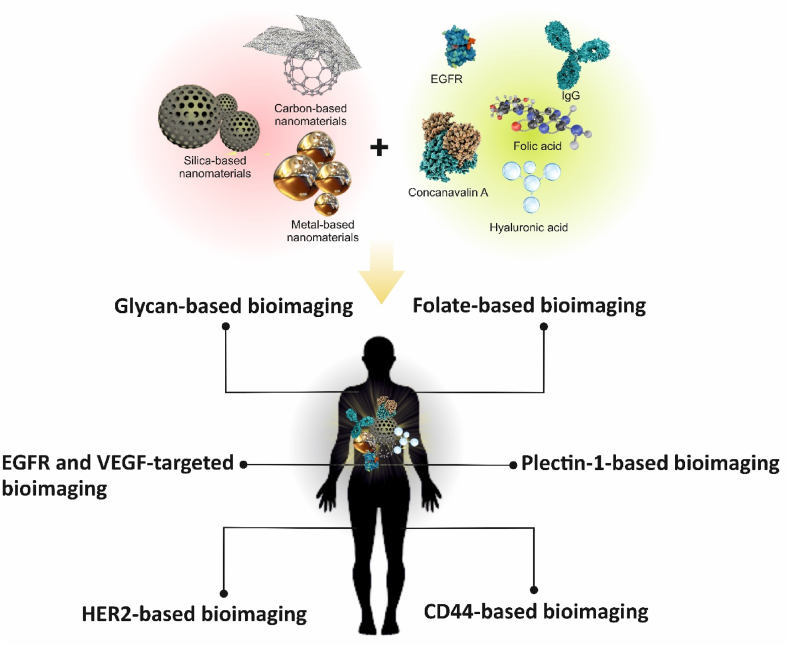
The investigation of changes in the membrane of cancer cells holds great potential for biomedical applications. Malignant cells exhibit overexpression of receptors, which can be used for targeted drug delivery, therapy, and bioimaging. Targeted bioimaging is one the most accurate imaging methods with a non-invasive nature, allowing for localization of the malignant cell without disrupting cellular integrity. Also, bioimaging has the potential to enhance the quality of established imaging techniques like magnetic resonance imaging (MRI). The utilization of nanoparticles in targeted bioimaging enhances the imaging quality and efficiency. Biocompatible nanoparticles can easily penetrate cell membranes, while they can be readily functionalized on their surfaces toward cell receptors. This study reviews reports on the application of new advanced photoluminescent materials for targeted bioimaging using the cell membrane receptors. Also, the limitations and advantages of the application of nanoparticles have been reviewed along with the clinical consideration of their uses in bioimaging.

Keywords: Bioimaging; Nanoparticles; Neoplasms; Targeted bioimaging; Targeted therapy; Theranostic nanomedicine.


Conflict of interest statement

The authors declare that they have no known competing financial interests or personal relationships that could have appeared to influence the work reported in this paper.

Figures

Scheme 1
Scheme 1
A graphical representation of the application of materials in targeted bioimaging.
Fig. 1
Fig. 1
(A) Attachment of QDs in the nucleus and (B) in the perinuclear region of cells. "Reprinted (adapted) with permission from Ref. [38]. Copyright 2024 American Chemical Society."
Fig. 2
Fig. 2
CS-CD hybrid nanogel loaded with silibinin, an anticancer drug, demonstrating selective penetration into MCF7 cells. “Reprinted from Ref. [19] with permission code of 5878130312616)”.
Fig. 3
Fig. 3
PB-dot functionalization and CTX conjugation. “Reprinted from Ref. [48] with permission code of 5878140737561)”.
Fig. 4
Fig. 4
Schematic representation of application of DOX-loaded TiO2/zinc phthalocyanine-folic acid for bioimaging and photodynamic therapy. “Reprinted from Ref. [53] with permission code of 5880081084183)”.
Fig. 5
Fig. 5
Schematic representation of the synthesis process for MFCQDs using a microwave-assisted hydrothermal method. “Reprinted (adapted) with permission from Ref. [58]. Copyright 2024 American Chemical Society."
Fig. 6
Fig. 6
Schematic representation illustrating the fabrication process of COS/DEx NGs. "Reprinted (adapted) with permission from Ref. [70]. Copyright 2024 American Chemical Society."
Fig. 7
Fig. 7
Successful accumulation of SiNPs in EGFR-positive colorectal (CaCo-2) cancer cells compared to normal colon epithelial cells. "Reprinted (adapted) with permission from Ref. [77]. Copyright 2024 American Chemical Society."
Fig. 8
Fig. 8
Schematic representation of the (A) synthesis and (B) application of PFOB@IR825-HA-Cy5.5 NPs for triple imaging (fluorescent, photoacoustic, and computed tomography) and tumor ablation in HT-29 mice cells. “Reprinted from Ref. [74] with permission code of 5880090791756)”.
Fig. 9
Fig. 9
A) Schematic illustration depicting the preparation of ND-PTX-Cet. B) Preparation of ND-PTX-Cet involved a simple mixture at 4 °C, followed by centrifugation at 12,000 rpm for 10 min at 4 °C. C) Evaluation of Cet loading efficiency on ND by comparing the OD values of ND-PTX-Cet with the standard curve to assess binding affinity. “Reprinted from Ref. [82] with permission code of 5880070179259)”.
Fig. 10
Fig. 10
Schematic representation illustrating the process of Her-Au nanoclusters (NCs) formation and their potential applications in HER2-Targeted imaging for cancer diagnosis. "Reprinted (adapted) with permission from Ref. [89]. Copyright 2024 American Chemical Society."

References

    1. Zaimy M.A., Saffarzadeh N., Mohammadi A., Pourghadamyari H., Izadi P., Sarli A., Moghaddam L.K., Paschepari S.R., Azizi H., Torkamandi S., Tavakkoly-Bazzaz J. New methods in the diagnosis of cancer and gene therapy of cancer based on nanoparticles. Cancer Gene Ther. 2017;24:233–243. doi: 10.1038/cgt.2017.16. - DOI - PubMed
    1. Zubair M., Wang S., Ali N. Advanced approaches to breast cancer classification and diagnosis. Front. Pharmacol. 2020;11 doi: 10.3389/fphar.2020.632079. - DOI - PMC - PubMed
    1. Sharma P., Brown S., Walter G., Santra S., Moudgil B. Nanoparticles for bioimaging. Adv. Colloid Interface Sci. 2006;123:471–485. doi: 10.1016/j.cis.2006.05.026. - DOI - PubMed
    1. Wolfbeis O.S. An overview of nanoparticles commonly used in fluorescent bioimaging. Chem. Soc. Rev. 2015;44:4743–4768. doi: 10.1039/c4cs00392f. - DOI - PubMed
    1. Yao J., Yang M., Duan Y. Chemistry, biology, and medicine of fluorescent nanomaterials and related systems: new insights into biosensing, bioimaging, genomics, diagnostics, and therapy. Chem. Rev. 2014;114:6130–6178. doi: 10.1021/cr200359p. - DOI - PubMed

LinkOut - more resources