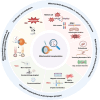
Mitochondrial transplantation: a promising strategy for the treatment of retinal degenerative diseases
- PMID: 39851134
- PMCID: PMC11974652
- DOI: 10.4103/NRR.NRR-D-24-00851
Mitochondrial transplantation: a promising strategy for the treatment of retinal degenerative diseases
Abstract
The retina, a crucial neural tissue, is responsible for transforming light signals into visual information, a process that necessitates a significant amount of energy. Mitochondria, the primary powerhouses of the cell, play an integral role in retinal physiology by fulfilling the high-energy requirements of photoreceptors and secondary neurons through oxidative phosphorylation. In a healthy state, mitochondria ensure proper visual function by facilitating efficient conversion and transduction of visual signals. However, in retinal degenerative diseases, mitochondrial dysfunction significantly contributes to disease progression, involving a decline in membrane potential, the occurrence of DNA mutations, increased oxidative stress, and imbalances in quality-control mechanisms. These abnormalities lead to an inadequate energy supply, the exacerbation of oxidative damage, and the activation of cell death pathways, ultimately resulting in neuronal injury and dysfunction in the retina. Mitochondrial transplantation has emerged as a promising strategy for addressing these challenges. This procedure aims to restore metabolic activity and function in compromised cells through the introduction of healthy mitochondria, thereby enhancing the cellular energy production capacity and offering new strategies for the treatment of retinal degenerative diseases. Although mitochondrial transplantation presents operational and safety challenges that require further investigation, it has demonstrated potential for reviving the vitality of retinal neurons. This review offers a comprehensive examination of the principles and techniques underlying mitochondrial transplantation and its prospects for application in retinal degenerative diseases, while also delving into the associated technical and safety challenges, thereby providing references and insights for future research and treatment.
Copyright © 2025 Neural Regeneration Research.
Conflict of interest statement
Figures


Similar articles
-
Mitochondrial biogenesis: pharmacological approaches.Curr Pharm Des. 2014;20(35):5507-9. doi: 10.2174/138161282035140911142118. Curr Pharm Des. 2014. PMID: 24606795
-
Essential roles of mitochondrial biogenesis regulator Nrf1 in retinal development and homeostasis.Mol Neurodegener. 2018 Oct 17;13(1):56. doi: 10.1186/s13024-018-0287-z. Mol Neurodegener. 2018. PMID: 30333037 Free PMC article.
-
Differential Susceptibility of Retinal Neurons to the Loss of Mitochondrial Biogenesis Factor Nrf1.Cells. 2022 Jul 14;11(14):2203. doi: 10.3390/cells11142203. Cells. 2022. PMID: 35883647 Free PMC article.
-
Targeting Mitochondrial Dysfunction and Oxidative Stress to Prevent the Neurodegeneration of Retinal Ganglion Cells.Antioxidants (Basel). 2023 Nov 17;12(11):2011. doi: 10.3390/antiox12112011. Antioxidants (Basel). 2023. PMID: 38001864 Free PMC article. Review.
-
Mitochondrial Dysfunction and Oxidative Stress in Liver Transplantation and Underlying Diseases: New Insights and Therapeutics.Transplantation. 2021 Nov 1;105(11):2362-2373. doi: 10.1097/TP.0000000000003691. Transplantation. 2021. PMID: 33577251 Free PMC article. Review.
Cited by
-
Mitochondrial dysfunction in epilepsy: mechanistic insights and clinical strategies.Mol Biol Rep. 2025 May 20;52(1):470. doi: 10.1007/s11033-025-10577-1. Mol Biol Rep. 2025. PMID: 40392243 Review.
-
Update on the correlation between mitochondrial function and osteonecrosis of the femoral head osteocytes.Redox Rep. 2025 Dec;30(1):2491846. doi: 10.1080/13510002.2025.2491846. Epub 2025 Apr 18. Redox Rep. 2025. PMID: 40249372 Free PMC article. Review.
-
Tryptophan metabolism and ischemic stroke: An intricate balance.Neural Regen Res. 2026 Feb 1;21(2):466-477. doi: 10.4103/NRR.NRR-D-24-00777. Epub 2025 Jan 13. Neural Regen Res. 2026. PMID: 40326980 Free PMC article.
-
Harnessing stem cell-derived exosomes: a promising cell-free approach for spinal cord injury.Stem Cell Res Ther. 2025 Apr 17;16(1):182. doi: 10.1186/s13287-025-04296-4. Stem Cell Res Ther. 2025. PMID: 40247394 Free PMC article. Review.
-
Biotechnological approaches and therapeutic potential of mitochondria transfer and transplantation.Nat Commun. 2025 Jul 1;16(1):5709. doi: 10.1038/s41467-025-61239-6. Nat Commun. 2025. PMID: 40593725 Free PMC article. Review.
References
-
- Aboobakar IF, Kinzy TG, Zhao Y, Fan B, Pasquale LR, Qassim A, Kolovos A, Schmidt JM, Craig JE, Cooke Bailey JN, Wiggs JL; NEIGHBORHOOD Consortium Mitochondrial TXNRD2 and ME3 genetic risk scores are associated with specific primary open-angle glaucoma phenotypes. Ophthalmology. 2023;130:756–763. - PMC - PubMed
-
- Angelova PR, Abramov AY. Role of mitochondrial ROS in the brain: from physiology to neurodegeneration. FEBS Lett. 2018;592:692–702. - PubMed
LinkOut - more resources
Full Text Sources

