. 2024 Dec 28;12(1):15.
doi: 10.3390/bioengineering12010015.

Spectral Analysis of Light-Adapted Electroretinograms in Neurodevelopmental Disorders: Classification with Machine Learning

Affiliations

Spectral Analysis of Light-Adapted Electroretinograms in Neurodevelopmental Disorders: Classification with Machine Learning

Paul A Constable et al. Bioengineering (Basel). .

Abstract

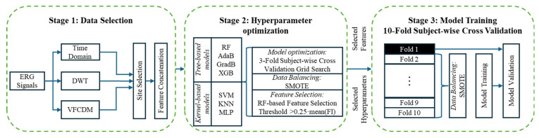
Electroretinograms (ERGs) show differences between typically developing populations and those with a diagnosis of autism spectrum disorder (ASD) or attention deficit/hyperactivity disorder (ADHD). In a series of ERGs collected in ASD (n = 77), ADHD (n = 43), ASD + ADHD (n = 21), and control (n = 137) groups, this analysis explores the use of machine learning and feature selection techniques to improve the classification between these clinically defined groups. Standard time domain and signal analysis features were evaluated in different machine learning models. For ASD classification, a balanced accuracy (BA) of 0.87 was achieved for male participants. For ADHD, a BA of 0.84 was achieved for female participants. When a three-group model (ASD, ADHD, and control) the BA was lower, at 0.70, and fell further to 0.53 when all groups were included (ASD, ADHD, ASD + ADHD, and control). The findings support a role for the ERG in establishing a broad two-group classification of ASD or ADHD, but the model's performance depends upon sex and is limited when multiple classes are included in machine learning modeling.

Keywords: attention deficit hyperactivity disorder; autism; biomarker; feature selection; medication; retina; sex.


Conflict of interest statement

Aleksei Zhdanov is employed by the company VisioMed.AI which had no role in the design of the study; in the collection, analyses, or interpretation of data; in the writing of the manuscript, or in the decision to publish the results. All other authors declare no conflicts of interest.

Figures

Figure A1
Figure A1
Light-adapted electroretinogram illustrating the main features of the waveform. A vertical line indicates the onset of flash stimulus. The a-wave negative deflection is mainly formed by the hyperpolarization of the cone photoreceptors. The b-wave is a result of depolarizing bipolar cells and is modulated by amacrine and glial cells. The photopic negative response is mainly driven by retinal ganglion cells.
Figure A2
Figure A2
(a) Light-adapted electroretinogram (ERG) signal with a-wave, b-wave, and oscillatory potentials indicated. The waveform is shaped by the contributions of the cone photoreceptors and horizontal, bipolar, amacrine, and retinal ganglion cells as they respond to the flash of light. (b) Representative light-adapted ERG waveforms recorded from control, ASD, ADHD, and ASD + ADHD participants. In this series, the b-wave amplitude is largest in the ADHD participant and smallest in the ASD participant.
Figure A3
Figure A3
Time–frequency spectrogram of the ERG signal. The coefficients a20 and a40 correspond to the a-wave components, while b20 and b40 correspond to the b-wave. The OP80 and OP160 relate to the early and late oscillatory potentials. The ON retinal pathway is characterized by the 20 Hz frequency and the OFF retinal pathway is characterized by the 40 Hz frequency band.
Figure A4
Figure A4
VFCDM decomposition of the ERG signal: The top plot displays the ERG signal alongside its power spectral density. The bottom plot illustrates the first 8 VFCDM components along with their corresponding spectral information.
Figure 1
Figure 1
Model training procedure flowchart incorporating three stages. One: the data selection of the electroretinogram signal with time–frequency analysis (discrete wavelet transform (DWT) and variable-frequency complex demodulation (VFCDM)) and standard time domain features. Two: hyperparameter optimization, including machine learning model optimization, data balancing with synthetic minority oversampling (SMOTE), and optimal feature selection before. Three: model training with a 10-fold subject-wise cross validation. Models: RF (Random Forest); AdaB (Adaptive Boosting); GradB (Gradient Boosting); XGB (Extreme Gradient Boosting); SVM (Support Vector Machine); KNN (K-nearest neighbor); MLP (multi-layer perceptron).
Figure 2
Figure 2
Shapley summary of the ten most important features for the XGB classifier for ASD (A) and the RF classifier for ADHD (B). For ASD, the time of the b-wave peak at 446 Td.s (left eye) (Tb_L446) was the most important feature in the model in terms of differentiating the ASD group from the control group. For the ADHD group, the kurtosis of the 3rd band at 113 Td.s (left eye) (vfcdm_kt_3_L113) had the highest Shapley feature importance for classification. A red asterisk (*) refers to statistically non-significant differences (p ≥ 0.05), as assessed based on the Kruskal–Wallis test, between the groups for these features.
Figure 3
Figure 3
A Shapley summary of the top 10 features for KNN classifier for the 3-group classification: (A) control, (B) ASD, and (C) ADHD. Feature importance for the group classification is indicated by the red scale. The most important features were as follows: for control, it was the DWT OP160 component; for ASD, it was the kurtosis of the 6th VFCDM sub-band; and for ADHD, it was the DWT OP80 component. The red asterisk (*) indicates a statistically non-significant difference (p ≥ 0.05) between the groups for these features based on the Kruskal–Wallis test.
Figure 4
Figure 4
A Shapley summary of the top 10 features of the RF classifier for the 4-group classification: control (A), ASD (B), ADHD (C), and ASD + ADHD (D). When classifying between the four groups, the most important features were as follows: for the control, it was the high-frequency DWT range; for ASD, it was the interquartile range of the 8th VFCDM sub-band; for ADHD, it was the sum of the OP80 DWT components; for ASD + ADHD, it was also the high-frequency DWT range. Red markers in the Shapley plots have greater importance for classifying the group. The colored boxes highlight the common important features for each of the group’s classifications highlighted vfcdm_kt_4 (green), vfcdm_kt_7 (red) and dwt_sum_OP80 (yellow). The red asterisk (*) refers to statistically non-significant (p ≥ 0.05) differences between the groups for these features based on the Kruskal–Wallis test.
Figure 5
Figure 5
A Shapley summary of the best models for male or female 2-group classification (ASD vs. control or ADHD vs. control). (A,B) show the male participants for ASD or ADHD vs. control, respectively, and (C,D) show the female participants for ASD or ADHD vs. control, respectively. For males, the high-frequency features from the VFCDM were more important for ASD and ADHD classification (sub-bands 7 and 8). In contrast, for female participants, the lower-frequency features were more important for ASD or ADHD classification (sub-bands 2 and 3). A red asterisk (*) refers to statistically non-significant differences (p ≥ 0.05) based on the Kruskal–Wallis test between the groups for these features.
Figure 6
Figure 6
(A) Shapley values of the SVM classifier that achieved the best 2-group (ASD vs. control) classification performance. Waterfall plots for an individual control (C) and an individual ASD (B) participant with the relative contributions to a positive classification for control (blue) and ASD (red). At the left side of each feature name, the actual value of the feature from that subject is presented. A red asterisk (*) refers to statistically non-significant differences (p ≥ 0.05) based on the Kruskal–Wallis test between the groups for these features.
Figure 7
Figure 7
(A) Shapley values of the SVM classifier that achieved the best 2-group (ADHD vs. control) classification performance. Waterfall plots for an individual control (C) and an individual ADHD (B) participant with the relative contributions to a positive classification for control (blue) and ADHD (red). At the left side of each feature name, the actual value of the feature from that subject is presented. A red asterisk (*) refers to statistically non-significant differences (p ≥ 0.05) based on the Kruskal–Wallis test between the groups for these features.

Similar articles

Cited by

References

    1. Parellada M., Andreu-Bernabeu Á., Burdeus M., San José Cáceres A., Urbiola E., Carpenter L.L., Kraguljac N.V., McDonald W.M., Nemeroff C.B., Rodriguez C.I., et al. In search of biomarkers to guide interventions in autism spectrum disorder: A Systematic Review. Am. J. Psychiatry. 2023;180:23–40. doi: 10.1176/appi.ajp.21100992. - DOI - PMC - PubMed
    1. London A., Benhar I., Schwartz M. The retina as a window to the brain—From eye research to CNS disorders. Nat. Rev. Neurol. 2013;9:44–53. doi: 10.1038/nrneurol.2012.227. - DOI - PubMed
    1. Hébert M., Mérette C., Gagné A.M., Paccalet T., Moreau I., Lavoie J., Maziade M. The electroretinogram may differentiate schizophrenia from bipolar disorder. Biol. Psychiatry. 2020;87:263–270. doi: 10.1016/j.biopsych.2019.06.014. - DOI - PubMed
    1. Asanad S., Felix C.M., Fantini M., Harrington M.G., Sadun A.A., Karanjia R. Retinal ganglion cell dysfunction in preclinical Alzheimer’s disease: An electrophysiologic biomarker signature. Sci. Rep. 2021;11:6344. doi: 10.1038/s41598-021-85010-1. - DOI - PMC - PubMed
    1. Schwitzer T., Le Cam S., Cosker E., Vinsard H., Leguay A., Angioi-Duprez K., Laprevote V., Ranta R., Schwan R., Dorr V.L. Retinal electroretinogram features can detect depression state and treatment response in adults: A machine learning approach. J. Affect. Disord. 2022;306:208–214. doi: 10.1016/j.jad.2022.03.025. - DOI - PubMed

LinkOut - more resources