. 2024 Dec 31;15(1):35.
doi: 10.3390/brainsci15010035.

Role of Virtual iMRI in Glioblastoma Surgery: Advantages, Limitations, and Correlation with iCT and Brain Shift

Affiliations

Role of Virtual iMRI in Glioblastoma Surgery: Advantages, Limitations, and Correlation with iCT and Brain Shift

Erica Grasso et al. Brain Sci. .

Abstract

Background: Elastic image fusion (EIF) using an intraoperative CT (iCT) scan may enhance neuronavigation accuracy and compensate for brain shift. Objective: To evaluate the safety and reliability of the EIF algorithm (Virtual iMRI Cranial 4.5, Brainlab AG, Munich Germany, for the identification of residual tumour in glioblastoma surgery. Moreover, the impact of brain shift on software reliability is assessed. Methods: This ambispective study included 80 patients with a diagnosis of glioblastoma. Pre-operative MRI was elastically fused with an intraoperative CT scan (BodyTom; Samsung-Neurologica, Danvers, MA, USA) acquired at the end of the resection. Diagnostic specificity and the sensitivity of each tool was determined. The impact of brain shift on residual tumour was statistically analysed. An analysis of accuracy was performed through Target Registration Error (TRE) measurement after rigid image fusion (RIF) and EIF. A qualitative evaluation of each Virtual MRI image (VMRI) was performed. Results: VMRI identified residual tumour in 26/80 patients (32.5%), confirmed by post-operative MRI (true positive). Of these, 5 cases were left intentionally due to DES-positive responses, 8 cases underwent near maximal or subtotal resection, and 13 cases were not detected by iCT. However, in the other 27/80 cases (33.8%), VMRI reported residual tumour that was present neither on iCT nor on post-operative MRI (false positive). i-CT showed a sensitivity of 56% and specificity of 100%; VMRI demonstrated a sensitivity of 100% and specificity of 50%. Spearman correlation analysis showed a moderate correlation between pre-operative volume and VMRI tumour residual. Moreover, tumour involving insula or infiltrating more than one lobe displayed higher median values (p = 0.023) of virtual residual tumour. A statistically significant reduction towards lower TRE values after EIF was observed for test structures. Conclusions: Virtual iMRI was proven to be a feasible option to detect residual tumour. Its integration within a multimodal imaging protocol may provide neurosurgeons with intraoperatively updated imaging.

Keywords: Virtual iMRI; brain shift; brain tumour surgery; elastic image fusion; glioblastoma; intraoperative CT; rigid image fusion.


Conflict of interest statement

The authors have no personal, financial, or institutional interest in any of the drugs, materials, or devices described in this article.

Figures

Figure 1
Figure 1
Identification of corresponding anatomic landmarks used for TRE calculation after RIF and EIF. (A) Pre-operative MRI with corresponding cortical points. (B) iCT scan image with corresponding cortical point. (C) Virtual iMRI with corresponding cortical points. (D) Pre-operative MRI with AComm set as reference point. (E) iCT scan image with corresponding AComm. (F) Virtual iMRI with corresponding AComm.
Figure 2
Figure 2
Method adopted for assessing the accuracy of Virtual iMRI. (AC) Pre-operative MRI in axial, sagittal, and coronal view. (DF) Post-contrast i-CT scan in axial, sagittal, and coronal view. (GI) Virtual iMRI image with axial, sagittal, and coronal view. (JL) Post-operative MRI in axial, sagittal, and coronal view. Red arrows indicate residual tumour on Virtual iMRI which is not included in the resection cavity (grey area).
Figure 3
Figure 3
Heatmap illustrating the correlations among the quantitative variables under investigation. The figure presents Spearman correlation coefficients ranging from −1 (depicted in blue) to +1 (depicted in red). Correlations that are statistically significant (p-values < 0.05) are highlighted in bold.
Figure 4
Figure 4
Heatmap illustrating the differences among the quantitative variables according to tumour location, patient positioning, and ventricular opening. The figure displays the p-values obtained through Mann–Whitney or Kruskal–Wallis tests. Statistically significant p-values (<0.05) are highlighted in bold.
Figure 5
Figure 5
Factors potentially associated with residual tumour on Virtual iMRI. Scatter plots in Figures (AC) show the relationship of Virtual iMRI residual with pre-operative T1 + Gd tumour volume, craniotomy, and NTR; in Figure (A), the solid line and dashed lines represent the linear regression line and its confidence intervals, respectively, for the statistically significant relationship between Virtual iMRI residual and pre-operative T1 + Gd tumour volume. Box plots in Figures (DF) illustrate the variations in Virtual iMRI residual based on tumour location, patient positioning, and ventricular opening; the p-value in Figure (D) is determined by the Kruskal–Wallis test.
Figure 6
Figure 6
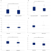
Comparison of lateral (A) and cranio-caudal shift (B), anterior communicating artery (C), Basilar apex (D), midline shift (E), and Evan’s index (F) after rigid and elastic fusion. Statistical significance is denoted as ** for p-value < 0.01, and *** for p-value < 0.001, based on the Wilcoxon signed-rank test for paired samples.

References

    1. Reinges M.H.T., Nguyen H.-H., Krings T., Hütter B.-O., Rohde V., Gilsbach J.M. Course of Brain Shift during Microsurgical Resection of Supratentorial Cerebral Lesions: Limits of Conventional Neuronavigation. Acta Neurochir. 2004;146:369–377. doi: 10.1007/s00701-003-0204-1. - DOI - PubMed
    1. Gerard I.J., Kersten-Oertel M., Petrecca K., Sirhan D., Hall J.A., Collins D.L. Brain Shift in Neuronavigation of Brain Tumors: A Review. Med. Image Anal. 2017;35:403–420. doi: 10.1016/j.media.2016.08.007. - DOI - PubMed
    1. Ganau M., Ligarotti G.K., Apostolopoulos V. Real-Time Intraoperative Ultrasound in Brain Surgery: Neuronavigation and Use of Contrast-Enhanced Image Fusion. Quant. Imaging Med. Surg. 2019;9:35058–35358. doi: 10.21037/qims.2019.03.06. - DOI - PMC - PubMed
    1. Ohue S., Kumon Y., Nagato S., Kohno S., Harada H., Nakagawa K., Kikuchi K., Miki H., Ohnishi T. Evaluation of Intraoperative Brain Shift Using an Ultrasound-Linked Navigation System for Brain Tumor Surgery. Neurol. Med. Chir. 2010;50:291–300. doi: 10.2176/nmc.50.291. - DOI - PubMed
    1. Becker S., Mang A., Toma A., Buzug T.M. In-Silico Oncology: An Approximate Model of Brain Tumor Mass Effect Based on Directly Manipulated Free Form Deformation. Int. J. CARS. 2010;5:607–622. doi: 10.1007/s11548-010-0531-7. - DOI - PubMed

LinkOut - more resources